Metastatic Disease | Mirels' Score | IMN vs Arthroplasty | Palliative Treatment
- Mirels' score 9 or more supports prophylactic fixation (33% fracture risk at 6 months, 10 or more = 50%); 8 is borderline. Its positive predictive value is only 23-70%, so a high score means assess further, not operate automatically
- Life expectancy over 3-6 months = surgery beneficial (use Katagiri or PATHFx score to estimate)
- Surgical principles: Fix normal bone to normal bone, bypass lesion by ≥2 cortical diameters, cement augmentation, construct must outlast patient
- IMN for diaphyseal lesions, arthroplasty for periarticular - cemented implants for immediate stability
- Postoperative radiation essential - 30 Gy in 10 fractions at 2-3 weeks postop for local control
- “Mirels' score 9 or more = prophylactic fixation (8 borderline) - Site (1-3), Pain (1-3), Lesion (1-3), Size (1-3)
- “Most common primaries: Breast, Lung, Prostate, Kidney, Thyroid (BLT with Kosher Pickle - BLT K P)
- “Fix normal bone to normal bone, bypass lesion by ≥2 cortical diameters, cement augmentation for lytic lesions
- “Nonunion common (radiotherapy impairs healing) but not problematic if construct stable - durable fixation more important than biological healing
Overview and Epidemiology
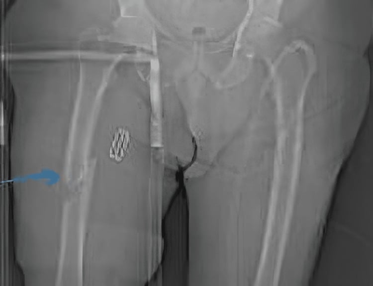
A pathological fracture of the femur runs through bone weakened by disease, most commonly metastatic cancer. Treatment is palliative: the aim is to restore function, relieve pain and improve the quality of the life that remains. Surgery is indicated when life expectancy is over 3-6 months and, for an impending fracture, when the Mirels' score is 9 or more.
Who. Between 5 and 10% of patients with bone metastases develop a pathological fracture, and the femur is the most common site. The peak age is 50-70 years, the age of the cancer population, and the sex distribution follows the primary tumour: breast in women, prostate in men. The fracture is usually unilateral, but bilateral disease is possible.
The primaries. Breast, lung, thyroid, kidney and prostate account for the large majority of skeletal metastases, and breast is the most common. The memory aid also carries whether each is lytic or blastic.
BLT K PMost Common Primaries
Hook:BLT with a Kosher Pickle: Breast, Lung, Thyroid, Kidney, Prostate - the most common primaries metastasising to bone.
Median survival is 6-12 months, so fixation must last 12-24 months at minimum.
Anatomy and Pathophysiology
The femur. Proximally it has the head, the neck and the greater and lesser trochanters; the shaft is the diaphyseal region; distally lie the metaphysis and the condyles. Its blood supply is endosteal and periosteal.
Where the lesions are. The proximal femur accounts for 50% of pathological fractures, the shaft for 30% and the distal femur for 20%. The proximal femur is the most vulnerable because it is weight-bearing and under high stress. Shaft fractures arise from diaphyseal lesions and distal fractures from metaphyseal ones.
Spread. Tumour cells reach bone through the bloodstream, and certain tumours prefer bone: breast, prostate, lung, kidney and thyroid. Whether a deposit is lytic or blastic depends on the primary; melanoma is also lytic, and breast deposits may be mixed.
Bone destruction. In a lytic lesion, osteoclast activation destroys bone and weakens the cortex. In a blastic lesion, osteoblast activation forms abnormal bone, which may be weaker. Mixed lesions combine the two.
Why it breaks. Normal bone needs high energy to fracture. Diseased bone fractures with low energy (a pathological fracture) or under normal activity (an impending fracture). Loss of circumferential cortex converts physiological loading into local bending failure.
Classification Systems
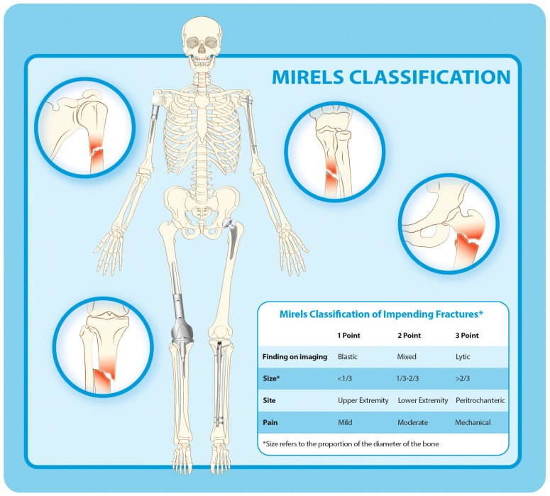
Mirels' score grades an impending fracture, a lesion that has not yet broken, and guides the decision to fix it prophylactically. Four features each score 1 to 3, for a total of 4 to 12.
- 1 point
- Upper limb
- 2 points
- Lower limb
- 3 points
- Peritrochanteric
- 1 point
- Mild
- 2 points
- Moderate
- 3 points
- Functional (affects activities)
- 1 point
- Blastic
- 2 points
- Mixed
- 3 points
- Lytic
- 1 point
- Less than 1/3
- 2 points
- 1/3 to 2/3
- 3 points
- Over 2/3
- 7 or less: can be irradiated
- 8: borderline and individualised; integrate functional pain, lesion geometry, prognosis and structural imaging
- 9 or more: prophylactic fixation generally recommended; approximately 33% fracture risk within 6 months
- 10 or more: approximately 50% fracture risk within 6 months
Reading the score. Its negative predictive value is 86-100%, but its positive predictive value is only 23-70%. A low score reliably reassures, while a high score flags a group in which most lesions still would not have fractured. Treat 9 or more as a trigger to assess further, not as an automatic operation. Mirels' original paper recommended fixation at 8 or more (see the Evidence Base); here 8 is borderline and 9 the threshold.
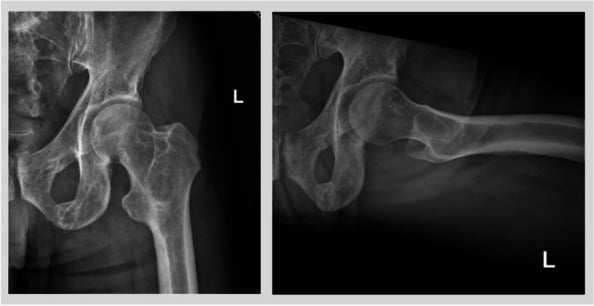
Location determines the operation.
- Lesion
- Femoral head or neck involvement
- Treatment
- Cemented arthroplasty
- Success
- 85-90%
- Lesion
- Diaphyseal
- Treatment
- IMN with cement
- Success
- 80-85%
- Lesion
- Metaphyseal
- Treatment
- IMN or arthroplasty
- Success
- 80-85%
Life expectancy determines whether to operate at all. A prognosis over 6 months supports surgical fixation, with good outcomes. Under 3 months, consider non-operative treatment: outcomes are poor and surgery may not benefit. Breast, prostate, kidney and thyroid primaries carry the better prognosis; lung, melanoma and GI primaries the poorer.
Clinical Assessment
History. Start with the cancer: the primary tumour, its stage and treatment so far; known bone and visceral metastases; systemic therapy, whether chemotherapy, radiation or immunotherapy; and performance status on the ECOG or Karnofsky scale. Then the limb. The mechanism is low energy in a pathological fracture, or normal activity in an impending one. Record where the pain is, how severe it is and what it stops the patient doing, and whether they walk and bear weight.
Associated disease. Look for visceral metastases in the liver, lung and brain, other skeletal lesions, ongoing chemotherapy, and previous radiation to the affected area.
Examination.
- Look for deformity if the fracture is complete, swelling, and the skin: previous radiation, surgical scars
- Feel for tenderness over the lesion or fracture, crepitus if the fracture is complete, and a soft-tissue mass if the tumour is large
- Move the hip if the lesion is proximal and the knee if it is distal, noting pain with movement
- Check distal pulses, sensation and motor function
Assess performance status and goals of care - surgery is palliative. If patient is bedbound with weeks to live, surgery may not benefit. If ambulatory with months to live, surgery can restore function and improve quality of life.
Investigations
Radiographs. AP and lateral views of the full length of the femur. They show whether the fracture is complete or impending, whether the lesion is lytic, blastic or mixed, how much cortex is destroyed for the Mirels' size component, and where the lesion sits. Full-length views are essential because the whole femur must be assessed for other lesions.
CT is essential for surgical planning and staging. At the lesion it measures cortical destruction for the Mirels' size component, shows bone quality, plans the fixation and finds other lesions. CT of the chest, abdomen and pelvis stages the metastatic burden, including visceral metastases.
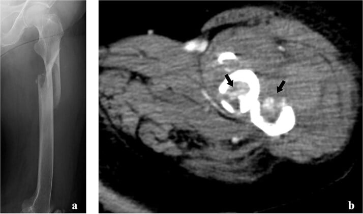
MRI shows the extent of the tumour: the soft-tissue mass, any neurovascular involvement, and the surgical approach these dictate. It also identifies other bone metastases and skip lesions, and is essential for planning when soft-tissue extension or neurovascular involvement is suspected.
Bone scintigraphy needs care in lytic disease, because a purely lytic deposit may not be intensely avid.
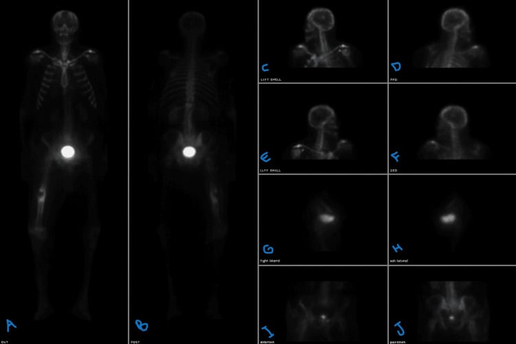
Biopsy. Without a known primary, a tissue diagnosis must be obtained before surgery. CT-guided biopsy is preferred, with open biopsy if that is not feasible, and either follows oncological biopsy principles. Treating an undiagnosed primary bone sarcoma as a metastasis is a catastrophic error. With a known primary and imaging consistent with metastasis, biopsy is usually not needed, though atypical features may justify one.
Differential Diagnosis of a Destructive Femoral Lesion
- Typical Age
- Over 40, known/likely primary
- Key Discriminators
- Multiple lesions, known cancer, lung/breast/prostate/kidney/thyroid
- Action Before Fixation
- Confirm primary; biopsy if solitary or no primary
- Typical Age
- 50-70
- Key Discriminators
- Multiple punched-out lytic lesions, paraprotein, raised ESR, anaemia
- Action Before Fixation
- SPEP/serum free light chains, marrow - medical, not always surgical
- Typical Age
- Bimodal / any
- Key Discriminators
- Solitary, aggressive periosteal reaction, soft-tissue mass, no known primary
- Action Before Fixation
- STOP - refer to tumour unit, biopsy along future resection track. NEVER nail
- Typical Age
- Any
- Key Discriminators
- Permeative lesion, relatively well patient, B-symptoms
- Action Before Fixation
- Biopsy - chemo/radiotherapy responsive
- Typical Age
- Elderly
- Key Discriminators
- No discrete lesion, generalised osteopenia, low-energy
- Action Before Fixation
- Treat as fragility fracture; investigate if atypical
- Typical Age
- Over 55
- Key Discriminators
- Cortical thickening, bony expansion, raised ALP, bowing
- Action Before Fixation
- Bisphosphonates; pathological fracture risk in lytic phase
- Typical Age
- On long-term antiresorptive
- Key Discriminators
- Lateral cortical beaking, transverse subtrochanteric/diaphyseal fracture
- Action Before Fixation
- Distinct entity - prophylactic/therapeutic nailing, stop drug
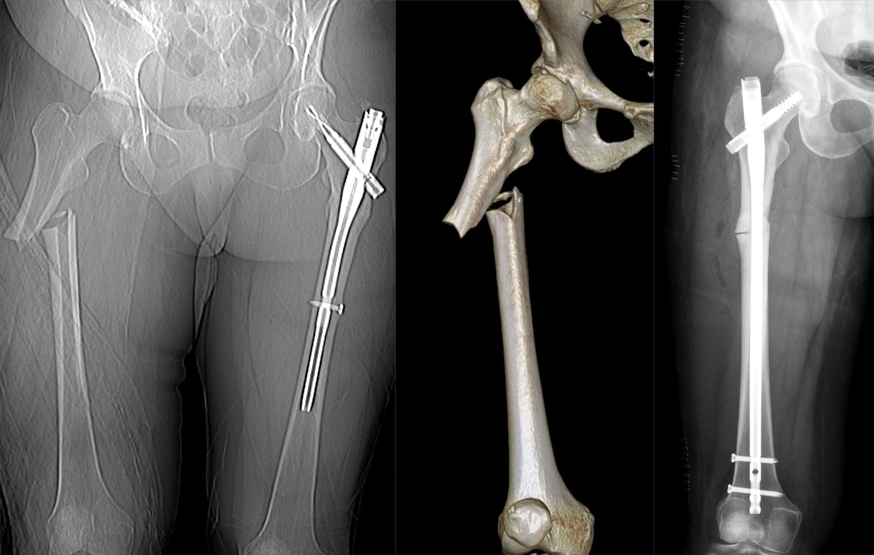
A solitary destructive femoral lesion in a patient without an established cancer diagnosis must be treated as a possible primary bone sarcoma until proven otherwise. Biopsy through an inappropriate track or intramedullary nailing of a sarcoma contaminates the canal, converts a limb-salvageable tumour into an amputation, and worsens survival. Refer to a specialist musculoskeletal oncology unit for staging and biopsy before any fixation.
Management Algorithm
The decision turns on two questions: how long the patient is likely to live and, for an unbroken lesion, how likely it is to fracture.
Pathological Fracture Management
Confirm metastatic disease (history, imaging, biopsy if needed). Stage with CT chest/abdomen/pelvis. Estimate life expectancy (Katagiri or PATHFx score). Calculate Mirels' score if impending fracture.
If life expectancy over 3-6 months and Mirels' score ≥9 (impending) or complete fracture, proceed with surgery. If under 3 months, consider non-operative (palliative care).
IMN with cement augmentation for a diaphyseal lesion, cemented arthroplasty for a periarticular one (see Surgical Technique).
Postoperative radiotherapy, coordinated with radiation oncology (see Postoperative Care).
Non-operative treatment is for life expectancy under 3 months, very poor performance status, patient or family preference, or medical contraindications to surgery. It consists of pain management with opioids and radiation, bed rest or minimal mobilisation, supportive care and a palliative care consultation. Function is poor, but it may be appropriate when life expectancy is very short.
Operative indications. The absolute indications both require life expectancy over 3-6 months; the relative ones stretch the Mirels' threshold for a patient who needs more from the limb or whose lesion is large.
Absolute
- Complete pathological fracture with life expectancy over 3-6 months
- Impending fracture with Mirels' score 9 or more (8 borderline) and life expectancy over 3-6 months
Relative
- Impending fracture with Mirels' score 7 and high functional demand
- Large lytic lesion (over 50% of the cortex) despite a lower Mirels' score, if symptomatic
Timing. Surgery is semi-urgent, within 1-2 weeks. It is not an emergency unless the fracture is complete and the pain severe.
Surgical Technique
Principles. Fix normal bone to normal bone, bypass the lesion by at least two cortical diameters, augment lytic lesions with cement, and build a construct that outlasts the patient and allows immediate weight-bearing.
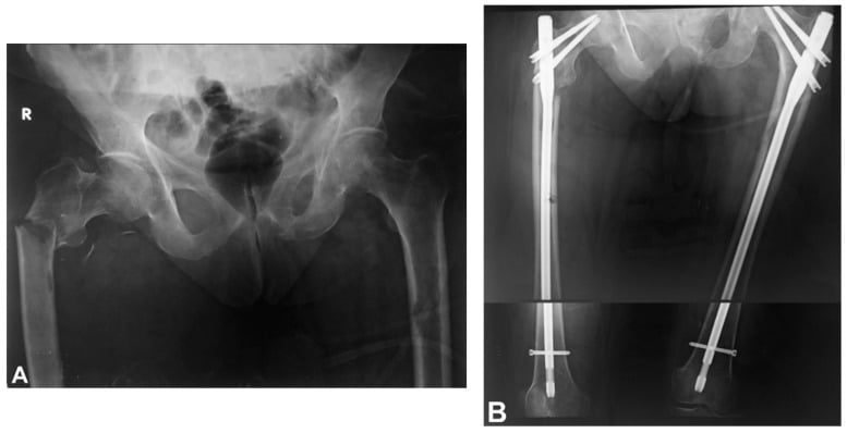
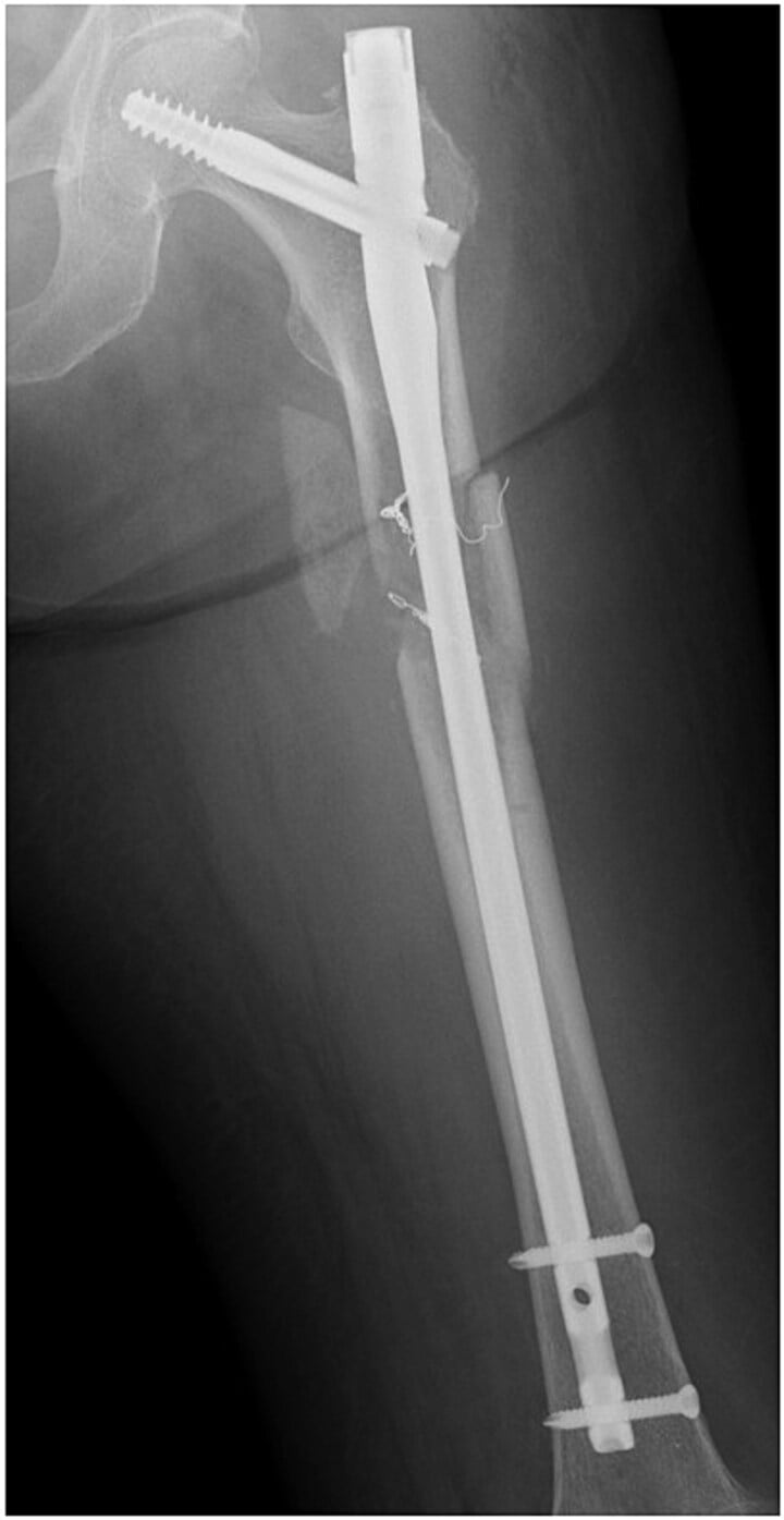
Intramedullary nailing is the gold standard for diaphyseal pathological fractures, and also serves proximal and distal shaft lesions. It is less invasive, gives good stability and allows immediate weight-bearing.
- A long nail, from the proximal to the distal metaphysis
- Bypass of at least 2 cortical diameters beyond the lesion
- Static interlocking at both ends
- PMMA injected through a cortical window at the lesion site, after the nail is inserted or before it (the cement-first technique)
Arthroplasty is preferred for periarticular lesions with articular involvement: femoral head or neck disease, or destruction of the articular surface. The implant is a hemiarthroplasty or THA, cemented for immediate stability, with a long stem bypassing the lesion by at least 2 cortical diameters, inserted through a standard posterolateral or anterolateral approach with standard cementing technique. It allows immediate weight-bearing, gives a durable construct and relieves pain well.
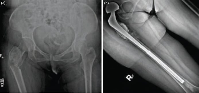
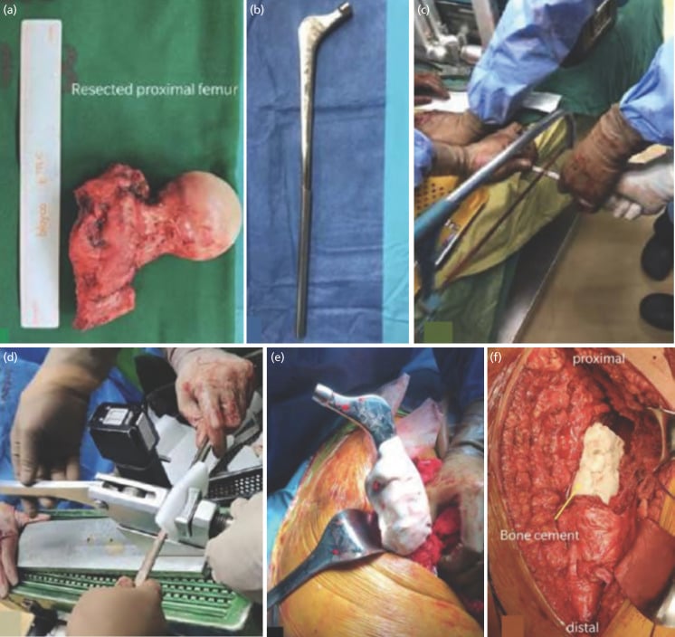
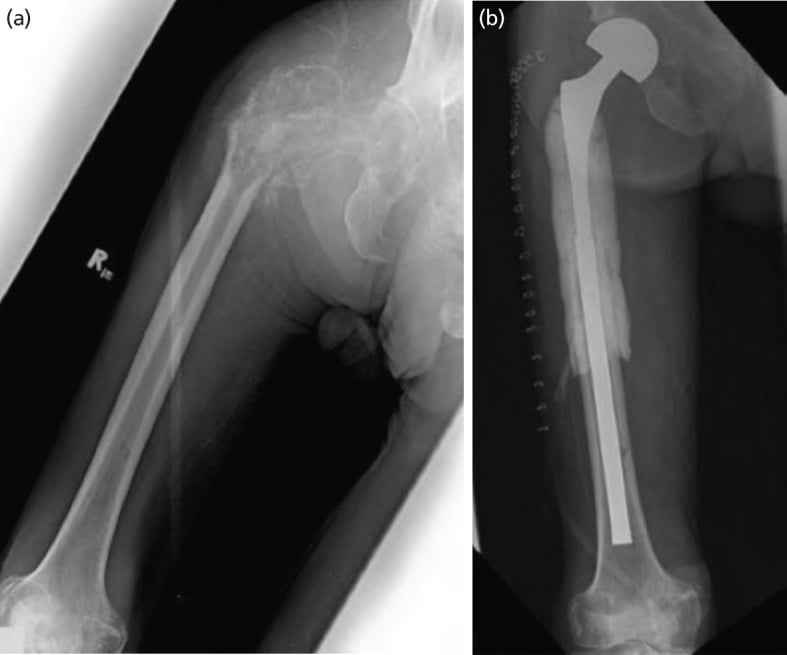
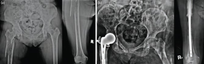
Cement augmentation is essential for lytic lesions. It is indicated for lytic lesions with poor bone quality and for large defects after curettage, and it fills the void the tumour leaves, adds structural support and improves fixation stability.
- Curette the tumour, if the approach is open
- Achieve haemostasis; tumour can be very vascular
- Mix PMMA to the doughy phase
- Inject it through a cortical window or into the curetted cavity, 40-80 g depending on the size of the cavity
Minimise tumour spillage during surgery - use gentle handling, copious irrigation. If open approach, consider curettage through limited window. Postoperative radiation provides local control, but minimising spillage reduces risk.
Distal and Periarticular Femoral Reconstruction
In the distal femur the choice between fixing and replacing is driven by how much of the distal metaphysis and condyles is destroyed, and whether the joint surface is involved.
Intact condyles. A distal-third or supracondylar lesion with a preserved articular block and enough distal bone to hold fixation is reconstructed with a retrograde intramedullary nail, spanning to the proximal metaphysis and bypassing the lesion by two cortical diameters, or a distal femoral locking plate, with cement augmentation of the lytic defect. The nail protects the whole diaphysis; a locked plate or condylar construct is preferred when the canal cannot be safely engaged distally. An isolated locked plate across a lytic distal femoral lesion in poor bone can fatigue and fail, so combine it with cement, or choose a load-bypassing nail or an endoprosthesis when destruction is extensive.
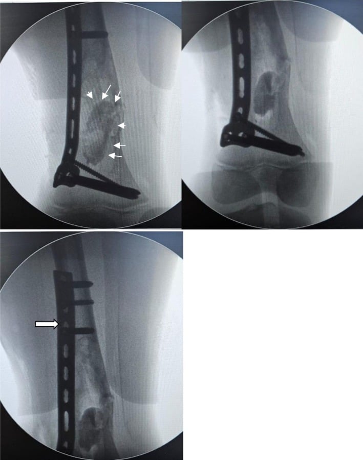
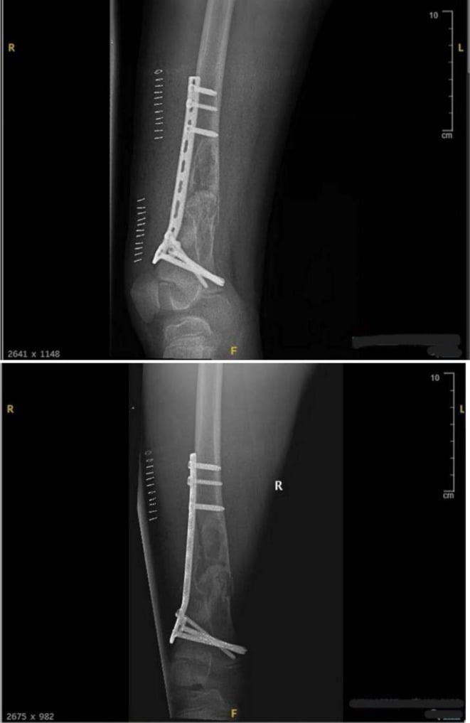
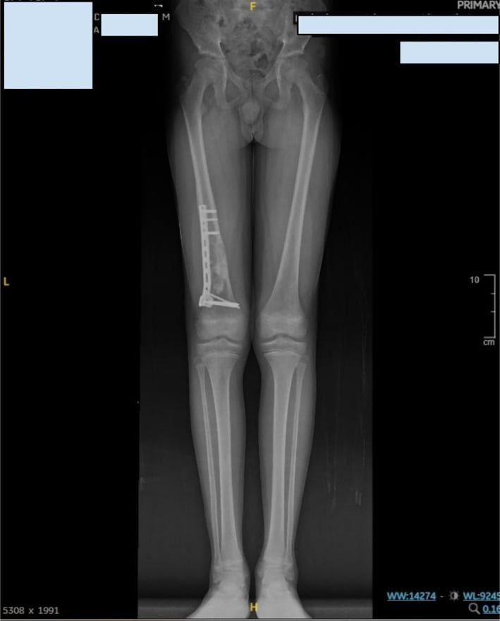
Destroyed condyles or an involved joint. Fixation then has nothing durable to purchase, and a distal femoral endoprosthetic replacement, a tumour megaprosthesis with a hinged knee, is the reliable option. It resects the disease, gives immediate stability and weight-bearing, and outlasts the patient. The trade-off is a much larger operation with its own infection and mechanical-failure profile.
The same first principles. Confirm the diagnosis, and never treat a solitary distal femoral lesion as a metastasis without excluding sarcoma. Estimate survival, protect the whole bone and allow immediate weight-bearing. Postoperative radiotherapy is given to a fixation, and not usually to a megaprosthesis.
The Hypervascular and Embolic Femur: Bleeding and Marrow-Embolism Control
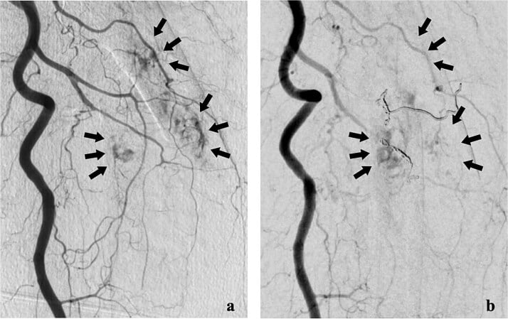
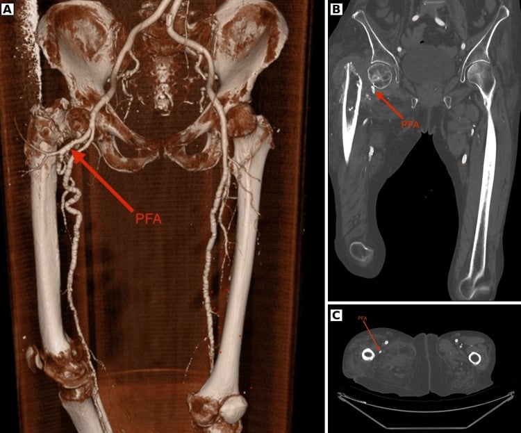
Embolise first. Renal cell and thyroid femoral metastases, and sometimes hepatocellular ones, can bleed catastrophically during curettage or nailing. Trans-arterial embolisation within about 24-48 hours before surgery substantially reduces intraoperative blood loss and transfusion, and is strongly considered for large, vascular lesions, especially when open debulking or arthroplasty is planned.
Renal cell disease. Embolisation does not remove the higher local-progression risk of renal cell carcinoma, an odds ratio of about 5 after nailing (Arpornsuksant and Levin). That risk is a separate reason to consider resection or an endoprosthesis over a nail.
Marrow and tumour embolism. Passing a long reamed intramedullary device down a tumour-laden femoral canal pressurises marrow, fat and tumour into the venous system. It can cause intraoperative hypoxia, hypotension and, rarely, fatal embolism; the non-fatal embolic-event rate of about 12% is the price of whole-bone protection. Mitigate it with:
- Anaesthetic vigilance and communication
- Venting or decompressing the canal
- Slow, gentle reaming, or the reamer-irrigator-aspirator to lower intramedullary pressure
- Avoiding over-pressurisation of cement
In a frail patient with a short prognosis, this embolic risk is a legitimate reason to accept a shorter construct rather than reflexively spanning the whole bone.
Complications
- Incidence
- Common (radiation-impaired healing)
- Risk Factors
- Tumour progression, radiotherapy, poor bone
- Prevention/Management
- Not problematic if construct stable
- Incidence
- Reoperation ~6%
- Risk Factors
- Inadequate bypass, no cement
- Prevention/Management
- Bypass ≥2 diameters, cement augmentation
- Incidence
- ~5% at 12 months (higher in RCC)
- Risk Factors
- Renal cell primary, older age, no adjuvant RT
- Prevention/Management
- Postoperative radiotherapy, embolise RCC
- Incidence
- Low
- Risk Factors
- Tumour progression, short implant
- Prevention/Management
- Long implant, bypass lesion widely
- Incidence
- ~12%
- Risk Factors
- Long reamed intramedullary devices
- Prevention/Management
- Vent canal, slow reaming, anaesthetic vigilance
- Incidence
- Increased vs elective
- Risk Factors
- Immunosuppression, prior radiation
- Prevention/Management
- Prophylactic antibiotics, careful technique
Nonunion. Tumour at the fracture site, radiotherapy-impaired osteogenesis and poor bone stock make bony union the exception rather than the rule, particularly after a completed rather than a prophylactically fixed fracture. It is not a problem if the construct is stable: the construct, not bony healing, carries the patient, and radiographic nonunion is acceptable if the patient is comfortable and functional. Prevention is fixing impending lesions before they fracture, with durable, cement-augmented, load-sharing constructs.
Fixation failure. Symptomatic failure requiring reoperation is uncommon, about 6% with a bone-protecting long-implant strategy (Alvi and Damron). Failure follows inadequate bypass, absent cement augmentation or disease progression, and is prevented by the surgical principles with a long implant protecting the whole bone. It is managed by revision fixation or conversion to (revision) arthroplasty.
Local tumour progression after intramedullary nailing is lower than historically taught (Arpornsuksant and Levin), and new disease distant from the index lesion is rare. Omitting adjuvant radiotherapy is a risk factor alongside a renal cell primary and older age. Prevention is postoperative radiotherapy, with the renal cell measures above, and symptomatic progression is treated with additional radiotherapy or surgical revision.
Postoperative Care
Mobilise at once. No immobilisation is needed: the construct is designed for immediate weight-bearing as tolerated, with early hip and knee range of motion from the start and physiotherapy for ambulation training and strengthening.
Radiotherapy. Postoperative radiation is essential, commonly 30 Gy in 10 fractions around 2-3 weeks after surgery, coordinated with radiation oncology. It provides local control and pain relief, and adjuvant radiotherapy improves and maintains function and reduces reoperation to the treated site (Townsend).
Rehabilitation Protocol
Weight-bearing as tolerated, ambulation training, hip and knee range-of-motion exercises, pain management.
Postoperative radiotherapy, continued ambulation, progressive activity.
Full activity as tolerated, continued monitoring, systemic therapy if indicated.
Goals. Pain relief, restored ambulation, a better quality of life, and function maintained until death. The goal is a rapid return to function, not a return to sport.
Outcomes and Prognosis
Results of surgery. Fixation succeeds in 80-90%, measured as pain relief and restored function, and 70-80% return to walking. Surgery gives immediate pain relief in 80-90%, which radiation maintains in the long term. Complications occur in 20-30%: nonunion, failure and recurrence.
What drives the result. Return to walking depends on performance status, life expectancy and rehabilitation; pain relief depends on tumour type and radiation response.
Survival depends on the primary tumour. Median survival is 12-24 months with breast or prostate primaries, the better prognosis, and 3-6 months with lung or melanoma, the poorer.
- Favourable
- Good
- Unfavourable
- Poor
- Favourable
- Over 6 months
- Unfavourable
- Under 3 months
- Favourable
- Single or few
- Unfavourable
- Multiple
- Favourable
- -
- Unfavourable
- Present
- Favourable
- Responsive
- Unfavourable
- Poor response
Prevention
Primary prevention is early detection and treatment of the primary cancer, systemic therapy for metastatic disease, and bisphosphonates or denosumab to prevent skeletal-related events.
Secondary prevention, once bone metastases are known, means monitoring them with imaging and scoring each impending lesion with Mirels' score to select those for prophylactic fixation.
Guidelines, Registries & Global Practice
Global Epidemiology
- Bone is the third most common site of metastasis after lung and liver; breast, prostate, lung, kidney and thyroid account for the large majority of skeletal metastases worldwide.
- The femur is the most frequently affected long bone, and the proximal femur is the commonest site of long-bone pathological/impending fracture requiring surgery.
- With rising cancer incidence and improving systemic therapy (longer survival with metastatic disease), the absolute number of patients living with skeletal metastases - and at risk of pathological fracture - is increasing globally.
Side-by-Side Guidance
- Emphasis
- Multidisciplinary, durable single-operation construct
- Practical Recommendation
- Stabilise the whole bone, allow immediate weight-bearing, adjuvant radiotherapy
- Emphasis
- Metastatic bone disease pathway, urgent specialist referral
- Practical Recommendation
- Discuss at MDT; suspected primary sarcoma must NOT be operated on locally - refer to a bone tumour centre
- Emphasis
- Biomechanically sound, load-sharing fixation
- Practical Recommendation
- Bypass lesion, cement augmentation for defects, protect the entire bone
- Emphasis
- Survival-adapted, function-focused palliation
- Practical Recommendation
- Use prognostic scores; reserve major reconstruction for adequate predicted survival
- Emphasis
- Skeletal-related event prevention
- Practical Recommendation
- Antiresorptives (bisphosphonate or denosumab) to reduce skeletal-related events
Registry and Practice Variation
- National sarcoma/bone-tumour referral pathways (e.g. UK) consistently warn against local fixation of unbiopsied solitary lesions.
- Arthroplasty registries (NJR, AOANJRR, SHAR) capture tumour/endoprosthetic and long-stem revision arthroplasty used for metastatic proximal femoral disease.
- Contemporary cohorts show low local progression (~5% at 12 months) and reoperation (~6%) after appropriate fixation.
- High-resource settings: routine MDT, prognostic scoring (Katagiri/PATHFx), endoprosthetic options, preoperative embolisation for renal cell, ready adjuvant radiotherapy.
- Limited-resource settings: later presentation with completed fractures, restricted access to endoprostheses, embolisation and radiotherapy - intramedullary nailing with cement is often the pragmatic durable option.
- Universal principles: confirm diagnosis, protect the whole bone, allow early weight-bearing, and coordinate oncological care where available.
Pathological fractures are a common viva topic. Know that treatment is palliative (not curative), Mirels' score ≥9 = prophylactic fixation, life expectancy over 3-6 months = surgery beneficial, surgical principles (fix normal to normal, bypass ≥2 diameters, cement augmentation), IMN for diaphyseal vs arthroplasty for periarticular, and postoperative radiation essential. Be prepared to discuss Mirels' score calculation and life expectancy estimation.
Related pages: Pathologic Fracture Management for the general principles, decision scores and spinal disease this femoral page applies; Pathological Fractures - Humerus for the upper-limb counterpart, where the load is lower, nailing dominates and plate fixation fails disproportionately; Metastatic Bone Disease for the underlying disease and its systemic treatment; Multiple Myeloma for the commonest primary in several of the series cited here, and the one diagnosis in which a solitary lucency may be marrow rather than metastasis; Osteosarcoma for the primary sarcoma that must be excluded before any nail enters the canal - biopsy precedes fixation; Femoral Shaft Fractures and Subtrochanteric Fractures for the non-pathological versions of these constructs and the reduction problems the proximal fragment creates; Fat Embolism Syndrome for the cardiopulmonary event that reaming a metastatic canal precipitates, quantified on this page at about 12%; Total Hip Arthroplasty Indications for the periarticular alternative to nailing; and Radiotherapy for Musculoskeletal Tumours for the adjuvant whose functional benefit is established and whose apparent survival benefit is selection.
Controversies and Areas of Uncertainty
Mirels' system is sensitive but not very specific, so strict application over-predicts fracture and can lead to over-treatment. Many units now individualise the decision using lesion location, functional pain, axial versus bending load, response to systemic therapy and survival estimate rather than the threshold of 8 alone. CT-based structural rigidity analysis is an emerging, more specific alternative still being validated.
For peritrochanteric/subtrochanteric metastatic lesions there is genuine debate between cephalomedullary nailing (faster, less invasive, but relies on diseased bone healing) and proximal femoral endoprosthetic replacement (resects the disease, durable, immediate stability, but bigger operation). Choice depends on bone destruction, primary tumour, survival estimate and surgeon experience - no high-level RCT exists.
Fixing an impending fracture is technically easier, allows shorter stay and better function than fixing after the bone breaks - but a proportion of prophylactically fixed bones would never have fractured, so some patients are over-treated. Balancing over-treatment against the morbidity of a completed pathological fracture remains judgement-based.
Protecting the whole bone with long intramedullary implants reduces reoperation, but reaming/insertion carries a real fat/tumour embolic risk (around 12% non-fatal physiologic events). Whether to always span the entire bone, or to accept a shorter construct in frail patients, is unresolved (Alvi & Damron).
Postoperative radiotherapy improves function and reduces reoperation, but the optimal regimen (single fraction vs 30 Gy in 10, exact timing, whether to irradiate the whole implant) is not standardised and varies between guidelines and registries.
Hypervascular renal and thyroid metastases bleed heavily and carry higher local progression risk after nailing. The roles of preoperative embolisation and of resection/endoprosthesis versus simple fixation are debated and depend on local resources.
MCQ Practice Points
Q: What Mirels' score indicates prophylactic fixation? A: Score ≥9 - Mirels' score ≥9 = prophylactic fixation recommended (33% fracture risk); 8 is a borderline score; ≤7 can be irradiated. Components: Site (1-3), Pain (1-3), Lesion (1-3), Size (1-3).
Q: What is the minimum life expectancy for surgery to be beneficial in pathological fractures? A: 3-6 months - Surgery beneficial if life expectancy over 3-6 months. Use Katagiri or PATHFx score to estimate. If under 3 months, consider non-operative (palliative care).
Q: What are the key surgical principles for pathological fractures? A: Fix normal bone to normal bone, bypass lesion by ≥2 cortical diameters, cement augmentation for lytic lesions, construct must outlast patient - IMN for diaphyseal, arthroplasty for periarticular. Allow immediate weight bearing.
Q: What are the most common primary tumors metastasizing to bone? A: Breast, Lung, Prostate, Kidney, Thyroid (BLT with Kosher Pickle - BLT K P) - Breast most common. Lytic: lung, kidney, thyroid. Blastic: prostate. Mixed: breast.
Q: What is the postoperative radiation protocol for pathological fractures? A: Commonly 30 Gy in 10 fractions, around 2-3 weeks postop - Adjuvant radiotherapy independently improves and maintains functional status and reduces reoperations to the treated site (Townsend). Coordinate with radiation oncology.
Q: What is the nonunion rate in pathological fractures? A: Common (radiotherapy impairs bony healing) - but not problematic if construct stable. Durable fixation matters more than biological healing; radiographic nonunion is acceptable if the patient is comfortable and functional.
Exam Viva Scenarios
Practise clinical reasoning and management decisions out loud
“A 60-year-old woman with known breast cancer and bone metastases presents with increasing right thigh pain. She has difficulty walking due to pain. X-ray shows a large lytic lesion in the femoral shaft involving 70% of the cortex. The lesion is in the lower limb, and she has moderate pain that affects her activities.”
“A 65-year-old man with known prostate cancer and multiple bone metastases presents after a fall. He has a complete pathological fracture through the proximal femur with femoral head and neck involvement. He is otherwise healthy and ambulatory. CT shows blastic metastases throughout skeleton but no visceral metastases.”
“A 45-year-old previously well man presents with 3 months of progressive thigh pain and a limp. There is no history of cancer. AP and lateral radiographs show a single destructive lesion in the proximal femoral diaphysis with cortical destruction, a hint of periosteal reaction and an associated soft-tissue shadow. The on-call team asks whether they should book him for an intramedullary nail.”
Key Concepts
- Pathological fracture = fracture through abnormal bone weakened by disease
- Treatment is palliative (not curative) - restore function, relieve pain, improve quality of life
- Most common primaries: Breast, Lung, Prostate, Kidney, Thyroid (BLT K P)
- Lytic: lung, kidney, thyroid, breast. Blastic: prostate. Mixed: breast
Mirels' Score
- Site: Upper limb 1, Lower limb 2, Peritrochanteric 3
- Pain: Mild 1, Moderate 2, Functional 3
- Lesion: Blastic 1, Mixed 2, Lytic 3
- Size: Under 1/3 cortex 1, 1/3-2/3 is 2, over 2/3 is 3
- Score 8 is borderline; ≥9 generally supports prophylactic fixation, with approximately 33% risk at 9 and 50% at 10
Treatment Algorithm
- Life expectancy over 3-6 months = surgery beneficial (use Katagiri or PATHFx score)
- Mirels' 9 or more (impending) or complete fracture = surgical fixation; 8 is borderline
- Diaphyseal lesion: IMN + cement augmentation
- Periarticular lesion: Arthroplasty (cemented) with long stem
Surgical Pearls
- Fix normal bone to normal bone
- Bypass lesion by ≥2 cortical diameters
- Cement augmentation for lytic lesions (40-80g PMMA)
- Long implant (proximal to distal metaphysis)
- Allow immediate weight bearing
Complications
- Nonunion common (not problematic if construct stable)
- Reoperation for fixation failure ~6% (prevent with adequate bypass, cement)
- Local progression ~5% at 12 months (higher in renal cell; reduced by radiotherapy)
- Embolic complications ~12% with long intramedullary implants
Evidence Base
Mirels' Score (Original Description)
- Components: Site (1-3), Pain (1-3), Lesion type (1-3), Size (1-3); range 4-12
- Mean score 7 in non-fracture group vs 10 in fracture group
- Score 8 or higher = prophylactic fixation recommended before irradiation
Revised Katagiri Prognostic Scoring System
- Six weighted factors; primary lesion, visceral/cerebral mets and abnormal labs carry the largest weight
- Score 7 or higher: 27% survival at 6 months, 6% at 1 year
- Score 3 or lower: 91% survival at 1 year, 78% at 2 years
PATHFx External Validation
- AUC 0.80-0.83 for 3-, 6- and 12-month survival predictions
- Validated across Western and Asian cohorts - generalisable decision-support tool
- Best used to guide surgical decisions at 3, 6 and 12 months rather than 1 month
Surgical Principles (Prophylactic Fixation)
- More than 50% cortical destruction (or a proximal femoral lesion over 2.5 cm) = high fracture risk
- Prophylactic fixation prevents secondary fracture and its high non-union risk
- Augment fixation with debulking plus methylmethacrylate to restore load-sharing and improve radiotherapy efficacy
Postoperative Radiotherapy After Stabilisation
- Postoperative radiotherapy independently predicted good functional status (53% vs 11.5%)
- Fewer reoperations to the treated site with adjuvant radiotherapy
- Median survival 12.4 vs 3.3 months (likely partly selection bias)
Length of Fixation and Reoperation Risk
- Reoperation for symptomatic failure 6.3% with a bone-protecting (long-implant) strategy
- New disease distant from the index lesion in only 1 of 96 bones
- Embolic-type complications from long implants (12.5%) must be weighed against reoperation risk
Local Progression After Intramedullary Nailing
- Local progression around the nail was low (4.9% at 12 months)
- Only 2% underwent reoperation for local progression
- Renal cell carcinoma carries higher progression risk - consider resection/alternative reconstruction