Metastatic Disease | Mirels Score | Prophylactic Fixation | Workup First
- Diagnosis before definitive fixation: stage and obtain a planned biopsy for an unknown destructive or solitary lesion unless an immediate life- or limb-saving intervention prevents this
- Mirels is a screen, not a humerus-specific mandate: combine it with functional pain, cortical geometry, lesion location and patient goals
- Renal and thyroid metastases may be hypervascular: angiography and embolisation are selective MDT decisions, not universal requirements
- Prognosis matters: choose a construct whose durability, morbidity and rehabilitation burden fit expected survival and function
- Construct selection is anatomical: nail, plate-cement or endoprosthesis according to segment, remaining bone, joint involvement and local-control plan
- “Always ask about constitutional symptoms (weight loss, night sweats)
- “Solitary lesion greater than 40y is Mets/Myeloma until proven otherwise
- “Solitary lesion less than 20y is Primary Bone Tumour until proven otherwise
- “Do not nail a primary bone sarcoma (contaminates whole compartment)
Overview
Pathological fractures of the humerus occur when bone weakened by disease (cystic, metabolic or neoplastic) fails under physiological loads. In adults over 40, metastatic disease and myeloma are the overwhelming causes. In children the proximal humerus is the classic site of a unicameral bone cyst, the commonest cause of a pathological fracture through benign bone.
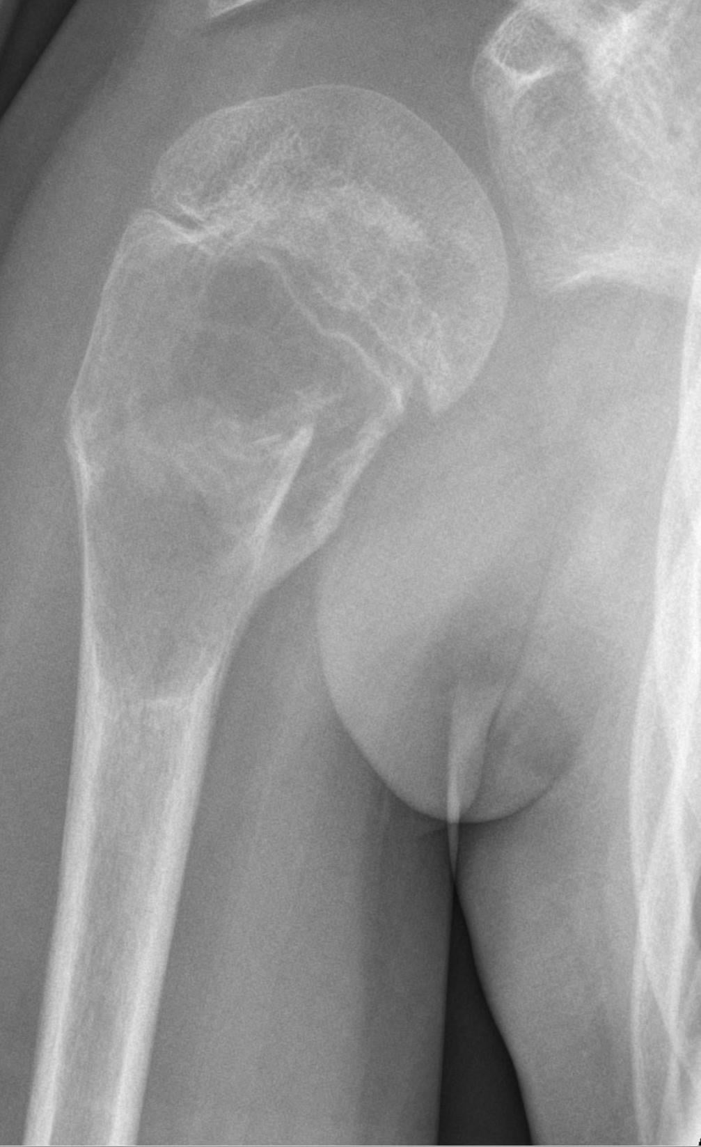
Epidemiology. Bone is the third most common site of metastatic disease, and the humerus is involved in 20% of bone metastases, second among long bones only to the femur. The number is increasing as cancer survival improves. The primaries to remember are breast, lung, thyroid, kidney and prostate: BLT-KP, a BLT with a Kosher Pickle for lunch.
Prognosis. Median survival depends on the primary, and the fixation must be durable enough for the survival expected.
- Prostate and breast: years (24-36 months)
- Renal and thyroid: intermediate (12 months)
- Lung: short (less than 6 months)
Anatomy and Pathophysiology
Spread. Metastasis to bone is haematogenous. Tumour emboli deposit in the vascular red marrow, and Batson's plexus, a low-pressure venous system, facilitates spread. Cytokines (RANKL and PTHrP) stimulate osteoclasts.
Lytic or blastic. Lytic lesions destroy bone and lead to fracture; they come from lung, renal and thyroid primaries and from myeloma, with breast giving mixed lesions. Blastic lesions, from prostate and again breast, form bone and leave it brittle. Lytic lesions are the more prone to fracture and the harder to fix.
Biomechanics. The humerus is a non-weight-bearing bone, loaded mainly in torsion and bending rather than axial compression, which allows non-operative management more often than in the femur. Functional demands such as lifting and toileting still require stability. A cortical defect greater than 50% reduces torsional strength by 60%.
Classification
Mirels score. Mirels predicts the risk of fracture in an impending pathological fracture from four variables, SPLS: site, pain, lesion and size. Each scores 1 to 3 and the sum runs from a minimum of 4 to a maximum of 12. The score applies to metastatic disease in long bones; it does not apply to primary bone tumours or spinal metastases.
- Score 1
- Upper Limb
- Score 2
- Lower Limb
- Score 3
- Peritrochanteric
- Score 1
- Mild
- Score 2
- Moderate
- Score 3
- Functional
- Score 1
- Blastic
- Score 2
- Mixed
- Score 3
- Lytic
- Score 1
- Less than 1/3
- Score 2
- 1/3 - 2/3
- Score 3
- Greater than 2/3
- Fracture Risk
- Conventionally lower risk
- Recommendation
- Clinical review; observation or radiotherapy may be appropriate
- Fracture Risk
- Indeterminate
- Recommendation
- Use pain, geometry, demand and prognosis
- Fracture Risk
- Conventionally high risk but low specificity
- Recommendation
- Consider prophylactic fixation; do not operate from score alone
The thresholds. Mirels's own paper recommended prophylactic fixation at 8 or more; the conventional teaching threshold is 9 or more for fixation and 7 or less for radiotherapy. A score of 8 remains a clinical-judgement grey zone in which functional demand and prognosis drive the decision.
Why the score over-treats. Mirels has high sensitivity but low specificity, so a score of 9 or more over-predicts fracture and may lead to over-treatment. A systematic review of fracture-risk methods found a negative predictive value of 86-100% but a positive predictive value of only 23-70%, and concluded that the score "leads to an overtreatment" (Benca, Bone Rep 2016, PMID 28326347). A low score therefore reliably reassures; a high score identifies a group in which most lesions still would not have fractured.
Harrington's criteria. The classic indications for prophylactic fixation of a lytic metastasis:
- Destruction of 50% or more of the cortex
- A lesion 2.5cm or larger
- Pain despite radiotherapy
Read them as femoral criteria. Harrington described them in 1982 for lytic lesions of the femur, in a paper on lower-extremity metastases. His third indication was pain specifically with weight-bearing, which cannot apply literally to the humerus, and his fourth, avulsion of the lesser trochanter, is a hip finding with no upper-limb equivalent. Because the humerus fails in torsion and bending, neither the 50% nor the 2.5cm figure has been validated here, and Mirels scores the upper limb lower than the lower limb precisely because the risk differs.
Using them in the humerus. Neither Harrington's criteria nor Mirels was derived in this bone. Use them to decide who needs a closer look, and let functional pain, lesion geometry on CT and the demands the patient actually places on the arm drive the decision.
History and Examination
History. Ask about a known history of cancer and about constitutional symptoms: weight loss, night sweats and fatigue. Separate mechanical pain from the fracture from biological pain at night, which typically indicates active tumour, and establish the functional demand on the arm.
Examination. The examination focuses on finding a primary source.
- A soft-tissue mass, which raises concern for sarcoma
- Previous mastectomy scars
- Thyroid enlargement
- Axillary and supraclavicular lymph nodes
The radial nerve. It is commonly involved in humeral shaft fractures. A pathological shaft lesion at the spiral groove, or the fracture through it, can cause a radial nerve palsy from tumour infiltration, compression or the fracture itself.
Document active wrist, finger and thumb extension and first-dorsal-web-space sensation before any intervention on a pathological humerus. It is both planning and medicolegal protection: never be left explaining a post-operative palsy you never checked for pre-operatively.
Investigations
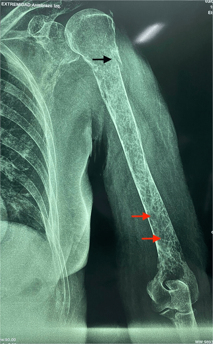
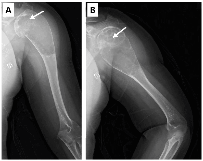
Radiographs. Look for a "moth-eaten" or permeative appearance and cortical destruction. The pathological fracture is transverse, after minimal trauma.
Staging. CT of the chest, abdomen and pelvis stages the disease, and CT defines the bone stock remaining. A bone scan or PET identifies other skeletal metastases, and whether the lesion is solitary or multiple is prognostic. Stage sufficiently to establish the diagnosis, the extent of disease and a safe treatment plan, tailoring imaging to urgency, known cancer history and the likely primary. A recent negative staging CT does not exclude disease beyond its field: review the scan coverage and image the entire symptomatic bone.
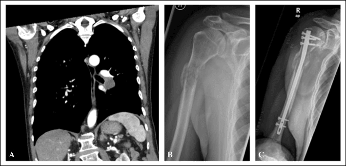
Laboratory. Rule out myeloma in every patient over 40: serum protein electrophoresis (SPEP), urine Bence-Jones protein and free light chains, with calcium, phosphate and ALP. A normal or equivocal serum electrophoresis does not exclude oligosecretory myeloma. The features of myeloma are CRAB:
- Calcium elevation: hypercalcaemia
- Renal insufficiency: raised creatinine
- Anaemia: normocytic
- Bone lesions: lytic, the "raindrop skull"
Tumour markers point to the primary: PSA for prostate, CEA and CA-125 for gastrointestinal and ovarian cancer, and thyroid function tests for thyroid.
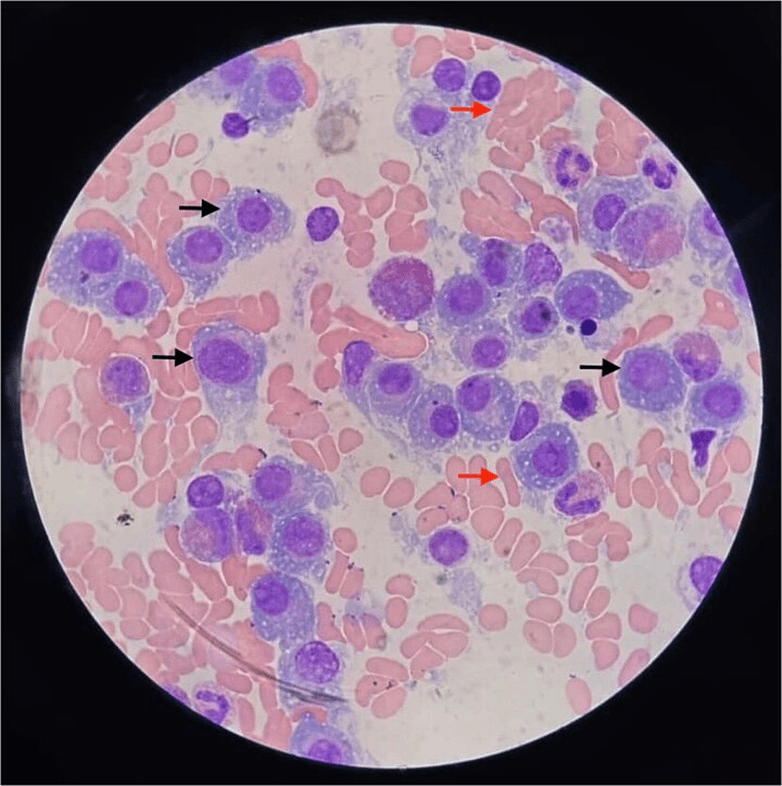
Biopsy. Biopsy provides the definitive tissue diagnosis. It is indicated when:
- The primary is unknown
- There is a history of cancer but more than 5 years disease free, to confirm the lesion is not a new primary
- The imaging is inconsistent
Core needle biopsy is the gold standard. Open biopsy with frozen section at the time of surgery is an option when fixation is impending.
The biopsy trap. An unplanned nail can contaminate the humeral canal and the soft-tissue compartments if the lesion is a primary sarcoma. Stage first, and place the biopsy tract so it can be excised with a definitive tumour operation.
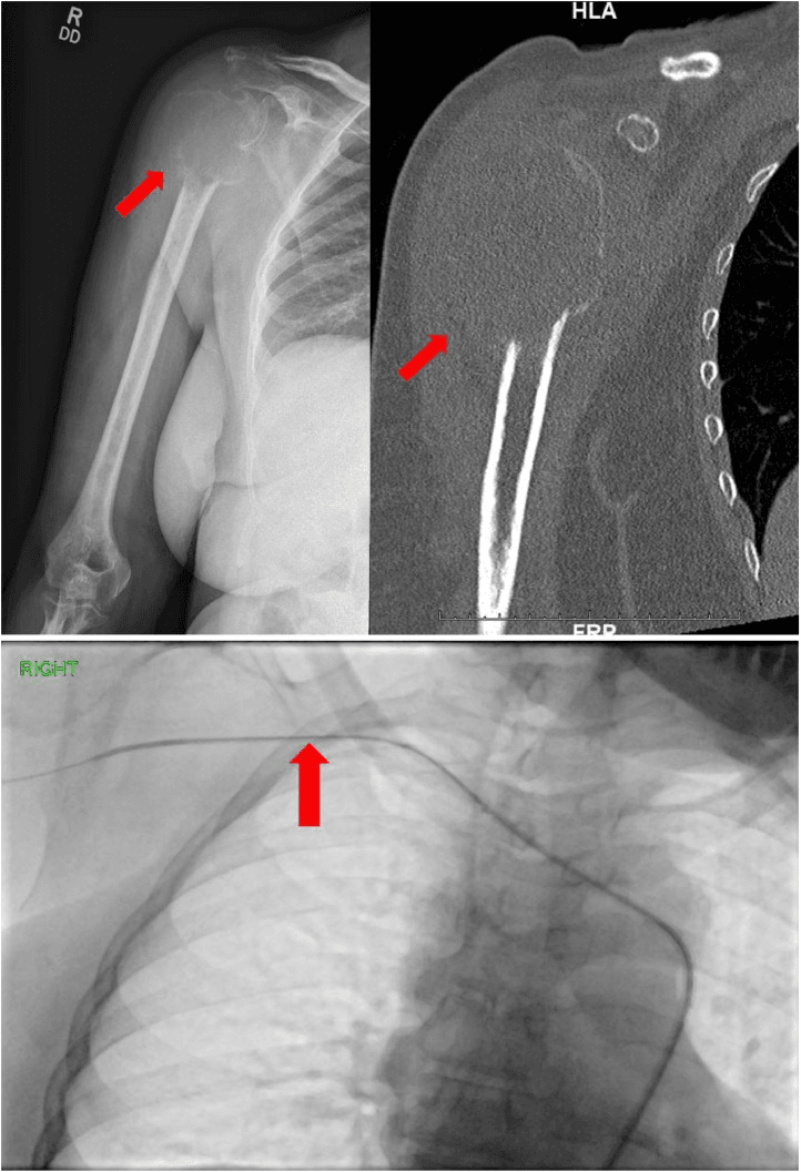
Vascular metastases. Renal and thyroid metastases may be hypervascular. Consider pre-operative angiography and embolisation when imaging or tumour biology suggests a substantial arterial supply and the planned procedure risks major bleeding. The evidence does not support a blanket mandate, and embolisation is a selective MDT decision.
Differential Diagnosis
A destructive humeral lesion in an adult is metastasis or myeloma until proven otherwise, but the differential changes dramatically with age and imaging. Distinguishing these before any fixation is the single most important decision.
- Typical Age
- Over 40
- Imaging Clue
- Multiple lytic/mixed lesions, moth-eaten
- Key Discriminator
- Known primary or constitutional symptoms
- Typical Age
- Over 50
- Imaging Clue
- Punched-out lytic lesions, cold on bone scan
- Key Discriminator
- Positive SPEP/free light chains, CRAB features
- Typical Age
- Under 30 (or any age)
- Imaging Clue
- Aggressive periosteal reaction, soft-tissue mass
- Key Discriminator
- Solitary aggressive lesion, no primary on staging
- Typical Age
- Any
- Imaging Clue
- Permeative lesion, large soft-tissue component
- Key Discriminator
- Tissue diagnosis; often radiosensitive
- Typical Age
- Under 30
- Imaging Clue
- Well-defined, non-aggressive margin
- Key Discriminator
- No cortical breach, no soft-tissue mass
The decisive distinction is metastasis/myeloma (fix it) versus primary bone sarcoma (do NOT touch it without an oncology MDT). When the primary is unknown, treat every solitary lesion as a potential sarcoma until staging and biopsy say otherwise.
Management Algorithm
Unknown diagnosis. A destructive or solitary lesion without a known diagnosis is staged and biopsied, by plan, before definitive fixation whenever clinically feasible.
Impending fracture. In a known diagnosis the Mirels bands open the discussion. A score of 7 or less calls for clinical review, and radiotherapy or observation may be appropriate; at 9 or more, consider prophylactic fixation after a humerus-specific assessment.
Complete fracture. In a known diagnosis, stabilise or reconstruct when the benefit outweighs the morbidity, and coordinate the adjuvant treatment.
Non-operative treatment. Consider it when:
- The lesion is mechanically stable and symptoms are controlled
- Surgery is unlikely to improve function or comfort relative to its burden
- Radiotherapy or systemic treatment is expected to control pain and local disease
- Bracing is tolerable and does not compromise skin or function
The options are analgesia, activity adaptation, selective functional bracing, radiotherapy, systemic anticancer treatment and bone-modifying therapy when oncologically indicated.
Operative treatment. Surgery is palliative, for quality of life, not cure. Consider it when:
- A completed fracture or impending structural failure causes pain or loss of useful function
- Immediate stability would restore transfers, self-care or use of walking aids
- The patient can tolerate the intervention and the construct matches expected survival
- Local progression requires resection or reconstruction rather than fixation alone
What the fixation must survive. The goals are immediate stability, immediate loading of the arm, and easier nursing care and transfers. Patients push through the humerus to rise from a chair, to transfer and to use a frame or crutches, and that is the load the fixation has to survive rather than gait.
Solitary renal or thyroid metastasis. For a solitary metastasis from a favourable primary, the balance between palliative fixation and wide resection with endoprosthetic reconstruction is unsettled, and it should be an oncology MDT decision based on prognosis.
Surgical Technique
Protect what is at risk. Span the diseased bone sufficiently for the observed distribution of disease and the planned local control. Whole-bone protection is not an automatic requirement for every lesion.
Intramedullary nail. The common diaphyseal option, used prophylactically or for an acute fracture. It is a load-sharing device, minimally invasive, preserves the soft tissues and gives immediate stability. Lock it statically, proximally and distally, or augment it with cement screws.
Reaming. Reaming improves nail fit, but it can spread tumour and raises intramedullary pressure, with a theoretical risk of tumour and fat embolism. Whether to ream remains debated; ream carefully, and many advocate venting or unreamed nails in patients with poor pulmonary reserve.
Plate and cement. Indicated for:
- Meta-diaphyseal fractures
- The distal humerus
- The need for open tumour debulking
Curette the lesion to create a cavity, fill it with PMMA cement (the "rebar" concept) and apply a locking plate over the cement. The cement restores compressive strength; the plate provides neutralisation.
Nail or plate. No randomised data favour intramedullary nailing over locked plating with cement augmentation for the humeral shaft. The nail protects the whole bone; the plate allows debulking and direct control of the lesion. The choice depends on lesion location and the surgeon.
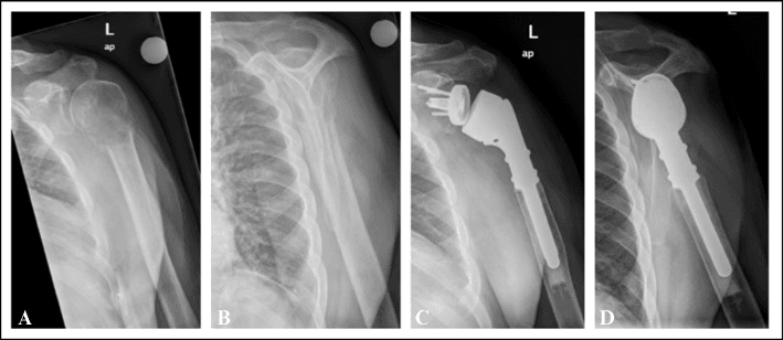
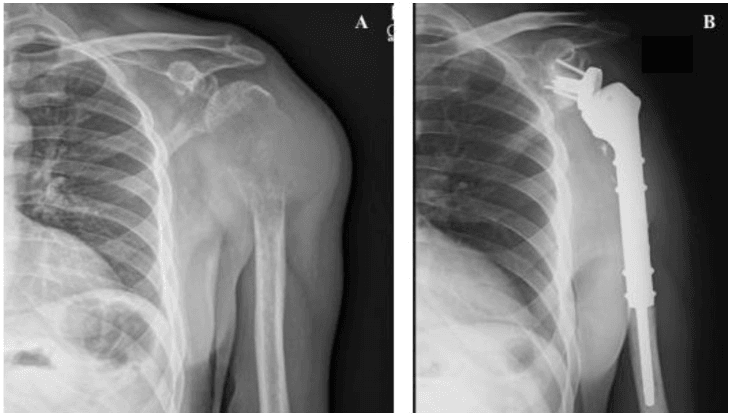
Resection arthroplasty. Indicated for:
- Extensive proximal humeral destruction
- Articular involvement
- Failed fixation
The implant is a proximal humeral replacement (endoprosthesis), or a reverse total shoulder if the cuff is compromised. Resection arthroplasty allows immediate function but carries a high dislocation risk.
The Radial Nerve: The Humerus-Specific Pitfall
The radial nerve is what makes fixing a pathological humerus different from any other long bone.
Iatrogenic risk. The nerve runs in the spiral groove on the posterolateral shaft. It is at risk during open plating, particularly posterior approaches and distal-third exposure, during percutaneous or distal interlocking screw placement, and from retraction, all made harder when tumour distorts the normal anatomy.
Plan the construct around the nerve. An antegrade intramedullary nail avoids open dissection over the nerve along the shaft, but distal interlocking screws still endanger it: place them with the nerve in mind, open if necessary. Open plating mandates identifying and protecting the radial nerve, and a posterior approach requires formally exposing it.
Complications
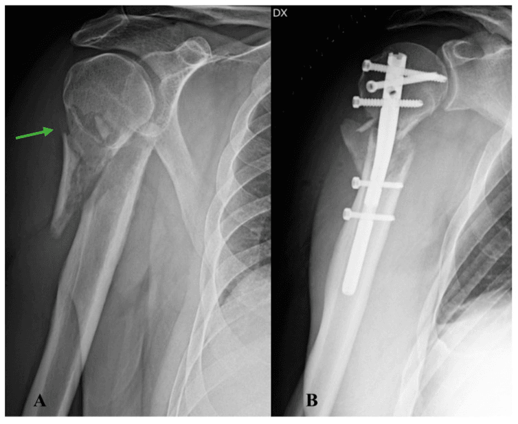
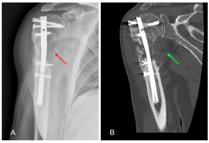
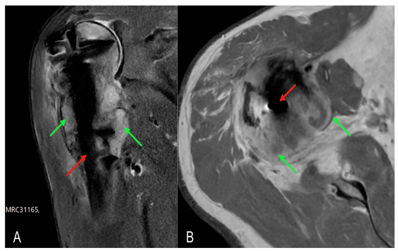
Local tumour progression. Progression can occur with or without postoperative radiotherapy, and it can compromise fixation despite a technically satisfactory initial reduction. Surveillance and the systemic and local-control plans must match the tumour biology and the chosen construct. When progression appears, re-stage, biopsy if the diagnosis remains uncertain, and reconsider whether fixation alone can provide durable control.
Haemorrhage. Intra-operative haemorrhage is the risk in renal, thyroid and other hypervascular lesions. Assess vascularity before operating on them, and use embolisation selectively when a target arterial supply and a meaningful bleeding risk are demonstrated.
Nonunion. Tumour, systemic therapy and radiotherapy can impair healing, so the construct should not depend on union when union is unlikely.
Embolic and cardiopulmonary risk. Reaming and cementation may increase intramedullary pressure. Plan anaesthesia, venting and implant technique according to the patient's cardiopulmonary reserve.
Postoperative Care
A sling for comfort. The arm is loaded at once and there are usually no restrictions, so the construct must be strictly stable.
Review the histology, the systemic treatment and the indication, target and timing of radiotherapy. Start bone-modifying therapy only when it is indicated for the underlying malignancy and the renal and calcium status allow.
Surveillance for hardware failure, and systemic therapy for the underlying disease.
Post-operative Radiotherapy: Indication and Field
Coordinate, do not automate. Postoperative radiotherapy is not an automatic whole-implant rule. After fixation, request radiation-oncology review and state the residual disease, the implant extent and the operative contamination. Whether and how it is delivered then depends on tumour radiosensitivity, residual disease, surgical margins, prior irradiation, systemic therapy, wound status and expected survival.
The field. When radiotherapy is selected after stabilisation of metastatic long-bone disease, field design traditionally covered the instrumented bone because reaming may seed tumour cells along the canal. Contemporary retrospective data remain mixed: some series support broader coverage for local control, while others show no implant-failure benefit and substantial selection bias. The field must therefore be planned by the treating radiation oncologist, not prescribed by a fixed orthopaedic template.
Timing. Timing follows wound recovery, the urgency of systemic treatment and the radiotherapy technique rather than a universal two-to-three-week date. Build fixation that is durable without assuming either fracture union or radiotherapy failure.
Outcomes
Outcomes are driven by tumour biology, systemic disease, pre-fracture function and the match between lesion and construct. Report pain relief, useful hand-to-mouth function, ability to transfer or use walking aids, neurological status, local progression, revision and implant survival. Avoid quoting one universal success or failure percentage across nails, plates and endoprostheses.
Guidelines, Registries & Global Practice
- A destructive solitary lesion without an established concordant diagnosis is discussed with an orthopaedic oncology service before definitive fixation.
- Mirels and Harrington criteria are historical screens, not humerus-specific directives.
- Fixation or reconstruction is selected by lesion segment, remaining bone, articular involvement, tumour biology, systemic burden, function and prognosis.
- Pre-operative embolisation for hypervascular metastases and postoperative radiotherapy are selective multidisciplinary decisions.
- The chosen construct should remain mechanically useful for the patient's expected survival without imposing disproportionate morbidity.
metastatic humeral fracture series are mainly retrospective and heterogeneous. Sarcoma registries and arthroplasty registries do not supply a universal failure rate for palliative nails, plate-cement constructs and proximal humeral endoprostheses. Report outcomes by implant, tumour, indication and competing mortality.
limited access to embolisation, modular endoprostheses or advanced radiotherapy changes available techniques, but it does not change the requirement for diagnosis, deliberate biopsy planning and multidisciplinary prioritisation.
Related pages: Pathological Fractures - Femur for the lower-limb counterpart; Pathologic Fracture Management for general principles and prognostic scores; Metastatic Bone Disease for systemic treatment; Humeral Shaft Fractures and Radial Nerve Anatomy for construct and nerve planning; Multiple Myeloma and Osteosarcoma for critical differentials; and Radiotherapy for Musculoskeletal Tumours for indication, timing and field planning.
MCQ Practice
Self-Assessment Questions
Q: Which factor is weighted most heavily (highest score) in the Mirels scoring system for "Site"?
- A) Humerus
- B) Radius
- C) Femoral Shaft
- D) Peritrochanteric region
- E) Tibia
A: D - The Peritrochanteric region is assigned a score of 3 due to the high biomechanical loads and consequences of failure. Upper limb is 1, Lower limb (shaft) is 2.
Q: What is the most common source of bone metastasis in women?
- A) Lung
- B) Thyroid
- C) Breast
- D) Kidney
- E) Cervix
A: C - Breast cancer is the most common source of bone metastases in women (about 70%). Prostate is most common in men.
Q: Which primary tumor gives rise to highly vascular bone metastases requiring pre-operative embolization?
- A) Breast
- B) Prostate
- C) Renal Cell Carcinoma
- D) Lung
- E) Melanoma
A: C - Renal Cell and Thyroid carcinomas are classically highly vascular. Pre-operative embolization is recommended to control bleeding.
Q: A Mirels score of 10 indicates:
- A) Low risk of fracture (less than 4%)
- B) Moderate risk, Observation indicated
- C) High risk, Prophylactic fixation indicated
- D) Imminent death
- E) Need for amputation
A: C - A score of 9 or greater indicates a high risk of fracture (greater than 33%) and is the threshold for recommending prophylactic fixation.
Q: Internal fixation of a pathological fracture is potentially contraindicated if:
- A) The patient has multiple metastases
- B) The primary tumor is unknown (solitary lesion)
- C) The Mirels score is 12
- D) The fracture is displaced
- E) The patient is on bisphosphonate therapy
A: B - If the primary is unknown and the lesion is solitary, internal fixation (nailing) is contraindicated until a primary bone sarcoma is ruled out via biopsy/staging, to avoid compartment contamination.
Viva Scenarios
Practise clinical reasoning and management decisions out loud
“You are called to ED for a 60-year-old male with a solitary lytic lesion of the humerus and a fracture. He has no history of cancer. What is your plan?”
“Describe the Mirels Score and how you use it.”
“How do you manage a pathological humerus fracture from Renal Cell Carcinoma?”
Key Facts
- Common primaries: breast, lung, renal, thyroid and prostate
- Mirels 9 or more = concern, not an automatic operation
- Unknown solitary destructive lesion = stage and planned biopsy
- Renal/thyroid vascularity = assess; embolise selectively
- Fixation: durable and anatomy-matched
- Coordinate systemic therapy and radiotherapy through the MDT
Mirels Score (SPLS)
- Site (Upper/Lower/Troch)
- Pain (Mild/Mod/Func)
- Lesion (Blastic/Mixed/Lytic)
- Size (less than 1/3, 1/3-2/3, greater than 2/3)
Surgical Steps
- Positioning (Beach chair/Supine)
- Approach (Deltopectoral/Split)
- Biopsy (If needed, frozen section)
- Reaming (Gentle/Unreamed if pulmonary risk)
- Implant Placement (protect the diseased segment and planned local-control field)
- Cement Augmentation (If proximal/distal voids)
- Closure (Layered)
Common Pitfalls
- Nailing a primary sarcoma
- Missing hypercalcemia
- Underestimating bleeding in Renal mets
- Inadequate fixation length for the demonstrated disease and local-control plan
- Ignoring radial nerve in percutaneous nailing
Examiner Favorites
- Calculate Mirels for this X-ray...
- Workup of solitary lesion
- Role of radiotherapy vs surgery
- Mechanism of bisphosphonates
- What to do if fixation fails?
Evidence Base
Key Studies
Mirels - Scoring System for Impending Fractures
- Retrospective analysis of 78 irradiated metastatic long-bone lesions (27 fractured, 51 did not)
- Four weighted variables: site, pain, lesion type, size (range 4-12)
- Mean score 7 in non-fracture group vs 10 in fracture group; risk rises progressively above 7
- Lesions scoring 7 or lower safely irradiated; 8 or higher recommended for prophylactic fixation
Capanna & Campanacci - Appendicular Metastasis Treatment
- Comprehensive framework for surgical treatment of appendicular skeletal metastases
- Stratifies fixation by life expectancy and solitary vs multiple disease
- Promotes durable load-bearing constructs (cement augmentation, endoprosthesis) over biological union
- Solitary metastasis from favourable primaries (renal, thyroid) considered for wide resection
Katagiri et al. - Prognostic Scoring
- 350 patients with skeletal metastases; Cox model identified 5 survival factors
- Factors: primary site, poor performance status (ECOG 3-4), visceral/cerebral mets, prior chemotherapy, multiple bone mets
- Score 6 or more: 31% survival at 6 months, 11% at 1 year; score 2 or less: 98% at 6 months, 89% at 1 year
- Guides choice between simple palliative fixation and durable reconstruction
Patchell et al. - Surgery + Radiation vs Radiation
- Randomised trial (n=101) in metastatic spinal cord compression; principle applies to mechanical instability generally
- Surgery plus RT vs RT alone: 84% vs 57% able to walk after treatment (OR 6.2, p=0.001)
- Surgery group retained ambulation far longer (median 122 vs 13 days) and needed fewer steroids/opioids
- Trial stopped early at interim analysis for surgical superiority
Harrington - Criteria for Prophylactic Fixation
- Established the classic indications for prophylactic fixation of lytic metastatic lesions
- Lesion 2.5 cm or larger in size
- Destruction of 50% or more of the cortex
- Persistent local pain with loading despite adequate radiotherapy
Arpornsuksant et al. - Local Progression After IM Nailing
- 122 patients with long-bone metastases (humerus 27%) treated by intramedullary nail
- Cumulative local tumour progression only 4.9% at 12 months; reoperation in 2%
- Renal cell carcinoma (OR 5.1) and older age were the only factors linked to progression
- No survival difference between those with and without local progression