Superior Peroneal Retinaculum Tear | Fibular Groove Instability | Recurrent Dislocation
- SPR is PRIMARY restraint preventing peroneal subluxation (not fibular groove depth)
- Acute injury = forced dorsiflexion + reflex peroneal contraction (classic: skiing, soccer)
- Chronic: Recurrent painful 'snap' over lateral malleolus with eversion/dorsiflexion
- Grade IV (habitual) subluxation with peroneal flat fibula is rare variant
- Surgery: SPR repair to fibular periosteum, groove deepening if shallow under 3mm
- “Diagnostic clinical test: Passive dorsiflexion + eversion reproduces painful snap
- “50-80% are missed acutely - often misdiagnosed as lateral ankle sprain
- “Always assess fibular groove depth on axial MRI/CT (normal greater than 3mm)
- “Don't confuse with peroneal tendon tears (longitudinal splits) - different pathology
Peroneal Tendon Subluxation and Dislocation
Superior Peroneal Retinaculum. Fibrous band from lateral malleolus to calcaneus, holds peroneus longus/brevis in fibular groove. Primary restraint.
Forced Dorsiflexion + Contraction. Sudden dorsiflexion with reflex peroneal contraction (edge catch in skiing). SPR tears, tendons dislocate anterolaterally.
Passive DF + Eversion. Examiner holds ankle in dorsiflexion and everts the foot. Palpable/audible snap over fibula reproduces patient's symptoms.
Type-Specific Repair. Type I (periosteal) = drill holes. Type II (mid-substance) = groove deepening. Type III (avulsion) = fix or excise fragment.
- Key Feature
- Painful snap, forced DF mechanism
- Test
- Passive DF + Eversion (reproduces snap)
- Management
- SPR repair
- Key Feature
- Inversion injury, ATFL tenderness
- Test
- Anterior Drawer Test
- Management
- Functional rehab
- Key Feature
- Chronic lateral pain, no snap, MRI split
- Test
- Resisted eversion weakness
- Management
- Debridement/Tubularization
- Key Feature
- Deep lateral pain, post-sprain
- Test
- Sinus tarsi injection test
- Management
- Injection/Arthroscopy
BLABPeroneal Tendon Anatomy (Fibular Groove Relations)
Hook:BLAB = Brevis is Lateral, And Behind the malleolus (when SPR intact).
Overview and Epidemiology
Peroneal tendon subluxation is an uncommon but frequently missed cause of lateral ankle pain in young athletes. The superior peroneal retinaculum (SPR) tears, allowing the peroneus longus and brevis tendons to dislocate anterolaterally out of the fibular groove. Patients describe a painful "snap" or "pop" over the lateral malleolus, which is pathognomonic when reproduced on examination.
Acute peroneal subluxation presents with lateral ankle pain and swelling, mimicking a lateral ankle sprain. The tendons may relocate spontaneously after injury, and without a high index of suspicion and specific examination maneuvers, the diagnosis is delayed until recurrent subluxation develops.
Pathophysiology and Mechanisms
- Origin: Lateral ridge of fibula (posterior aspect)
- Insertion: Lateral calcaneus (superior aspect)
- Function: Primary restraint to anterior subluxation
- Thickness: 2-4mm fibrous band, reinforced by fascia
- Critical: SPR failure (not groove depth) is primary pathology
- Location: Posterior aspect of lateral malleolus
- Normal depth: greater than 3mm (measured on axial imaging)
- Shallow groove: Predisposing factor (under 2mm)
- Flat fibula: Convex groove (rare variant) predisposes to habitual subluxation
- Fibrocartilage ridge: Lateral margin of groove, often torn in Type II injury
In chronic peroneal subluxation, the peroneus brevis tendon is at risk of longitudinal split tears as it rubs against the fibular edge. Up to 30% of chronic subluxation patients have associated tendon pathology requiring debridement or tubularization at surgery.
SURFSPR Restraining Force Components
Hook:Tendons SURF behind the fibula when SPR holds them.
Classification Systems
SPR Tear Pattern (Surgical Guidance)
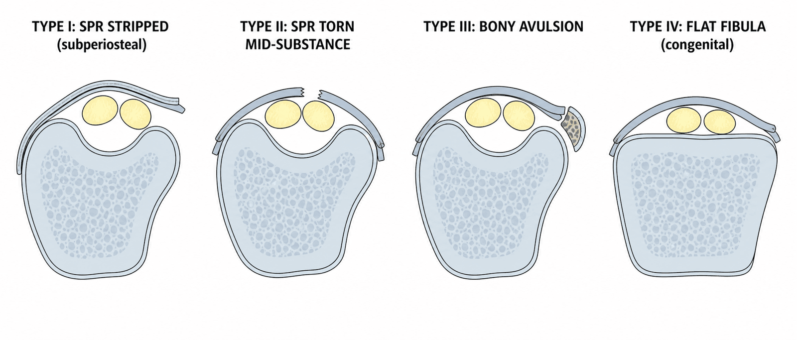
- Pathology
- SPR stripped from fibula (subperiosteal)
- Clinical Finding
- Tendons dislocate between periosteum and fibula
- Surgical Repair
- Drill holes in fibula, re-attach SPR
- Pathology
- SPR torn mid-substance, fibrocartilage ridge avulsed
- Clinical Finding
- Tendons dislocate over intact periosteum
- Surgical Repair
- Groove deepening + SPR repair
- Pathology
- SPR torn with bony avulsion (flake fracture)
- Clinical Finding
- Bone fragment at fibular insertion
- Surgical Repair
- Fix fragment if large, excise if small + repair
- Pathology
- Congenital flat fibula, habitual subluxation
- Clinical Finding
- No trauma, voluntary subluxation
- Surgical Repair
- Groove deepening + retinacular reconstruction
Type I (50-60%) is most common. Type IV is rare and usually congenital.
Intrasheath (Intra-Sheath) Subluxation: the 'Normal-MRI Snapper'
The controversies section mentions tendoscopic treatment of intrasheath subluxation — this distinct, easily-missed entity deserves separating from frank (subsheath) dislocation.
- What it is. In intrasheath subluxation the superior peroneal retinaculum is intact and the tendons never leave the retromalleolar groove — instead they snap against each other within the sheath. It is the reason a patient can have a convincing painful snap yet a normal static MRI and no visible dislocation over the fibula.
- The Raikin classification. Type A - no tendon tear: the peroneus longus and brevis reverse their positions (flip past each other) within an intact sheath. Type B - with a tear: the peroneus longus subluxes through a longitudinal split in the peroneus brevis, snapping in and out of the split.
- Diagnosis is dynamic. Because static imaging is typically normal, the key test is dynamic ultrasound (or dynamic MRI) during active circumduction/eversion, which shows the tendons flipping or the longus moving through the brevis split in real time — the same reason it is under-recognised.
- Management. A conservative trial as for frank subluxation; when refractory, tendoscopic or open groove deepening (with repair of any brevis split) addresses the crowded sheath — an isolated SPR repair does not help, because the SPR was never torn.
Q: A patient has a convincing painful ankle snap but a normal MRI and no tendons visibly dislocating over the fibula - what is it? A: Intrasheath subluxation - the SPR is intact and the tendons stay in the groove but snap within the sheath. Raikin type A = longus and brevis flip past each other (no tear); type B = longus subluxes through a peroneus brevis split. Diagnose on dynamic ultrasound; treat refractory cases with groove deepening (± brevis-split repair), not SPR repair.
Clinical Presentation
History
Acute Presentation
- Mechanism: Forced dorsiflexion with reflex peroneal contraction (skiing, soccer, basketball)
- Sensation: Audible/palpable "pop" over lateral ankle
- Immediate: Lateral ankle pain, swelling, inability to weight bear
- Relocation: Tendons may relocate spontaneously with plantarflexion
Chronic Presentation
- Recurrent episodes: Painful snapping with eversion/dorsiflexion activities
- Instability: "Giving way" sensation (different from ATFL instability)
- Activity limitation: Unable to run, cut, pivot
- Apprehension: Fear of subluxation with certain movements
The classic chronic patient is a young athlete with recurrent painful lateral ankle "snapping" that was initially misdiagnosed as ankle sprain.
Examination
Inspection
- Standing: May see fullness over lateral malleolus (chronic thickening)
- Gait: Antalgic, avoids eversion
- Swelling: Localized to lateral malleolus (acute), minimal (chronic)
Palpation
- Tenderness: Over lateral malleolus, posterior to fibula
- Palpable subluxation: Tendons may be palpable anteriorly during provocation
Dynamic Testing
- Passive dorsiflexion + eversion test (Diagnostic): With ankle dorsiflexed, examiner everts foot. Palpable snap as tendons dislocate over fibula reproduces symptoms.
- Resisted eversion: May reproduce snap if tendons sublux with contraction
- Circumduction test: Ankle circumduction may elicit snap
A positive snap test is pathognomonic for peroneal subluxation.
Investigations
Imaging Protocol
Ankle AP, Lateral, Mortise. Rule out avulsion fracture (Type III), assess fibular groove. Lateral view may show small flake fracture off posterior fibula.
Axial T2-weighted sequences critical. Shows SPR discontinuity, tendon position (may sublux on imaging if provoked), groove depth measurement, peroneus brevis tear (30% association).
Real-time assessment: Can visualize subluxation with dorsiflexion/eversion maneuver. Operator-dependent but useful for dynamic confirmation.
Axial CT for groove depth: If considering groove deepening procedure, CT with 3D reconstruction quantifies groove depth (normal greater than 3mm, shallow under 2mm).
SPAREMRI Findings in Peroneal Subluxation
Hook:SPARE no detail on MRI - look for all signs of subluxation.
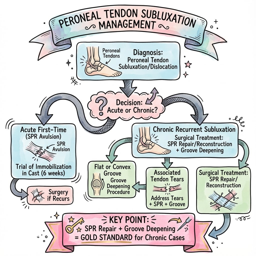
Management Algorithm
Indications
- Acute injury (first-time subluxation)
- Patient unfit for surgery
- Low-demand patient willing to modify activities
Protocol
Non-Operative Treatment
CAM walker boot in plantarflexion (20 degrees). Plantarflexion relaxes peroneals, allows SPR healing. Non-weight bearing for 2 weeks, then protected weight bearing.
Progressive ROM and strengthening. Avoid aggressive eversion exercises initially. Ankle stabilization exercises, proprioception training.
Lateral ankle brace or taping for sports. Prevents dorsiflexion/eversion extremes. Activity modification.
Success rate for conservative management is 30-50% for acute injuries. Chronic recurrent subluxation usually requires surgery.
Surgical Technique
Patient Positioning
- Position: Lateral decubitus with operative leg up
- Tourniquet: Thigh tourniquet (250-300 mmHg)
- Draping: Leg free draped to allow foot manipulation
Incision
- Location: Curvilinear incision posterior to fibula, centered over lateral malleolus
- Length: 6-8 cm from fibula tip to 4-5 cm proximal
- Plane: Through subcutaneous tissue, identify sural nerve (retract posteriorly)
Exposure
- Identify torn SPR (usually visible as frayed tissue)
- Inspect peroneal tendons (assess for brevis tear)
- Assess fibular groove depth (measure with probe)
- Confirm Oden classification type intraoperatively
The sural nerve crosses the field posteriorly and must be protected.
Complications
- Incidence
- 5-10%
- Risk Factors
- Inadequate repair, shallow groove not addressed
- Management
- Revision with groove deepening
- Incidence
- 2-5%
- Risk Factors
- Inadequate identification/protection
- Management
- Observation (usually neuropraxia), neurolysis if persistent
- Incidence
- 3-5%
- Risk Factors
- Aggressive deepening, thin fibula
- Management
- ORIF if displaced, non-operative if stable
- Incidence
- 3-5%
- Risk Factors
- Thin skin over lateral malleolus, diabetes
- Management
- Local wound care, VAC therapy if severe
- Incidence
- 10-15%
- Risk Factors
- Prolonged immobilization, aggressive rehab too early
- Management
- Physiotherapy, manual mobilization
Outcomes and Prognosis
Risk factors for failure:
- Missed associated peroneus brevis tear (requires concomitant debridement)
- Shallow fibular groove not addressed (under 2mm depth)
- Early return to sport (before 6 months)
- Habitual subluxation (Type IV) - congenital flat fibula difficult to correct
Habitual and Voluntary Subluxation, and the Flat (Convex) Fibula
The classification lists a Type IV / habitual variant and the outcomes section flags it as hard to correct — the atraumatic subluxer behaves differently enough to manage on its own terms.
- A different patient. Unlike the traumatic dislocator (a discrete injury), the habitual subluxer has no significant trauma; the tendons sublux repeatedly with ordinary activity, often bilaterally and relatively painlessly, and some patients can voluntarily sublux the tendons on demand. The underlying problem is usually a congenitally shallow, flat, or even convex (retro-fibular) groove rather than an acute SPR tear.
- Why SPR repair alone fails. With a flat or convex fibula there is no competent groove to contain the tendons, so an isolated retinacular repair predictably re-fails — this is the one scenario where bony work is mandatory, not optional.
- Management. Groove deepening (or a fibular osteotomy to create or rotate a groove) combined with retinacular reconstruction is the mainstay; historical sling/rerouting or bone-block procedures are reserved for the rare failed reconstruction.
- A counselling caveat. A voluntary or psychogenic subluxer with minimal pain may do poorly after surgery and needs careful selection — operate for genuinely disabling, involuntary symptoms, not for the "party trick" alone.
Q: How does habitual (Type IV) peroneal subluxation differ from the traumatic form, and why can't you just repair the SPR? A: The habitual subluxer has no trauma - often bilateral, relatively painless, sometimes voluntary - and the cause is a shallow, flat or convex fibular groove, not an SPR tear. With no competent groove to hold the tendons, isolated SPR repair fails, so groove deepening (or a fibular osteotomy) plus retinacular reconstruction is mandatory; select carefully, as a voluntary/painless subluxer may do poorly after surgery.
Guidelines, Registries & Global Practice
Peroneal tendon dislocation is rare (roughly 0.3-0.5% of ankle injuries) and there are no formal disease-specific society guidelines or arthroplasty/implant registries — it is a soft-tissue tendon problem managed by surgeon judgement and small-series evidence. Practice is shaped by the foot-and-ankle literature and by sports-medicine consensus rather than by national protocols.
- Position
- Anatomic SPR repair ± selective groove deepening; tear-based 50% rule for associated brevis tears
- Practical Emphasis
- Tailor to intraoperative pathology
- Position
- Non-operative trial for first-time low-demand injury; surgery for recurrent or high-demand
- Practical Emphasis
- Avoid routine groove deepening
- Position
- Early repair favoured in elite athletes to expedite reliable return to sport
- Practical Emphasis
- Endoscopic/tendoscopic options where expertise exists
- Position
- High index of suspicion in skiers and field-sport athletes; dynamic ultrasound for confirmation
- Practical Emphasis
- Reduce the high missed-diagnosis rate
- Accounts for ~0.3-0.5% of ankle injuries; true incidence likely under-reported because many are missed acutely
- Strong association with snow sports (skiing edge-catch) and pivoting field sports (soccer, basketball, rugby)
- Predominantly young, athletic adults; men slightly more affected in most series
- Up to ~30% of chronic cases develop an associated peroneus brevis longitudinal split
- High-resource: MRI plus dynamic ultrasound for diagnosis; option of endoscopic/tendoscopic repair; early surgery for elite athletes
- Limited-resource: diagnosis rests on history and the dorsiflexion-eversion snap test; plain radiographs to exclude the rim-avulsion (fleck) fracture; open anatomic SPR repair is low-cost and reliable
- Dynamic ultrasound is an inexpensive, high-value tool where MRI access is limited
- Core principle is universal: restore the retinacular restraint and address any tendon tear
Controversies and Areas of Uncertainty
The central debate. A systematic review favoured adding groove deepening for return to sport (van Dijk 2015), yet a Level II comparative study (Cho 2014) and a 36-patient series (Park 2021) found isolated SPR repair gave equivalent outcomes with shorter operating time when groove anatomy is adequate. Pragmatic position: deepen only for a genuinely shallow or convex groove, not routinely.
No randomised data. Conservative care fails in roughly half of acute dislocations, so early repair is increasingly offered to high-demand athletes (allowing faster, more reliable return), while a non-operative trial in plantarflexion remains reasonable for lower-demand patients. The threshold is judgement, not evidence-based consensus.
Endoscopic SPR reconstruction and tendoscopic treatment of intrasheath subluxation are described with less soft-tissue dissection and lower sural nerve risk, but evidence is limited to technical notes and small series. Open anatomic repair remains the reference standard.
Historical bone-block, tendon-rerouting and sling (Jones-type) reconstructions are now largely abandoned in favour of anatomic retinacular repair, as they alter biomechanics and risk stiffness. They retain a niche only for failed anatomic repair or true flat/convex fibula.
The intraoperative SPR-tear classification (Types I-III) was originally described by Eckert and Davis; Oden later added the bony-avulsion and habitual variants. Examiners may use either eponym — describe the pattern (periosteal stripping vs mid-substance tear vs bony avulsion vs flat-fibula/habitual) rather than relying on the number alone.
MCQ Practice Points
Q: What is the PRIMARY restraint preventing peroneal tendon subluxation? A: Superior peroneal retinaculum (SPR) - The SPR is the primary soft tissue restraint, accounting for 70% of restraining force. Fibular groove depth is a secondary restraint. This is why SPR repair is effective even with normal groove depth.
Q: In the Oden classification, Type I peroneal subluxation involves which pathology? A: SPR stripped from fibula with subperiosteal dissection - Tendons dislocate between the fibular periosteum and bone. Surgical repair requires drill holes in fibula to reattach SPR to bone.
Q: What clinical test is pathognomonic for peroneal tendon subluxation? A: Passive dorsiflexion plus eversion test - With ankle dorsiflexed, examiner everts the foot. A palpable or audible snap over the lateral malleolus that reproduces the patient's symptoms confirms subluxation.
Q: What is the optimal position for immobilization in acute peroneal subluxation? A: Plantarflexion (20 degrees) - Plantarflexion relaxes the peroneal tendons, allows the SPR to heal, and reduces the tendons into the fibular groove. Neutral or dorsiflexion positions maintain tension and prevent healing.
Q: What is the indication for fibular groove deepening in peroneal subluxation surgery? A: Shallow groove depth less than 3mm on CT - Normal groove depth is greater than 3mm. Grooves less than 2mm are predisposing factors. Routine groove deepening is not necessary if depth is adequate (greater than 3mm).
Exam Viva Scenarios
Practise clinical reasoning and management decisions out loud
“A 22-year-old competitive skier presents to the emergency department with acute lateral ankle pain after a forced dorsiflexion injury when catching an edge. She describes a 'pop' over the lateral ankle. On examination, you elicit a painful snap with passive dorsiflexion and eversion. How would you manage this patient?”
“You are performing SPR repair for chronic recurrent peroneal subluxation in a 28-year-old footballer. Intraoperatively, you find the SPR is stripped from the fibula (Oden Type I) and the fibular groove depth is 4mm. Walk me through your surgical technique.”
“A patient returns 12 months after SPR repair with recurrent subluxation. Review of the original surgery shows SPR repair was performed but groove depth was not assessed. CT now shows groove depth of 1.5mm. How would you manage?”
Key Anatomy
- SPR = Superior Peroneal Retinaculum (fibula to calcaneus) = Primary restraint (70%)
- Fibular groove normal depth greater than 3mm (shallow if under 2mm)
- Peroneus brevis is lateral tendon (against fibula), longus is medial
- Tendons dislocate anterolaterally (over fibula tip) when SPR tears
- Sural nerve posterior to field (protect during surgery)
Classification (Oden)
- Type I (50-60%) = SPR stripped from fibula (subperiosteal) = Drill holes repair
- Type II = SPR torn mid-substance + fibrocartilage ridge = Groove deepening + repair
- Type III = SPR avulsion with bone fragment = Fix or excise fragment + repair
- Type IV (rare) = Congenital flat fibula, habitual subluxation = Groove deepening
Diagnosis
- Mechanism: Forced dorsiflexion + reflex peroneal contraction (skiing, soccer)
- Snap test: Passive DF + eversion reproduces painful snap (pathognomonic)
- MRI: SPR discontinuity, tendon position, groove depth, brevis tear (30%)
- 50-80% missed acutely (often misdiagnosed as ankle sprain)
Management Algorithm
- Acute: Boot in plantarflexion (20 degrees) × 6 weeks, conservative trial
- Chronic/Recurrent: Surgical SPR repair indicated (failed conservative)
- SPR repair: Drill holes in fibula, suture fixation with foot in plantarflexion
- Groove deepening: Only if shallow (under 3mm), deepen by 4-5mm
- Postop: Non-weight bearing 6 weeks in plantarflexion boot
Surgical Pearls
- Protect sural nerve (posterior to incision)
- Inspect peroneal tendons (30% have associated brevis tear - debride if present)
- Measure groove depth intraoperatively (normal greater than 3mm)
- Tension repair with foot in plantarflexion (avoid over-tightening)
- Non-weight bearing 6 weeks critical for SPR healing to bone
Complications
- Recurrence: 5-10% (shallow groove not addressed, inadequate repair)
- Sural nerve injury: 2-5% (inadequate protection)
- Fibula fracture: 3-5% (during groove deepening, aggressive technique)
- Wound healing: 3-5% (thin lateral malleolus skin)
- Stiffness: 10-15% (prolonged immobilization)
Evidence Base and Key Studies
Return to Sport and Outcomes After Surgical Treatment — Systematic Review
- Systematic review and best-evidence synthesis of 14 studies on peroneal tendon dislocation
- Surgery significantly improved postoperative AOFAS scores with high satisfaction rates
- Redislocation rate was less than 1.5% at long-term follow-up
- Combined groove deepening plus SPR repair gave higher return-to-sport rates than SPR repair alone (p = 0.022)
Retinaculum Repair With vs Without Fibular Groove Deepening — Comparative Study
- Prospective non-randomised comparison of 29 patients with recurrent traumatic dislocation
- Group A (repair + groove deepening, n=13) AOFAS improved 59.3 to 92.2
- Group B (isolated SPR repair, n=16) AOFAS improved 58.5 to 91.3 — no significant difference
- Tourniquet time significantly shorter without deepening; mean return to sport ~3 months in both groups
Retinaculum Reattachment Without Groove Deepening — Larger Series
- 36 patients with recurrent peroneal tendon dislocation; anatomic SPR reattachment without groove deepening
- 34 of 36 fully recovered with no resubluxation
- Two recurrences (both during sport, at 6 and 20 months); one transient sural nerve injury
- Significant improvement in AOFAS, VAS and Foot Function Index; no evertor weakness or motion loss
Recurrent Dislocation — SPR Reconstruction With Groove Deepening
- 15 patients with post-traumatic recurrent dislocation (skiing and soccer) treated by SPR reattachment/reinforcement plus retrofibular groove deepening
- No redislocations and no neurovascular injuries at mean 3.5-year follow-up
- Satisfactory functional result in 13/15 (87%) with full motion and no pain
- Authors conclude conservative treatment is not an option for recurrent dislocation due to persistent instability and pain
Operative Treatment of Peroneal Tendon Disorders — Review
- Comprehensive review: peroneal disorders are rare, frequently missed, and diagnosed primarily on history and examination
- Subluxation usually requires operative SPR repair/reconstruction with or without retromalleolar groove deepening
- For associated tears: primary repair/tubularisation for tears involving under 50% of tendon
- Tenodesis indicated for tears involving over 50% of the tendon
Dislocation/Subluxation — Tailored Surgical Technique Case Series
- 23 patients (5 acute, 18 late) operated for symptomatic dislocating/subluxing peroneal tendons
- Procedure selected intraoperatively: SPR repair, reattachment, groove deepening, or combination
- Mean postoperative VAS 1.5 and AOFAS 85 at mean 53 months; 16/20 rated excellent
- No single technique proved superior with respect to chronicity, stage or satisfaction