Extra Digit Formation
- Preaxial: Thumb duplication - use Wassel classification (I-VII).
- Postaxial: Small finger - Type A (well-formed) vs Type B (rudimentary).
- Wassel IV: Most common thumb type - duplicated proximal phalanx.
- Bilhaut-Cloquet: Combine two equally hypoplastic thumbs into one (Wassel I/II).
- Retain the more functional digit: Usually the ulnar thumb.
- “Wassel IV = Most common thumb duplication
- “Postaxial more common in African descent
- “Type B postaxial can ligate in nursery
- “Bilhaut-Cloquet for equal hypoplastic thumbs
- “Reconstruct collateral ligaments - critical
Overview and Epidemiology
Polydactyly is the congenital duplication of digits and one of the most common congenital hand anomalies. It is classified by position: preaxial (radial, the thumb), postaxial (ulnar, the small finger) or central.
How common. The overall incidence is 1-2 per 1,000 live births, and the distribution varies by type:
- Preaxial - more common in Caucasians and Asians
- Postaxial - 10 times more common in people of African descent, at 1 in 300
- Central - rare, about 1 in 100,000
Anatomy and Biomechanics
The thumb. A normal thumb has two phalanges, against three in the fingers, so the three phalanges of a Wassel VII thumb are abnormal. FPL inserts on the distal phalanx and is critical to thumb function. The thenar muscles, APB, FPB and OP, provide opposition, which is unique to the thumb and enables pinch grip.
The MCP joint. The UCL and RCL provide MCP joint stability. Asymmetric ligament support produces angular deformity, and unequal growth can cause progressive deformity.
The duplicated thumb. The metacarpal may be single (Wassel I-V) or duplicated (Wassel VI), and each thumb has its own digital nerves and arteries. The physes are preserved at surgery to maintain growth.
The small finger. Its UCL is critical for power grip, and the hypothenar muscles, ADM, FDM and ODM, provide small-finger function.
Pathophysiology
Polydactyly is a disorder of anterior-posterior (radial-ulnar) axis patterning in the developing limb bud. It is not a mechanical splitting of a normal digit. It arises during limb-bud formation, at 4-8 weeks of gestation, from abnormal signalling within the bud.
The SHH-ZPA axis. The zone of polarising activity (ZPA), at the posterior (ulnar) margin of the limb bud, secretes Sonic Hedgehog (SHH). SHH establishes the radial-ulnar gradient that specifies digit identity and number.
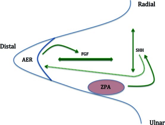
The genes. Each maps to a pattern of duplication:
- ZRS - SHH expression is controlled by a distant cis-regulatory element, the ZPA regulatory sequence, within intron 5 of LMBR1 at 7q36.3. Point mutations or copy-number gains of the ZRS cause ectopic anterior (radial) SHH activity, producing preaxial polydactyly and triphalangeal thumb.
- GLI3 - a downstream SHH effector and repressor. Loss of function underlies postaxial and central polydactyly, including Greig and Pallister-Hall syndromes.
- HOXD13 - expansion or mutation produces synpolydactyly, a central duplication fused with syndactyly.
From signal to phenotype. Ectopic or excess SHH activity expands the digit field and generates a supernumerary ray. The level at which the extra skeletal element separates, from distal phalanx to metacarpal, determines the Wassel type of a thumb duplication. Whether the elements are shared (bifid) or separate (duplicated) reflects how early in ray condensation the duplication occurred.
Why neither thumb is normal. The duplicated ray shares tendons, ligaments and neurovascular structures with the dominant ray, so neither thumb is fully normal. That is why simple ablation without soft-tissue rebalancing fails.
Inheritance. Postaxial polydactyly is often autosomal dominant with variable penetrance, and isolated postaxial Type B typically is, often GLI3-related. Preaxial duplication is usually sporadic, but ZRS-related forms such as triphalangeal thumb-polysyndactyly are autosomal dominant.
Classification Systems
Wassel (preaxial). Thumb duplication is classified by Wassel into types I-VII by the level of bifurcation, from distal to proximal: the higher the number, the more proximal the duplication. Odd numbers are bifid, sharing an element; even numbers are duplicated, with separate elements. Type VII is the triphalangeal thumb component.
Wassel IV is the most common type, at 43%. It is a duplicated proximal phalanx: two complete proximal phalanges articulating with a single metacarpal.
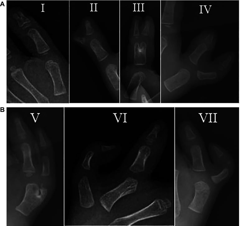
- Level of Bifurcation
- Bifid distal phalanx (shared)
- Frequency
- Rare (2%)
- Level of Bifurcation
- Duplicated distal phalanx
- Frequency
- 15%
- Level of Bifurcation
- Bifid proximal phalanx (shared)
- Frequency
- Rare (6%)
- Level of Bifurcation
- Duplicated proximal phalanx
- Frequency
- MOST COMMON (43%)
- Level of Bifurcation
- Bifid metacarpal (shared)
- Frequency
- Uncommon (10%)
- Level of Bifurcation
- Duplicated metacarpal
- Frequency
- Rare (4%)
- Level of Bifurcation
- Triphalangeal thumb component
- Frequency
- 20%
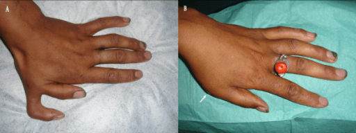
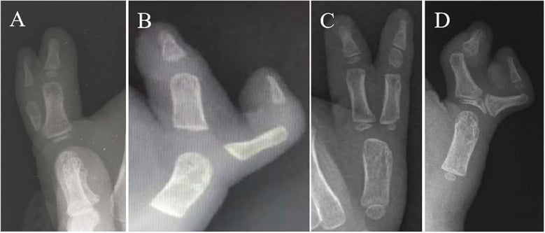
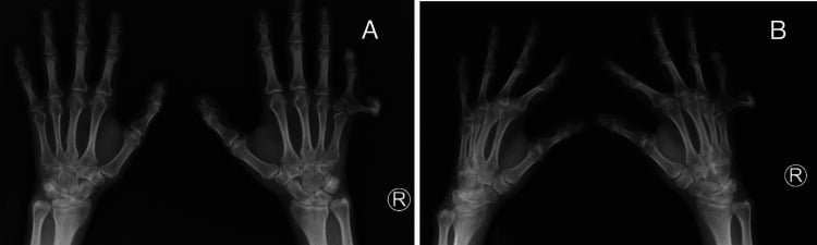
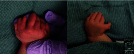
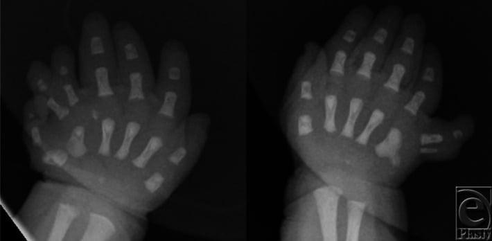
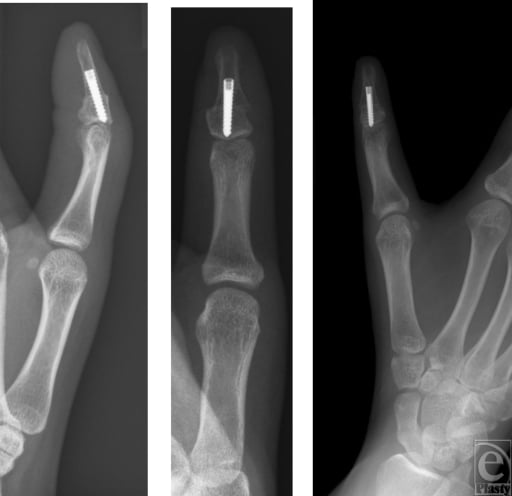
Triphalangeal thumb and the delta phalanx. A triphalangeal thumb has an extra (middle) phalanx in the thumb ray. It is the component of Wassel VII duplication, and it also occurs in isolation and in the five-fingered (non-opposable) hand. It is strongly linked to ZRS/SHH dysregulation and is autosomal dominant in many families.
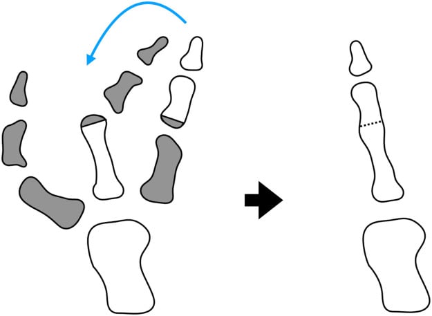
The delta phalanx. The extra phalanx is frequently a delta phalanx, a wedge or trapezoid-shaped bone with a longitudinally bracketed (C-shaped) epiphysis. The bracket tethers one side and drives progressive angulation (clinodactyly).
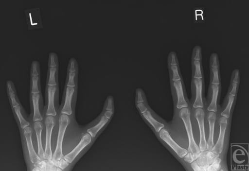
Treating it. Management depends on the phalanx:
- A small delta phalanx is excised in infancy
- A larger rectangular extra phalanx needs a reduction or closing-wedge osteotomy, or fusion to an adjacent phalanx, to shorten and realign the over-long thumb
- A non-opposable, finger-like thumb (the five-fingered hand) requires pollicisation or opponensplasty, not simple excision
Recognising a triphalangeal component is essential, because ablating the duplicate alone leaves a long, deviated, poorly opposing thumb.
Clinical Assessment
History. The questions that matter:
- Family history - especially for postaxial (autosomal dominant)
- Pregnancy - maternal diabetes, teratogens
- Syndromic features - cardiac and renal anomalies
- The parents' goals - functional and cosmetic concerns
Examination. Inspection establishes the location (preaxial, postaxial or central), whether the digit is well formed or rudimentary, the width of the skin bridge in a Type B digit, and any associated syndactyly. Function is judged from active movement of each thumb, thenar bulk, stability and grip pattern. Look beyond the hand for a cardiac murmur (Holt-Oram), syndactyly (central), other limb anomalies and facial dysmorphism.
Which thumb to keep. The dominant thumb is usually the ulnar one, and the radial thumb is often hypoplastic. The features that decide it are gathered below; assess MCP and IP joint stability alongside them.
THUMBTHUMB Selection for Retention
Hook:THUMB: The ulnar thumb is usually retained - Thenar bulk, Heavier, Ulnar, Motion, Better FPL
Investigations
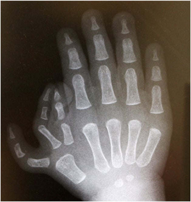
Radiographs are essential for surgical planning. They define:
- The level of bifurcation or duplication
- The bony anatomy of each digit
- Joint articulation
- Metacarpal morphology
- Any delta phalanx, whose bracketed physis is identified before choosing excision, osteotomy or fusion
Further investigation. Consider:
- Cardiac echo - Holt-Oram syndrome (radial anomalies with ASD/VSD)
- Renal ultrasound - associated genitourinary anomalies
- Genetic testing - if there are syndromic features
- Polydactyly Type
- Preaxial
- Key Features
- Radial dysplasia, ASD/VSD
- Polydactyly Type
- Postaxial
- Key Features
- Short stature, cardiac defects
- Polydactyly Type
- Postaxial
- Key Features
- Multiple malformations, poor prognosis
- Polydactyly Type
- Postaxial
- Key Features
- Obesity, retinitis pigmentosa, renal
Differential diagnosis. Not every extra or broad digit is a simple duplication. These entities must be distinguished, because they change the operation and the counselling.
- Key Distinguishing Feature
- Two thumb skeletal elements sharing or duplicating at one level
- Why It Matters
- Standard excision-plus-reconstruction
- Key Distinguishing Feature
- Three phalanges in the thumb ray (extra middle phalanx), often delta-shaped
- Why It Matters
- May coexist with duplication (Wassel VII); needs wedge/middle-phalanx excision, not simple ablation
- Key Distinguishing Feature
- Central duplication fused within a syndactyly (HOXD13)
- Why It Matters
- Shared tendons/neurovascular structures; staged complex reconstruction
- Key Distinguishing Feature
- One enlarged digit, not a duplicate (lipofibromatous/PIK3CA overgrowth)
- Why It Matters
- Debulking or epiphysiodesis, not duplication surgery
- Key Distinguishing Feature
- Multiple symmetrical fingers, no thumb, duplicated ulna
- Why It Matters
- Pollicisation and digit reduction, a wholly different plan
- Key Distinguishing Feature
- Type A has bone and a true joint; Type B is a skin pedicle
- Why It Matters
- Type A always needs formal excision; only true Type B is amenable to ligation
Management Algorithm
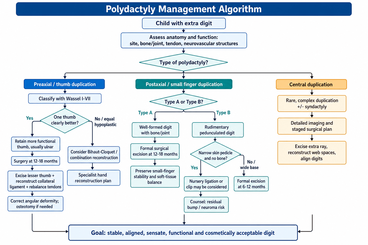
Timing. Preaxial duplication and postaxial Type A are operated on at 12-18 months. By then the hand is large enough for surgical manipulation, functional hand patterns are not yet established, and the child will not remember the operation. A postaxial Type B digit is either ligated in the nursery or formally excised at 6-12 months.
Planning. For a thumb duplication, decide which digit to retain, design the incision, and anticipate the need for ligament or tendon reconstruction. Consider Bilhaut-Cloquet if both thumbs are equally hypoplastic.
Non-operative management is rarely appropriate; most cases benefit from surgery. It is kept for families who decline surgery and children with significant medical comorbidities. The child is then monitored for functional limitation, any progression of angular deformity is documented, and the hand is reassessed at regular intervals.
Principles of thumb reconstruction. The aim is a functional, stable, cosmetically acceptable digit:
- Retain the more functional thumb, usually the ulnar one with the better FPL insertion
- Reconstruct the collateral ligaments, which are critical for stability
- Rebalance the tendons, transferring the intrinsics from the excised thumb
- Correct angular deformity, which may need an osteotomy
Surgical Technique
Thumb duplication. The sequence:
- Racquet incision around the hypoplastic thumb
- Identify and preserve the neurovascular bundle to the retained thumb
- Detach the collateral ligament from the hypoplastic thumb
- Detach FPL/EPL if they insert on the hypoplastic thumb
- Excise the hypoplastic thumb, with its metacarpal portion if needed
- Reconstruct the collateral ligament to the retained thumb
- Transfer and balance the tendons
- Correct any angular deformity
The key to successful surgery is meticulous soft-tissue reconstruction.
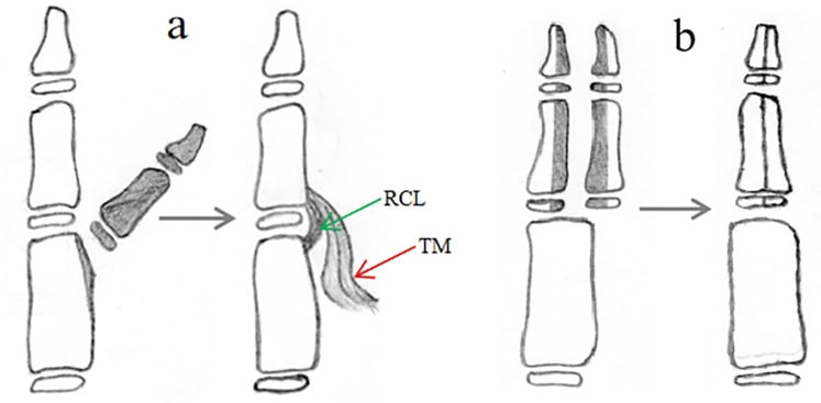
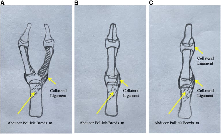
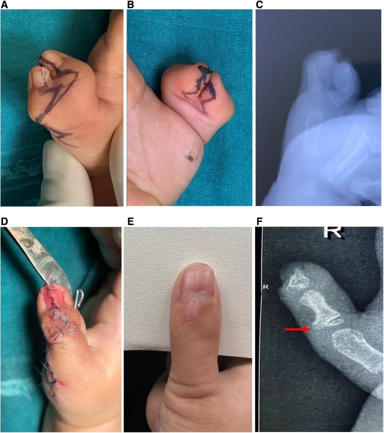
The zigzag deformity. The reason a duplicate thumb is never normal, and recurs after naive excision, is the zigzag (Z) collapse deformity of Wassel IV. The shared metacarpal head is broad and its articular surface often obliquely inclined, the FPL and EPL insert eccentrically and are bowstrung toward one side, and thenar (intrinsic) pull is imbalanced. Together these angulate the MCP joint in one direction and the IP joint in the opposite direction.
Correcting it. Excision plus collateral ligament reconstruction is not enough for a durable result. It also requires:
- Re-centralisation of the bowstrung FPL/EPL over the joint axis
- Rebalancing of the intrinsics
- A corrective (closing-wedge) osteotomy of the metacarpal or proximal phalanx to square an oblique articular surface
Failing to address the tendon eccentricity and bony obliquity is the commonest cause of recurrent deformity and the 15-20% revision rate.
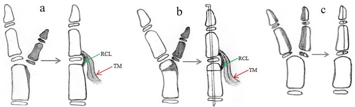
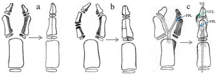
Must reconstruct collateral ligaments - failure to do so results in an unstable thumb. Always check joint stability intraoperatively after reconstruction. Consider osteotomy if angular deformity persists after ligament reconstruction.
Complications
Early. The risks in the first weeks:
- Wound infection - 1-2% incidence
- Flap necrosis - avoid excessive tension
- Nerve injury - the digital nerve is at risk
- Vascular compromise - ensure perfusion intraoperatively
Joint instability is the most common complication. It comes from inadequate ligament reconstruction and is associated with progression of angular deformity. It requires revision surgery, and meticulous technique is the prevention.
Angular deformity is progressive malalignment, related to growth and to asymmetric growth-plate injury. It is monitored during growth and may need corrective osteotomy.
Nail deformity is usually cosmetic only: the central ridge after Bilhaut-Cloquet, or a small hypoplastic nail. It may need nail-bed reconstruction.
Revision. 15-20% may need secondary procedures. Collateral ligament reconstruction is the most common; the others are corrective osteotomy for angular deformity, tendon rebalancing and web-space deepening.
Postoperative Care and Rehabilitation
The first two weeks. A thumb spica or volar splint, the wound kept clean and dry, and elevation to reduce swelling. Paracetamol is typically sufficient for pain.
Two to six weeks. Sutures are removed at 2 weeks, the splint is weaned gradually, and gentle range of motion begins, passive then active. Monitor for infection and stiffness.
Six weeks to six months. Full range-of-motion exercises and normal use are encouraged, with age-appropriate strengthening. Growth is monitored for angular deformity, and occupational therapy is added if there are functional concerns.
Long term. The child is monitored through growth, with annual assessment until skeletal maturity of ligament integrity, stability, and pinch and grip strength. Revision is considered for progressive deformity or instability.
Outcomes and Prognosis
Thumb duplication. Functional outcomes are good in most cases. Some residual instability is common, and the thumb may be slightly smaller than the normal side.
Postaxial polydactyly. Outcomes are excellent with proper technique. Type A excision has a low complication rate, while Type B ligation carries a risk of neuroma and a residual bump.
- Good Prognosis
- Simple duplication
- Poor Prognosis
- Complex with bone involvement
- Good Prognosis
- Ligament reconstruction done
- Poor Prognosis
- Ligaments not reconstructed
- Good Prognosis
- 12-18 months
- Poor Prognosis
- Delayed beyond 24 months
- Good Prognosis
- Paediatric hand specialist
- Poor Prognosis
- General surgeon
Satisfaction is high overall, and cosmetic concerns are more common than functional ones. Realistic expectations are important, including the possibility that secondary procedures may be needed.
Guidelines, Registries & Global Practice
Polydactyly is a clinical, not registry-tracked, condition, so practice is guided by national society reviews and IFSSH consensus rather than arthroplasty-style registries. The descriptive framework is converging worldwide on the Oberg-Manske-Tonkin (OMT) classification, endorsed by the IFSSH, with the Wassel (Wassel-Flatt) scheme retained for the thumb.
Global Epidemiology
- Overall: one of the most common congenital hand anomalies, roughly 1-2 per 1,000 live births worldwide.
- Postaxial (ulnar): markedly more frequent in populations of African ancestry (around 1 in 150-300), where it is often isolated and autosomal dominant; far less common (around 1 in 1,500-3,000) and more often syndromic in those of European ancestry.
- Preaxial (radial/thumb): more common in East Asian and white populations; usually sporadic and unilateral.
- Central: rare in all populations and frequently associated with syndactyly (synpolydactyly).
Side-by-Side Society Guidance
- Classification Emphasis
- OMT classification as unifying framework; Wassel retained for thumb
- Practice Position
- Promotes consistent terminology to enable cross-centre comparison
- Classification Emphasis
- Wassel-Flatt for thumb; Stelling-Turek A vs B for postaxial
- Practice Position
- Increasing shift from nursery suture ligation toward formal excision for Type B
- Classification Emphasis
- OMT and Wassel; specialist congenital hand network referral
- Practice Position
- Centralised paediatric hand surgery; collateral ligament reconstruction stressed
- Classification Emphasis
- OMT-based; emphasis on validated outcome scoring (JSSH/Tada)
- Practice Position
- Surgery in infancy by paediatric hand specialists
High- vs Limited-Resource Practice Variation
- High-resource settings: subspecialist paediatric hand surgery, day-case excision at 12-18 months, microsurgical capability for Bilhaut-Cloquet and on-top plasty, formal outcome scoring (JSSH/Tada), genetic counselling and cardiac/renal screening for syndromic cases.
- Limited-resource settings: nursery suture ligation or surgical-clip application remains widely used for narrow-based Type B digits because it avoids general anaesthesia and theatre access; families may present late, so reconstruction is performed at an older age. Awareness of the higher residual-bump and neuroma rate with ligation should still inform consent.
- Universal principle: regardless of resources, palpable bone or a broad base reclassifies a digit as Type A (or wide Type B) and mandates formal excision rather than ligation.
Controversies and Areas of Uncertainty
These are common viva escalation points. Take a position but acknowledge the evidence is limited.
Nursery suture ligation or formal excision for Type B postaxial. Ligation is convenient and avoids anaesthesia, but the systematic-review signal favours excision, with reported complication rates as high as 23.5% (residual bump, neuroma) for ligation versus around 3% for excision. Surgical-clip application is a middle ground for narrow-based digits. There is no high-level randomised evidence, so practice varies by resource setting and surgeon preference.
Near-symmetric Wassel IV thumbs. When the two thumbs are nearly equal in size, simple excision-plus-reconstruction yields worse JSSH outcomes. Whether Bilhaut-Cloquet, combination (on-top plasty) procedures, or accepting a smaller thumb is best remains debated; the symmetry index is an emerging predictive tool rather than a validated decision rule.
Bilhaut-Cloquet and its modifications. The classic procedure reliably creates a stable, wide thumb but is criticised for IP joint stiffness (it crosses the physis), a central nail ridge and a broad appearance. Modified Bilhaut, neurovascular island-flap pulp/nail augmentation and combination techniques aim to mitigate these, but comparative evidence is sparse.
Limits of the Wassel classification. It poorly captures the immature epiphysis, triphalangeal components and soft-tissue anatomy. The OMT framework addresses categorisation but the thumb-specific Wassel scheme persists for surgical planning, and intra- and inter-observer reliability of subtypes is imperfect.
Optimal timing. 12-18 months is conventional but not evidence-mandated. Some centres operate slightly earlier for technical convenience or later where access is constrained, with no clear functional penalty within the first few years; delay beyond 24 months nonetheless appears among the poor prognostic factors above.
MCQ Practice Points
Q: A 1-year-old child presents with bilateral thumb duplication. What is the most common Wassel classification type?
A: Wassel Type IV (duplicated proximal phalanx) - occurs in 43% of thumb duplications. Remember: even numbers = duplicated (separate elements), odd numbers = bifid (shared elements). Wassel IV has two complete proximal phalanges articulating with a single metacarpal.
Q: What is the incidence of postaxial polydactyly in people of African descent, and what is the inheritance pattern?
A: 1 in 300 in African descent (compared to 1 in 3,000 in Caucasians). Inheritance is typically autosomal dominant with variable penetrance. It is 10 times more common than in other populations.
Q: What is the optimal timing for surgical correction of preaxial (thumb) polydactyly and why?
A: 12-18 months is optimal. This timing allows adequate size for surgical manipulation, is performed before functional hand patterns are established, and occurs before the child's memory of surgery. Operating too early (less than 6 months) makes surgery technically difficult due to small structures.
Q: What is the most critical step in thumb duplication surgery that, if omitted, leads to the most common complication?
A: Reconstruction of the collateral ligaments. Failure to reconstruct the radial or ulnar collateral ligament from the excised thumb to the retained thumb results in joint instability - the most common complication requiring revision surgery.
Q: What is the indication for the Bilhaut-Cloquet procedure, and what is the main disadvantage?
A: Indicated for Wassel Type I or II with two equally hypoplastic thumbs where neither is adequate alone. The main disadvantage is IP joint stiffness because the procedure crosses the physis. Also expect a central nail ridge deformity and wide thumb appearance.
Q: A newborn has a rudimentary pedunculated extra digit (Type B postaxial polydactyly). What are the two management options and their respective risks?
A: (1) Suture ligation in nursery (traditional): Risks include incomplete removal, neuroma formation, and unsightly bump/scar. (2) Formal surgical excision at 6-12 months (preferred by many): Allows complete removal under vision with proper nerve division and better cosmetic result. Many hand surgeons now prefer formal excision despite the need for general anaesthetic.
Q: A child with preaxial polydactyly is found to have an atrial septal defect. What syndrome should you consider, and what other features would you look for?
A: Holt-Oram syndrome - characterized by radial ray anomalies (including preaxial polydactyly) with cardiac defects (ASD/VSD). Other features include radial dysplasia, hypoplastic or absent thumb, and other upper limb anomalies. Cardiac echo is essential in all patients with radial-sided upper limb anomalies.
Q: In Wassel IV thumb duplication, which thumb is typically retained and why?
A: The ulnar thumb is usually retained because it typically has: (1) Better FPL insertion and function, (2) Greater thenar muscle bulk, (3) Larger size, (4) Better active movement. The radial thumb is often more hypoplastic. However, always assess each case individually based on function.
Q: What percentage of patients require secondary surgery after thumb duplication correction, and what are the common reasons?
A: 15-20% require secondary procedures. Common reasons include: (1) Joint instability from inadequate ligament reconstruction, (2) Angular deformity from growth, (3) Tendon imbalance, (4) Web space narrowing. Parents should be counselled about this possibility preoperatively.
Q: How do you remember the difference between odd and even Wassel classification numbers?
A: ODD = bIfId (shared element) - Types I, III, V have a bifid bone where digits share a single element. EVEN = sEparatE (duplicated) - Types II, IV, VI have completely duplicated separate elements. Wassel IV (even) = duplicated PP, Wassel III (odd) = bifid PP.
Viva Scenarios
Practise clinical reasoning and management decisions out loud
“10-month-old child presents with bilateral thumb duplication. X-rays show Wassel IV bilaterally. Parents ask about treatment. What is your management?”
“Newborn of African descent has bilateral pedunculated extra digits on ulnar side of hands. Midwife asks about suture ligation in nursery. What is your advice?”
“6-month-old with Wassel I thumb duplication - two equally small thumbs with bifid distal phalanx. Neither appears adequate alone. What are your options?”
WASSEL CLASSIFICATION
- I-VII: Distal to proximal bifurcation
- IV = MOST COMMON (43%) - duplicated PP
- ODD (I, III, V) = BIFID (shared)
- EVEN (II, IV, VI) = DUPLICATED (separate)
- VII = Triphalangeal thumb
POSTAXIAL TYPES
- Type A: Well-formed with articulation
- Type B: Rudimentary/pedunculated
- 1:300 in African descent
- Autosomal dominant inheritance
- Type B ligation controversial
THUMB SURGERY PRINCIPLES
- Timing: 12-18 months
- Retain more functional (usually ulnar)
- RECONSTRUCT COLLATERAL LIGAMENTS
- Transfer intrinsics from excised thumb
- Correct angular deformity
BILHAUT-CLOQUET
- For Wassel I/II equal thumbs
- Combines both thumbs into one
- Expect IP stiffness
- Nail ridge deformity
- Reserve for specific cases
ASSOCIATED SYNDROMES
- Holt-Oram: Preaxial + ASD/VSD
- Ellis-van Creveld: Postaxial + cardiac
- Trisomy 13: Postaxial + poor prognosis
- Bardet-Biedl: Postaxial + obesity + retinal
COMPLICATIONS
- Joint instability (most common)
- Angular deformity
- 15-20% need secondary surgery
- Type B ligation: neuroma risk
- Bilhaut: IP stiffness expected
Evidence Base
Wassel HD
- Original classification of the duplicate thumb based on level of skeletal duplication
- Seven types described from distal (bifid distal phalanx) to proximal (duplicated metacarpal), with the triphalangeal variant designated Type VII
- Type IV (duplication at the metacarpophalangeal level) reported as the most common pattern
- Provided the anatomical framework that still underpins surgical planning
Tonkin MA
- Editorial synthesis of contemporary thumb duplication classification and terminology
- Highlights limitations of the original Wassel scheme, particularly misclassification of the immature epiphysis and failure to capture soft-tissue and triphalangeal variants
- Supports integration of duplication within the Oberg-Manske-Tonkin (OMT) framework adopted by the IFSSH
- Reinforces that classification should guide, not replace, individualised soft-tissue assessment
Perez-Lopez LM et al
- Comprehensive review of radial (preaxial) polydactyly genetics, classification and surgery
- Confirms Wassel as the universal classification and notes misclassification of the immature epiphysis as its key limitation
- Defines three surgical objectives: thumb alignment, stability and acceptable aesthetic appearance
- Identifies the Tada score and the JSSH assessment system as validated functional outcome measures
Chen PA et al
- Forty-seven radial polydactylies reviewed with JSSH scores at mean 4.6 years
- All fair or poor outcomes (score under 17) occurred in Wassel type IV thumbs
- Symmetry of the two thumbs (symmetric index) was the only independent predictor of outcome in type IV
- A symmetric index over 0.74 predicted greater risk of a fair or poor result
Chopan M et al
- PRISMA systematic review of suture ligation versus surgical excision for postaxial (ulnar) polydactyly
- Ten studies met inclusion criteria from 900 screened, with substantial heterogeneity
- Largest cohort reported a complication rate of 23.5% for suture ligation versus 3% for surgical excision
- Residual remnants and neuromas were the principal ligation complications
Mills JK, Ezaki M, Oishi SN
- Retrospective review of 231 hands (132 newborns) with Type B postaxial polydactyly treated by surgical clip application
- No wound complications recorded with minimum two-year follow-up
- Only 7 percent required later scar revision
- Clip application performed at the bedside or outpatient setting as a low-cost alternative
Shah A, Bohn DC, Van Heest AE, Hu CH
- Six-year literature review of congenital upper-limb differences
- Confirms the Oberg-Manske-Tonkin (OMT) classification, endorsed by the IFSSH, as the current standard framework
- Notes ongoing refinement as genetic and embryonic understanding improves
- Emphasises good peer relationships and adaptability in affected children
Liu Z et al
- Reports a 7q36.3 microduplication encompassing the ZRS, the long-range cis-regulator of SHH
- Associated with triphalangeal thumb-polysyndactyly syndrome (preaxial polydactyly with duplicated distal phalanx and hallux duplication)
- Confirms that copy-number gain of the ZRS dysregulates SHH expression in the zone of polarising activity (ZPA)
- A coincident 22q11.21 deletion accounted for the associated congenital heart disease