ATLS | Damage Control Orthopaedics | Second Hit
- ISS greater than 15 defines polytrauma (major trauma)
- ATLS primary survey (ABCDE) first
- Damage control orthopaedics (DCO) for unstable/borderline patients
- Early total care (ETC) for stable patients
- Avoid second hit (inflammatory surge from long surgery)
- “Lethal triad: Hypothermia, acidosis, coagulopathy
- “Borderline patient decision is most challenging
- “Femoral shaft: DCO with external fixator, convert to nail when stable
- “Pelvic binder at greater trochanters
Overview
Definition. Polytrauma is most often defined in practice by an Injury Severity Score (ISS) greater than 15, indicating multiple injuries with life-threatening potential. ISS is a purely anatomical score, which is why the 2014 Berlin definition (see Classification) adds physiology, and why the physiological categories, not the score, drive the fixation decision. ATLS principles prioritise life-saving interventions through a systematic primary survey.
Who. Polytrauma accounts for approximately 25-27% of major trauma admissions (all those with ISS greater than 15). [1,2] The age distribution is bimodal. Younger adults (21-30 years) are injured in high-energy trauma, road traffic accidents and falls [3], and prevalence is increasing in patients older than 65, from low-energy falls in fragile patients with comorbid conditions. [4]
How. Road traffic accidents are the predominant cause (~65%) in the working-age population. [1,5] Falls from height are a significant contributor to high-energy orthopaedic trauma. [5]
Anatomy and Pathophysiology
Every region at once. Polytrauma has no specific anatomical focus; it involves multiple body regions simultaneously, and each brings its own threat.
- Thorax: rib fractures, flail chest and pulmonary contusion affect respiratory function
- Abdomen: solid organ injury (liver, spleen) causes haemorrhage
- Pelvis: ring disruption causes major arterial and venous haemorrhage
- Femur: shaft fractures are associated with significant blood loss, 1-2 litres per femur
- Spine: associated in up to 10% of major trauma; assume it is unstable until cleared
The first hit. The initial injury triggers a systemic inflammatory response syndrome (SIRS), and the response is proportional to injury severity (ISS). Tissue damage releases damage-associated molecular patterns (DAMPs), and what follows is:
- An inflammatory cytokine cascade, with IL-1, IL-6 and TNF-α released
- Complement activation and neutrophil priming
- Endothelial dysfunction and capillary leak
The second hit. Further surgical insult during this inflammatory phase amplifies SIRS. An inflammatory surge from major surgery, long femoral nailing for example, can overwhelm an already primed patient and precipitate multi-organ dysfunction syndrome (MODS), ARDS and death; acute kidney injury and coagulopathy may also develop. Prolonged surgery (more than 2 hours) acts as a second hit, and reaming of long bones releases fat, marrow and cytokines.
The lethal triad. Hypothermia, acidosis and coagulopathy are inter-related and self-perpetuating, and the goal of resuscitation is to break the cycle: warm the patient, resuscitate, correct the coagulopathy with the massive transfusion protocol, and keep surgery short.
- Hypothermia: core temperature below 35°C, which impairs clotting and cardiac function
- Acidosis: base deficit greater than 6 and lactate greater than 2.5, reflecting tissue hypoperfusion
- Coagulopathy: consumptive, dilutional or hypothermia-induced, and causes ongoing bleeding
Why damage control. Damage control orthopaedics (DCO) aims to stabilise fractures with minimal physiological insult and so avoid the second hit. Temporary external fixation adds minimal trauma, and definitive surgery waits until the inflammation has resolved, with inflammatory markers (CRP, IL-6) monitored to guide the timing.
Trauma-Induced Coagulopathy: Beyond the Lethal Triad
Present on arrival. A quarter to a third of severely injured patients are already coagulopathic when they reach the resuscitation room, before large-volume fluids or hypothermia can be blamed. This early, endogenous acute traumatic coagulopathy (ATC, or acute coagulopathy of trauma-shock) is driven by tissue injury plus hypoperfusion (shock), not simply by consumption and dilution.
The mechanism. Shock and tissue trauma cause endothelial activation and glycocalyx shedding (the "endotheliopathy of trauma") and a surge of activated protein C, which inhibits factors Va and VIIIa and de-represses fibrinolysis. The result is a hypocoagulable, hyperfibrinolytic state. Hypothermia and acidosis then amplify it, and haemodilution, hypofibrinogenaemia and hypocalcaemia compound it, so the lethal triad is best seen as the endpoint of TIC, not its cause.
Fibrinogen falls first, and calcium matters. Fibrinogen is the first factor to become critically low in major haemorrhage; replace it with cryoprecipitate or concentrate to keep it above roughly 1.5-2 g/L. Ionised calcium must be actively maintained, because citrate in transfused blood chelates it; hypocalcaemia is "the fourth horseman", turning the triad into a lethal diamond.
Measure it, don't guess. Viscoelastic testing (ROTEM/TEG) gives rapid, goal-directed readouts: clot amplitude, the fibrinogen contribution, and a fibrinolysis trace that identifies the patients TXA helps most. It increasingly guides component therapy instead of fixed empirical ratios. Management is damage control resuscitation, early fibrinogen and calcium, TXA, and ROTEM/TEG-guided correction.
Classification
Injury Severity Score. The most widely used trauma scoring system. Each injury is graded on the Abbreviated Injury Scale (AIS), and the ISS is built from the squares of the AIS scores of the three most severely injured body regions, with only one injury counted per region. It ranges from 1 to 75, and AIS 6 in any region makes the ISS 75 automatically.
- Severity
- Minor
- Severity
- Moderate
- Severity
- Serious
- Severity
- Severe
- Severity
- Critical
- Severity
- Unsurvivable
The ISS body regions are head/neck, face, chest, abdomen, extremities and external. The thresholds:
- ISS greater than 15: major trauma (polytrauma)
- ISS greater than 25: severe trauma
- ISS greater than 40: critical, with high mortality
ISS = (highest AIS)² + (2nd highest AIS)² + (3rd highest AIS)². Example: Head AIS 4 + Chest AIS 3 + Extremity AIS 3 = 16 + 9 + 9 = ISS 34 (severe polytrauma).
New Injury Severity Score (NISS). The sum of the squares of the three highest AIS scores regardless of body region. It is particularly useful when there are multiple injuries in the same body region, and it may better predict mortality in certain injury patterns.
Trauma and Injury Severity Score (TRISS). Combines anatomical (ISS) and physiological (RTS) parameters to predict the probability of survival, and is used for trauma registry quality assessment.
The Berlin definition (2014). ISS alone lets a patient exceed 15 while remaining physiologically well, which is exactly the mismatch the patient categories exist to resolve. Berlin, the modern consensus, instead requires AIS 3 or greater in two or more body regions plus at least one of five physiological criteria:
- Systolic BP 90 mmHg or less
- GCS 8 or less
- Base excess -6 or worse
- INR 1.4 or more (or PTT 40 s or more)
- Age 70 or over
It builds the physiology into the definition rather than leaving it to a separate judgement, and it identifies a group with substantially higher mortality than ISS over 15 alone. Quote ISS over 15 as the working definition of major trauma, and Berlin as the consensus definition of polytrauma.
Clinical Assessment
The primary survey. The ATLS primary survey, ABCDE, deals with life-threatening injuries first.
- Airway with cervical spine protection. Chin lift or jaw thrust (avoid head tilt in trauma); clear debris, suction and insert an airway adjunct; a definitive airway if GCS is less than 8 or the patient cannot protect the airway; in-line immobilisation during intubation.
- Breathing and ventilation. Expose the chest and assess respiratory rate and effort. The life-threatening chest injuries are tension pneumothorax, open pneumothorax, massive haemothorax, and flail chest with pulmonary contusion; needle decompression or a chest tube as indicated.
- Circulation with haemorrhage control. Pulse, BP, capillary refill and skin colour; two large-bore IV lines and fluid resuscitation; direct pressure on external bleeding; a pelvic binder if a pelvic ring injury is suspected; the massive transfusion protocol if indicated.
- Disability. GCS, pupillary response and gross motor function.
- Exposure with environmental control. Fully undress the patient for complete examination, log roll for the back and spine, and actively prevent hypothermia with warm blankets and fluid warmers.
The secondary survey. A complete head-to-toe examination once the patient is stabilised: an AMPLE history (Allergies, Medications, Past history, Last meal, Events), a detailed neurological examination, and a complete musculoskeletal examination of all joints and the spine.
Missed injuries. These are common in polytrauma: the cervical spine (a 10% miss rate with radiographs alone), extremity fractures in intubated patients, posterior injuries of the spine and pelvis, and vascular injuries. Always log roll. Up to 10% of injuries are missed on the initial survey and detected on the tertiary survey.
The tertiary survey. Mandatory in polytrauma, at 24-48 hours, when the patient is awake and cooperative and can be examined better. Repeat imaging if new symptoms emerge, and document every injury in the trauma registry.
Physiological monitoring. The parameters and their targets:
- Base excess: greater than -6 mmol/L
- Lactate: less than 2.5 mmol/L, a marker of tissue perfusion
- Temperature: prevent a fall below 35°C
- Coagulation: INR, fibrinogen, platelets
- Urine output: greater than 0.5 mL/kg/hr
Investigations
Bloods. The first samples measure perfusion and coagulation and secure blood.
- Blood gas: pH, base excess and lactate, to assess tissue perfusion
- FBC: haemoglobin is often normal initially despite blood loss
- Coagulation: PT/INR, APTT and fibrinogen, to guide transfusion
- Cross-match: urgent type and screen; O-negative if exsanguinating
- TEG/ROTEM: point-of-care coagulation assessment, if available
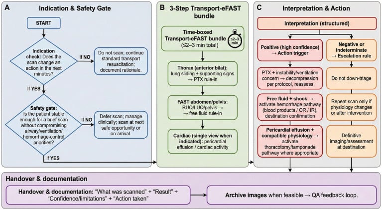
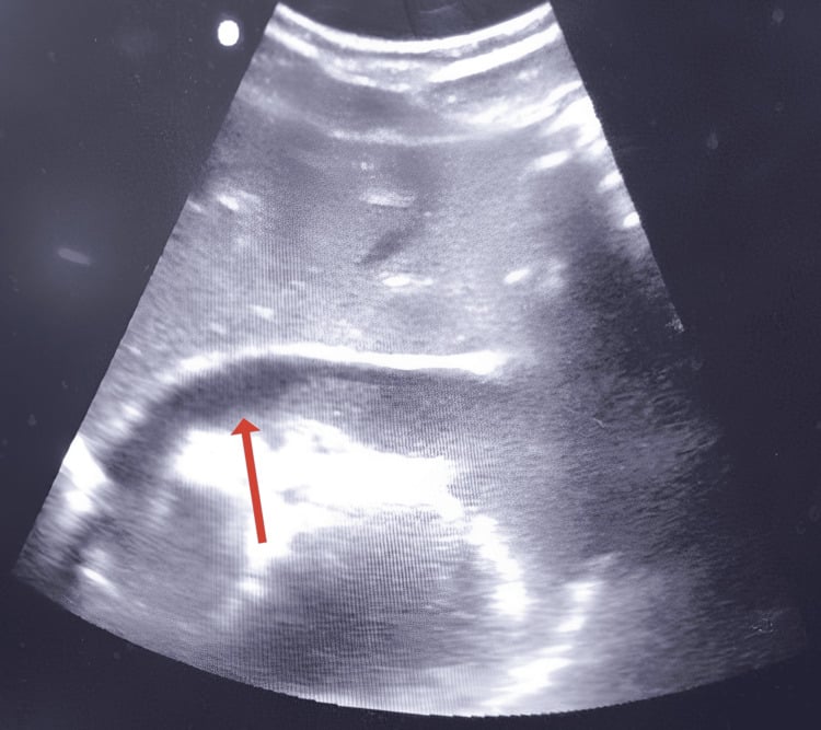
Imaging in the primary survey. A portable chest radiograph for pneumothorax, haemothorax and a widened mediastinum; a pelvic radiograph for pelvic ring disruption; and a FAST scan for free fluid in the abdomen or pericardium.
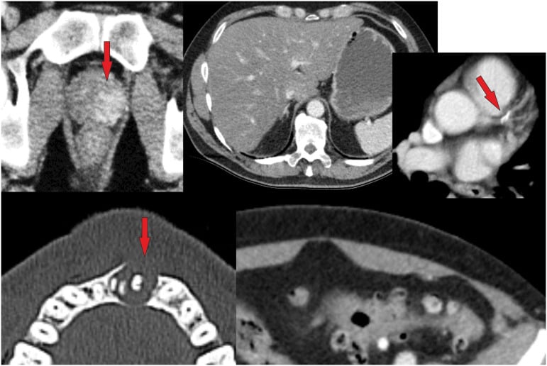
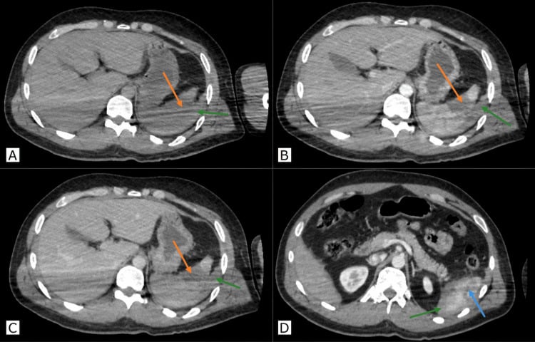
CT once stable. CT of the head shows intracranial haemorrhage and midline shift, and CT of the chest, abdomen and pelvis shows solid organ injury, aortic injury and spine fractures. Review the head, cervical spine, chest, abdomen, pelvis and reconstructed spine systematically, so that the musculoskeletal injury does not distract from intracranial, visceral or axial trauma.
Whole-body CT (head to pelvis with IV contrast) is standard for haemodynamically stable or stabilised major trauma (ISS greater than 15) in mature trauma systems worldwide. It reduces missed injury rate, is faster than sequential imaging and allows early operative planning. Perform after the primary survey and initial stabilisation; a haemodynamically unstable patient who does not respond to resuscitation belongs in theatre or interventional radiology, not the scanner.
Orthopaedic imaging. Once the patient is stabilised:
- Long bone radiographs
- CT spine (cervical, and thoracolumbar if indicated)
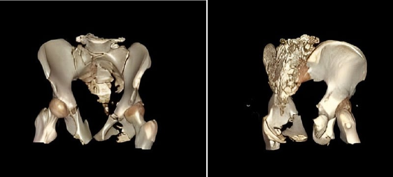
- CT pelvis with 3D reconstruction for complex pelvic ring injuries
- CT angiography if vascular injury is suspected
Patient Categories
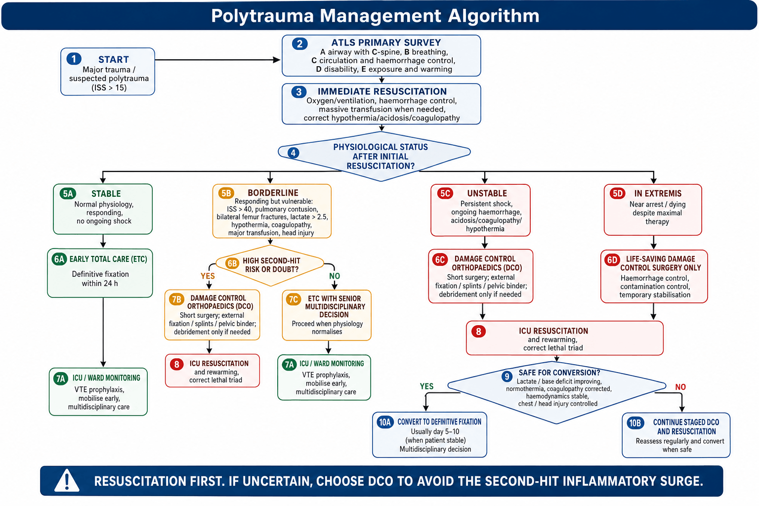
Fixation strategy is decided by physiological status, not injury severity alone.
- Definition
- Normal vital signs, responding to resuscitation, no evidence of ongoing shock
- Management goal
- Complete care
- Orthopaedic fixation
- Early total care (ETC), definitive
- Definition
- Initially responding but fragile, with risk factors
- Management goal
- Protect physiology
- Orthopaedic fixation
- DCO vs ETC, case specific
- Definition
- Persistent shock despite resuscitation, ongoing haemorrhage
- Management goal
- Life over limb
- Orthopaedic fixation
- Damage control orthopaedics (DCO)
- Definition
- Dying, near arrest, on maximal therapy
- Management goal
- Save life only
- Orthopaedic fixation
- Life-saving damage control surgery only
The borderline patient is the hard decision. They are initially responding to resuscitation but carry risk factors, and each of these favours DCO:
- ISS greater than 40
- Hypothermia (less than 35°C)
- Initial systolic BP less than 90 mmHg
- Coagulopathy
- Pulmonary injury (pulmonary contusion)
- Bilateral femoral fractures
- Massive transfusion (greater than 10 units PRBC)
- Lactate greater than 2.5 mmol/L
- Head injury with intracranial pressure issues
If in doubt, DCO is safer: the patient can always go on to definitive fixation later.
Damage Control Resuscitation and the Traumatic-Brain-Injury Exception
The resuscitation partner of DCO. Damage control resuscitation (DCR) rests on haemorrhage control first, permissive (hypotensive) resuscitation, haemostatic or component resuscitation, and minimal crystalloid. The aim is to keep the patient alive to definitive haemostasis without "popping the clot" or worsening TIC.
Permissive hypotension. In the actively bleeding patient before surgical or radiological control, resuscitating to a lower-than-normal blood pressure (a palpable radial pulse, a systolic around 80-90 mmHg, or a MAP near 50 mmHg) limits clot disruption and dilutional coagulopathy. It is a bridge, not a destination, and it is relaxed the moment haemorrhage is controlled.
Blood, not crystalloid. Give balanced blood products (or whole blood) rather than crystalloid; large-volume crystalloid worsens dilution, acidosis, hypothermia and clot instability and is avoided. The massive transfusion protocol:
- 1:1:1 ratio of PRBC, FFP and platelets
- Tranexamic acid early, within 3 hours of injury and ideally under 1 hour
- Fibrinogen supplementation, to the target given under trauma-induced coagulopathy
- Calcium replacement with every 4 units of blood
The TBI exception. Do not permit hypotension in traumatic brain (or spinal cord) injury. The injured brain has lost autoregulation, so hypotension is directly linked to secondary brain injury and death; the target shifts to normotension and adequate cerebral perfusion (avoid a systolic under about 110 mmHg in TBI) with strict avoidance of hypoxia.
When bleeding and TBI coexist, the brain wins the blood-pressure argument, and you resuscitate to a normal pressure. This competing physiology is a major reason a head-injured multitrauma patient is steered toward DCO: a short operation that avoids the intra-operative hypotension of a long definitive case.
Differential Diagnosis: The Shocked Trauma Patient
The exam question is rarely the fracture. It is why this patient is still shocked despite resuscitation. Haemorrhage is the assumed cause until excluded, but other shock states coexist and change management.
- Key clue
- Tachycardia, narrow pulse pressure, responds then relapses; rising lactate/base deficit
- Confirm
- FAST, pelvic XR, CT, fall in Hb
- Immediate action
- Control source, MTP 1:1:1, TXA, DCO/binder
- Key clue
- Distended neck veins, tracheal shift, absent breath sounds, hypoxia
- Confirm
- Clinical (do NOT wait for CXR)
- Immediate action
- Immediate needle/finger decompression then chest drain
- Key clue
- Distended neck veins, muffled heart sounds, PEA, penetrating chest
- Confirm
- FAST/echo (pericardial fluid)
- Immediate action
- Pericardiocentesis or thoracotomy
- Key clue
- Hypotension WITH bradycardia, warm peripheries, cord-level deficit
- Confirm
- Spinal injury on exam/CT
- Immediate action
- Exclude haemorrhage first; vasopressors, maintain MAP
- Key clue
- Arrhythmia, raised troponin, sternal fracture
- Confirm
- ECG, echo, troponin
- Immediate action
- Telemetry, inotropes, treat arrhythmia
- Key clue
- Multiple long-bone fractures each losing 1-2 L; under-resuscitation
- Confirm
- Sum blood loss, log roll, tertiary survey
- Immediate action
- Splint/stabilise fractures, continue resuscitation
Orthopaedic Management
Do not rush to fracture fixation in an unstable patient. Resuscitation and addressing life-threatening injuries takes priority. Orthopaedic DCO is designed to allow life-saving resuscitation to continue.
Damage control orthopaedics. For unstable and borderline patients, DCO stabilises fractures rapidly with minimal physiological insult and buys time for resuscitation. The operation is short, blood loss is minimal, and the second hit is avoided.
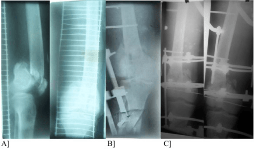
- External fixator for long bone fractures (femur, tibia)
- Pelvic binder or external fixator for pelvic fractures
- Splinting for other fractures
The evidence for DCO. Pape and colleagues' landmark studies showed that unstable and borderline polytrauma patients benefit from DCO with external fixation followed by delayed nailing. Those studies are credited with a reduced ARDS rate, but their randomised EPOFF trial (see Evidence Base) found a difference in IL-6 and IL-8 release and no significant difference in ARDS, sepsis or MOF.
Early total care. Definitive fixation within 24 hours in a stable patient with no evidence of physiological derangement. In stable patients ETC reduces pulmonary complications, DVT and fat embolism, shortens hospital stay and allows early mobilisation. The typical case is an isolated femoral fracture in an otherwise healthy patient, nailed antegrade within 24 hours. ETC suits:
- Isolated extremity fractures
- Stable vital signs throughout resuscitation
- No chest injury
- Lactate normalising
- No coagulopathy
Choose DCO if any one of these is present.
- ISS greater than 40
- ISS greater than 20 with thoracic injury (AIS greater than 2)
- Bilateral femoral fractures with shock
- Hypothermia less than 32°C
- Base deficit greater than 8
- Pulmonary contusion on CT
- Ongoing transfusion (greater than 10 units)
- Raised intracranial pressure
Physiological markers. The laboratory thresholds used in the DCO decision:
- DCO threshold
- Less than -6 to -8 mmol/L
- Significance
- Tissue hypoperfusion
- DCO threshold
- Greater than 2.5-4 mmol/L
- Significance
- Anaerobic metabolism
- DCO threshold
- Less than 35°C
- Significance
- Impaired coagulation
- DCO threshold
- Greater than 1.5
- Significance
- Coagulopathy
- DCO threshold
- Greater than 10 units PRBC
- Significance
- Massive blood loss
- DCO threshold
- Less than 7.25
- Significance
- Severe acidosis
The thresholds are not uniform: the borderline risk factors and this table put hypothermia at 35°C, and the table puts base excess at -6 to -8, while the any-one-present list uses 32°C and a base deficit greater than 8.
The three phases.
- Resuscitation (0-24 hours): ATLS primary survey and resuscitation; life-saving surgery (laparotomy, thoracotomy, craniotomy); the massive transfusion protocol if indicated; pelvic binder and pelvic external fixation; the DCO or ETC decision.
- Stabilisation (24-72 hours): ICU care with ventilation and organ support; correction of the lethal triad; serial lactate and base excess; reassessment of orthopaedic injuries; secondary and tertiary surveys.
- Definitive care: conversion of external fixators to definitive fixation (IM nail, ORIF) and scheduled orthopaedic procedures, with inflammatory markers (CRP, IL-6) monitored before surgery.
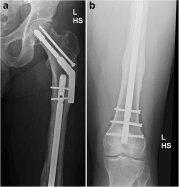
Conversion. The "window of opportunity" for converting the fixator is days 5-10 after injury, and conversion should wait until:
- The patient is haemodynamically stable, off vasopressors
- Lactate has normalised, below 2.5 mmol/L
- Temperature is greater than 36°C
- Coagulopathy is corrected
- CRP is trending down
- Pin sites are clean, with no active infection
Pin sites and timing. Pin site infection (5-10% with external fixation) increases the risk of deep infection when converting to intramedullary nailing: deep infection rises from 2% to 10-15% if the pin sites are colonised. The standard recommendation is to convert within 2 weeks, before biofilm formation; earlier conversion (day 5-7) has lower infection rates than conversion after 2 weeks. If the pin sites are infected, consider:
- A different entry point for the nail
- A staged procedure with an antibiotic spacer or antibiotic cement
- Plate fixation avoiding the pin sites
Surgical Technique
Femoral shaft. A spanning external fixator (hip to knee, or knee-sparing) with two proximal subtrochanteric and two distal supracondylar pins, restoring length and alignment, converted to an antegrade IM nail when the patient is stable. Restoring length and alignment at the first operation makes the conversion easier.
- 5 mm half-pins
- Proximal pins through an anterolateral approach, avoiding the femoral vessels
- Distal pins lateral and supracondylar, avoiding the knee joint
- A single bar or delta frame for stability
Tibial shaft. A spanning external fixator, with two proximal and two distal metaphyseal pins, or immediate IM nailing, which is less of a physiological insult than femoral nailing. Tibial nailing can often proceed even in borderline patients.
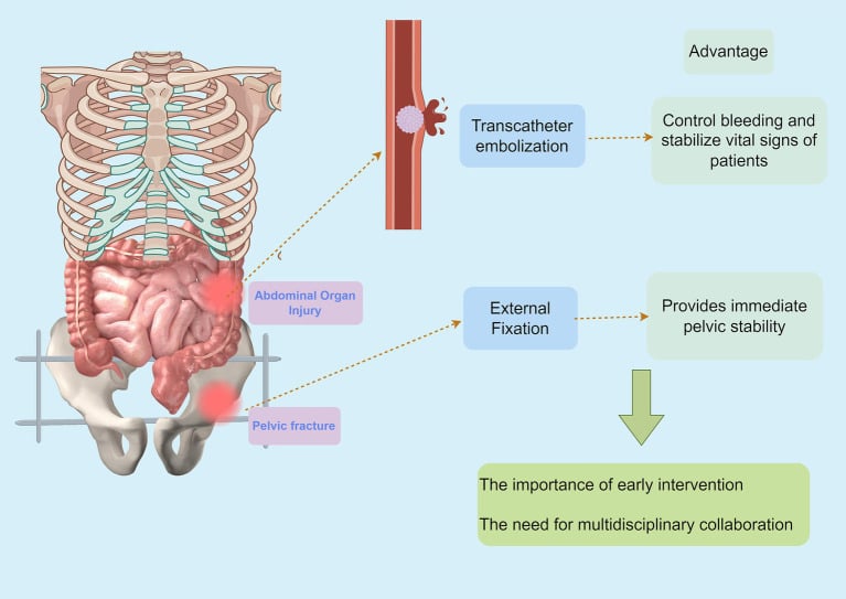
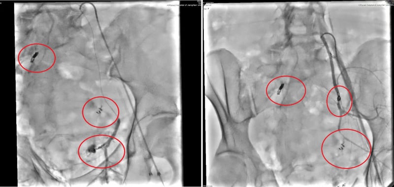
Pelvis. The pelvic binder goes on first, at the greater trochanters, not the iliac crests. A C-clamp addresses the posterior ring if the patient is haemodynamically unstable, and an anterior external fixator uses supra-acetabular or iliac crest pins. Persistent shock after mechanical pelvic stabilisation should prompt targeted haemorrhage control, with angiography and embolisation if arterial bleeding continues. The order and combination of embolisation and external fixation depend on physiology, the bleeding source and local hybrid-theatre capability.
- Indication
- Initial stabilisation
- Advantage
- Non-invasive, immediate
- Indication
- Open book injury
- Advantage
- Rapid, controls anterior ring
- Indication
- Posterior ring instability
- Advantage
- Posterior compression
- Indication
- Definitive fixation
- Advantage
- Minimally invasive if stable
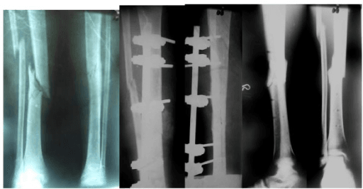
Open fractures. Debridement and washout, temporary external fixation, then delayed soft tissue coverage and conversion to internal fixation.
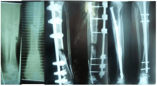
Nailing in polytrauma.
- Unreamed nailing may reduce the pulmonary insult (controversial)
- Femoral nailing causes more fat embolism than tibial nailing
- A vented nailing technique reduces intramedullary pressure
- Damage control nailing (a nail without final fixation) is an option to consider
Complications
Early (0-72 hours).
- Haemorrhagic shock: ongoing blood loss and coagulopathy
- ARDS: from pulmonary contusion, fat embolism or transfusion
- Compartment syndrome: keep a high index of suspicion in unconscious patients
- Missed injuries: see the tertiary survey
- Fat embolism syndrome
Late (days to weeks).
- MODS, from the inflammatory cascade
- Sepsis: nosocomial infection, open fractures
- VTE: high risk in immobile polytrauma patients
- Nonunion and malunion: inadequate initial stabilisation, and nonunion is higher with delayed treatment
- Heterotopic ossification: common with head injury plus extremity fracture
- Stiffness, from prolonged immobilisation
Preventing ARDS. Early stabilisation of long bone fractures reduces pulmonary emboli. Avoid fluid overload, use lung-protective ventilation, and choose DCO in high-risk patients to avoid the second hit.
Fat embolism syndrome. It occurs in 1-10% of long bone fractures and presents with the triad of hypoxia, confusion and petechiae. The mechanical theory holds that fat from the medullary canal enters the circulation; the biochemical theory holds that it is an inflammatory response to circulating fat. Prevention is early fracture stabilisation and avoiding hypoxia, and treatment is supportive (oxygen, ventilation), with no specific therapy.
VTE prophylaxis.
- Mechanical (SCDs, IPC) from admission
- Pharmacological LMWH (enoxaparin 40 mg daily or equivalent) when bleeding risk is acceptable, usually 24-48 hours after injury
- Continued until mobile, typically 2-4 weeks
- An IVC filter is considered if anticoagulation is contraindicated
Postoperative Care
Ongoing resuscitation in ICU. Goal-directed therapy follows lactate clearance and urine output, with blood product replacement per the MTP, active warming to greater than 36°C, and early enteral feeding when possible.
Monitoring. Serial lactate and base excess; daily FBC, coagulation and renal function; CRP and inflammatory markers to time conversion; and compartment checks in sedated patients.
Early mobilisation. Passive ROM once stable, chest physiotherapy (critical), early weight bearing according to the fracture pattern, and psychological support with PTSD screening.
Long-term rehabilitation.
- A multidisciplinary team approach
- A physical therapy programme
- Occupational therapy for ADL retraining
- Chronic pain management
- Return to work assessment
Outcomes
Survival. Mortality correlates strongly with ISS, and geriatric patients have significantly higher mortality for equivalent injury scores. [2,4]
- ISS 16-24: 5-10% mortality
- ISS 25-40: 15-25% mortality
- ISS greater than 40: 30-50% mortality
- ISS 50-75: mortality exceeding 50%
- ISS 75: near 100% mortality
What else decides survival. Proper patient selection is the most critical factor: inappropriate ETC in an unstable patient increases the risk of MODS. Beyond it:
- Age, with mortality increasing significantly over 65
- Injury pattern, head plus chest carrying the worst prognosis
- Time to definitive care
- Trauma centre volume and resources
- Comorbidities
Return to work. After ISS 16-24, 70-80% return to their previous occupation; after ISS 25-40, 50-60% return to work; above ISS 40, the result is often permanent disability.
Quality of life. Physical function is most affected. Psychological sequelae are common (PTSD in 20-40%), chronic pain affects 30-50%, and cognitive impairment follows a significant head injury.
The fractures. Healing is generally good with appropriate treatment, and the nonunion rate for long bones is 5-10%, higher with open fractures. Functional outcome depends on the associated injuries, not just the fracture.
Guidelines, Registries & Global Practice
Global Epidemiology
Trauma is a leading cause of death in those under 45 worldwide; road traffic injury alone causes roughly 1.2 million deaths per year (WHO), disproportionately in low- and middle-income countries (LMICs) where over 90% of road deaths occur despite those regions owning a minority of vehicles. The injury burden is bimodal: high-energy mechanisms in young adults and an expanding cohort of older patients sustaining major injury from low-energy falls.
Side-by-Side Guidelines
- Position on key issues
- ABCDE primary survey; permissive hypotension and haemorrhage control; the universal common language for resuscitation worldwide.
- Position on key issues
- Practice management guidelines support early appropriate fixation of femoral fractures and selective DCO; whole-body CT in stable major trauma.
- Position on key issues
- Open fracture and major trauma standards: senior decision-making, antibiotics within 1h, combined ortho-plastic care, definitive fixation only with soft-tissue cover.
- Position on key issues
- Provides the DCO vs ETC framework and the "safe definitive surgery / early appropriate care" concept tying the timing of fixation to the resuscitated physiological state.
- Position on key issues
- Major trauma pathway: networked care, damage control resuscitation, TXA early, restrictive crystalloid, whole-body CT in adults with suspected multiple injuries.
Antibiotics for Associated Open Fractures
A first-generation cephalosporin (e.g. cefazolin) given within 1 hour of injury is the global standard; gram-negative cover (e.g. an aminoglycoside) is added for higher-grade/contaminated wounds and high-dose penicillin where clostridial/soil contamination is a concern. Exact agents follow local microbiology and antimicrobial-stewardship policy.
Registry & System Evidence
National and regional trauma registries (e.g. UK TARN, US National Trauma Data Bank, the German TraumaRegister DGU, Victorian State Trauma Registry) consistently show that inclusive, networked trauma systems with direct transfer to a major trauma centre reduce mortality for ISS greater than 15 patients. Registry data also drive audit of "early appropriate care" timing and missed-injury rates.
High- vs Limited-Resource Practice
- High-resource: rapid whole-body CT, interventional radiology/embolisation, ROTEM/TEG-guided component therapy, ICU-supported staged conversion.
- Limited-resource: clinical and FAST-led triage, external fixation as both damage-control AND sometimes definitive treatment, judicious whole-blood or ratio-based transfusion, and earlier reliance on operative haemorrhage control where angio-embolisation is unavailable. DCO principles remain valid and are often MORE relevant where physiological monitoring is constrained.
Related pages: Damage Control Orthopaedics develops the staged-fixation strategy in full, including the conversion window and the borderline patient; ATLS Primary Survey for the ABCDE sequence this page assumes; Pelvic Ring Injuries for the fracture that most often drives the haemorrhage, and for binder, packing and embolisation decisions; Femoral Shaft Fractures for the injury at the centre of the entire ETC-versus-DCO literature - every landmark trial carded here is about the femur; Fat Embolism Syndrome for the complication that reaming a long bone in a physiologically marginal patient can precipitate; Open Fracture Management for the debridement and coverage pathway that runs alongside resuscitation; Compartment Syndrome for the diagnosis most easily missed in a sedated, intubated polytrauma patient who cannot report pain; and Spinal Cord Injury for neurogenic shock, the differential that changes the vasopressor decision and forbids permissive hypotension.
Controversies & Areas of Uncertainty
- Borderline thresholds are not absolute. No single lactate, base-deficit or ISS value cleanly separates DCO from ETC; the decision is a dynamic physiological judgement (response to resuscitation), not a number.
- DCO vs "Early Appropriate Care" (EAC). Modern data (Vallier and others) suggest many patients previously labelled borderline can undergo definitive fixation early once lactate, pH and base deficit are corrected — pushing practice from blanket DCO back toward earlier definitive surgery in the adequately resuscitated patient.
- Reamed vs unreamed nailing in chest trauma. The historical fear that reaming worsens pulmonary outcome in patients with thoracic injury is not strongly supported by later evidence; reaming improves union, and the chest injury itself, not the reaming, drives ARDS.
- PROPPR did not prove 1:1:1 saves lives. The primary mortality endpoint was negative; the rationale for balanced ratios rests on improved haemostasis and reduced exsanguination, with growing interest in whole blood.
- Whole-body CT ("pan-scan") radiation and overtriage. Survival benefit is debated in stable, lower-acuity patients (REACT-2 was neutral overall); selective imaging is reasonable when injury burden is clearly limited.
- Conversion timing window. The classic "5-10 day" window to convert ex-fix to nail balances inflammatory quiescence against pin-tract colonisation; some advocate conversion within 2 weeks before biofilm forms, but the ideal day is individualised.
MCQ Practice Points
Q: When should damage control orthopaedics (DCO) be used instead of early total care (ETC)?
A: DCO indicated: ISS greater than 40, ISS greater than 20 with additional thoracic injury (AIS greater than 2), bilateral femur fractures with shock, hypothermia less than 32°C, base deficit greater than 8, coagulopathy, ongoing transfusion requirements. ETC appropriate: Borderline patients stabilised by resuscitation, no chest trauma, ISS less than 20.
Q: What is the second hit phenomenon and how does DCO prevent it?
A: The first hit is the initial traumatic insult causing SIRS. The second hit is additional surgical trauma (e.g., prolonged orthopaedic surgery) that amplifies inflammation and can precipitate multi-organ dysfunction syndrome (MODS). DCO prevents this by using temporary stabilisation (external fixation) to minimise surgical stress during the inflammatory phase, with definitive fixation delayed 5-10 days when the patient is optimised.
Q: What is the optimal timing for femur fracture fixation in polytrauma?
A: In stable patients, early intramedullary nailing (within 24 hours) reduces pulmonary complications, ICU stay, and hospital stay. In unstable patients (shock, coagulopathy, base deficit greater than 6), use external fixation initially with conversion to IM nail at 5-10 days when inflammatory markers normalise and patient is optimised.
Q: How do you minimise fat embolism risk during IM nailing in polytrauma?
A: Techniques include: reaming cautiously or use unreamed nails in chest trauma, venting the femur during nailing, avoiding over-pressurisation of the canal, surgical stabilisation early (prevents ongoing marrow extravasation from mobile fracture). Monitor for fat embolism syndrome: petechial rash, hypoxia, confusion (classic triad).
Clinical Decision Scenarios
Practise clinical reasoning and management decisions out loud
“A motorcyclist arrives with bilateral femoral shaft fractures, pulmonary contusion, and initial BP 80/50. He has received 6 units of blood and now has BP 100/60. How do you manage his femoral fractures?”
“A 45-year-old male is brought in after a high-speed RTA. He is hemodynamically unstable (BP 70/40) with obvious pelvic deformity after an APC-III type injury. How do you proceed?”
“A 19-year-old female presents with a closed femoral shaft fracture and a minor chest injury (RIB AIS 1). She is hemodynamically stable, lactate 1.2, and pH 7.4. What is your management plan?”
ATLS
- ABCDE primary survey
- Life-threatening injuries first
- Resuscitate before fracture care
Lethal Triad
- Hypothermia
- Acidosis
- Coagulopathy
DCO vs ETC
- Stable → ETC (within 24h)
- Borderline/Unstable → DCO (ex-fix)
- In extremis → Life-saving surgery only
DCO Technique
- External fixators for long bones
- Pelvic binder for pelvis
- Short surgery, minimal blood loss
Evidence Base
Key Evidence for Polytrauma Management
- Bone et al. (1989): Landmark study showing early femoral stabilization (less than 24h) reduces ARDS, fat embolism, and hospital stay in stable patients
- Multiple subsequent studies confirmed benefit of early long bone fixation in stable patients
-
Pape et al. (2002): Defined DCO concept - external fixation for unstable patients avoids second hit
-
Scalea et al. (2000): Showed DCO reduces pulmonary complications in borderline patients
-
Conversion timing: Days 5-10 optimal (Pape et al.)
-
CRASH-2 Trial (2010): Tranexamic acid within 3 hours reduces mortality
Evidence-based protocols focus on physiological optimization and balanced resuscitation.