Shepherd Fracture | Cedell Fracture | FHL Impingement
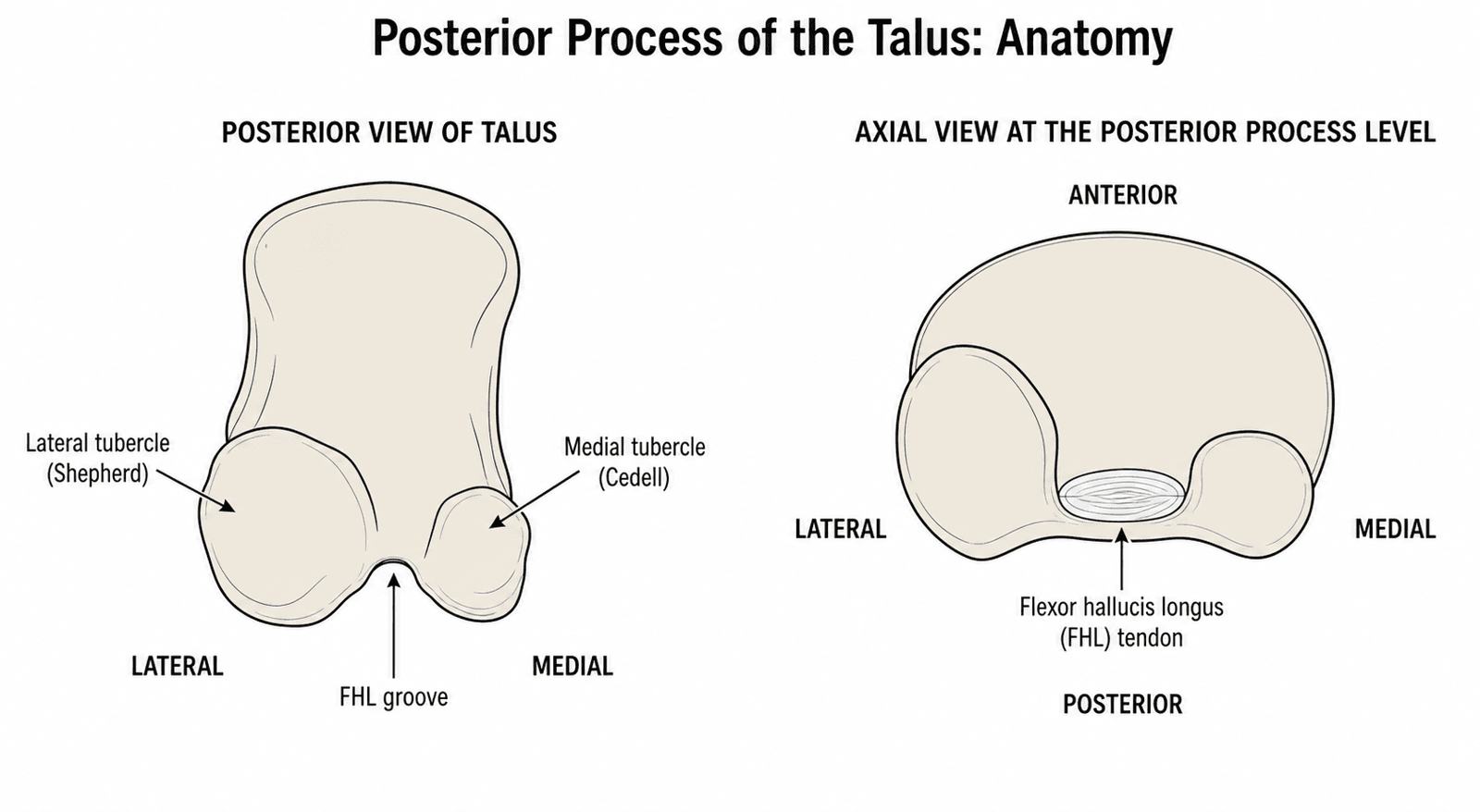
- Posterior process has 2 parts: Lateral tubercle (Shepherd fracture) and medial tubercle (Cedell fracture)
- FHL impingement: Lateral tubercle fracture can cause FHL tendon impingement - pain with great toe flexion
- Often missed: Subtle on X-ray, may be misdiagnosed as ankle sprain or os trigonum
- ORIF if displaced: about 2mm is the conventional trigger for ORIF (a working figure, not a validated cut-off), to prevent nonunion and FHL impingement
- Excision for small fragments: Small fragments causing impingement can be excised
- “Shepherd fracture = lateral tubercle of posterior process, FHL impingement risk
- “Cedell fracture = medial tubercle of posterior process, rare
- “FHL impingement causes pain with great toe flexion (hallux flexor hallucis longus)
- “Often missed - high index of suspicion, CT if suspected
Overview and Epidemiology
The posterior process of the talus has two parts, a lateral tubercle and a medial tubercle, with the groove for flexor hallucis longus (FHL) running between them. A fracture of the lateral tubercle is the Shepherd fracture and a fracture of the medial tubercle the Cedell fracture, named after Shepherd and Cedell. The Shepherd fracture is the more common and can cause FHL impingement; the Cedell fracture is rare. Both are often missed initially.
Incidence. Posterior process fractures are quoted at less than 1% of talus fractures. The 21% posterior tubercle figure in the CT evidence below is a share of talar body fractures in one trauma-centre cohort, not a population incidence. The peak age is 20-40 years, in sport and trauma, with no clear sex predominance.
Mechanism. Forced plantarflexion is the classic mechanism. Direct trauma to the posterior ankle and a ligament avulsion, by the posterior talofibular or deep deltoid ligament, are the others. Associated injuries are ankle injuries and other foot trauma, surveyed below.
Anatomy and Pathophysiology
The posterior process. It projects from the posterior aspect of the talar body, gives attachment to ligaments and forms the FHL groove. The lateral tubercle is the larger of the two and forms the lateral border of the groove, and its fracture is the more common. The medial tubercle is smaller and forms the medial border.
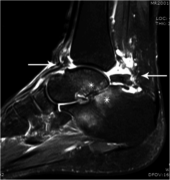
The tendon in the groove. FHL, the flexor of the great toe, runs between the two tubercles. A lateral tubercle fracture fragment can impinge on it, which is why great toe flexion hurts, and ORIF or excision of the fragment relieves the impingement.
Classification Systems
The fracture is described three ways. Location names it and determines the treatment approach, displacement decides between conservative and operative care, and fragment size decides between fixation and excision.
- Eponym
- Shepherd fracture
- Features
- More common
- Treatment
- ORIF or excision
- Complication
- FHL impingement
- Eponym
- Cedell fracture
- Features
- Rare
- Treatment
- ORIF if displaced
- Complication
- Rare
- Eponym
- -
- Features
- Complete posterior process fracture
- Treatment
- ORIF
- Complication
- FHL impingement
Displacement. A step-off of less than 2mm is treated conservatively, and one greater than 2mm by ORIF or excision. The 2mm figure is a working threshold borrowed from intra-articular fracture practice, not one validated for this fracture: no cited series defines a numeric cut-off, and fragment size, cartilage damage and subtalar instability weigh alongside it.
Fragment size. A large fragment is reconstructible and ORIF is preferred, with better outcomes than excision. A small fragment may not be reconstructible, and excision is then acceptable, with a good outcome if there is no impingement.
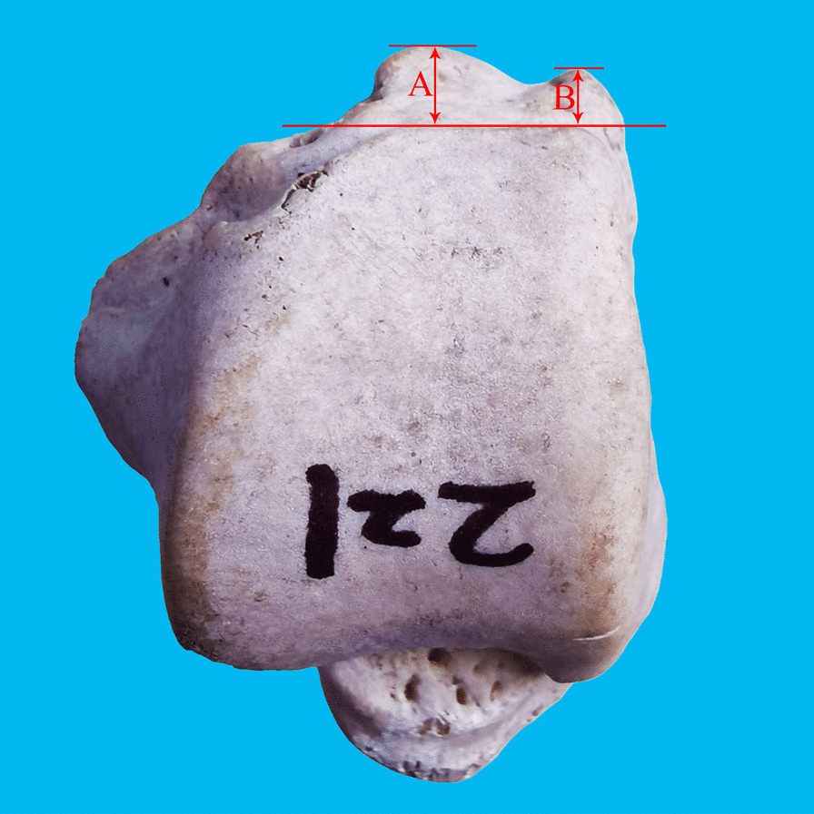
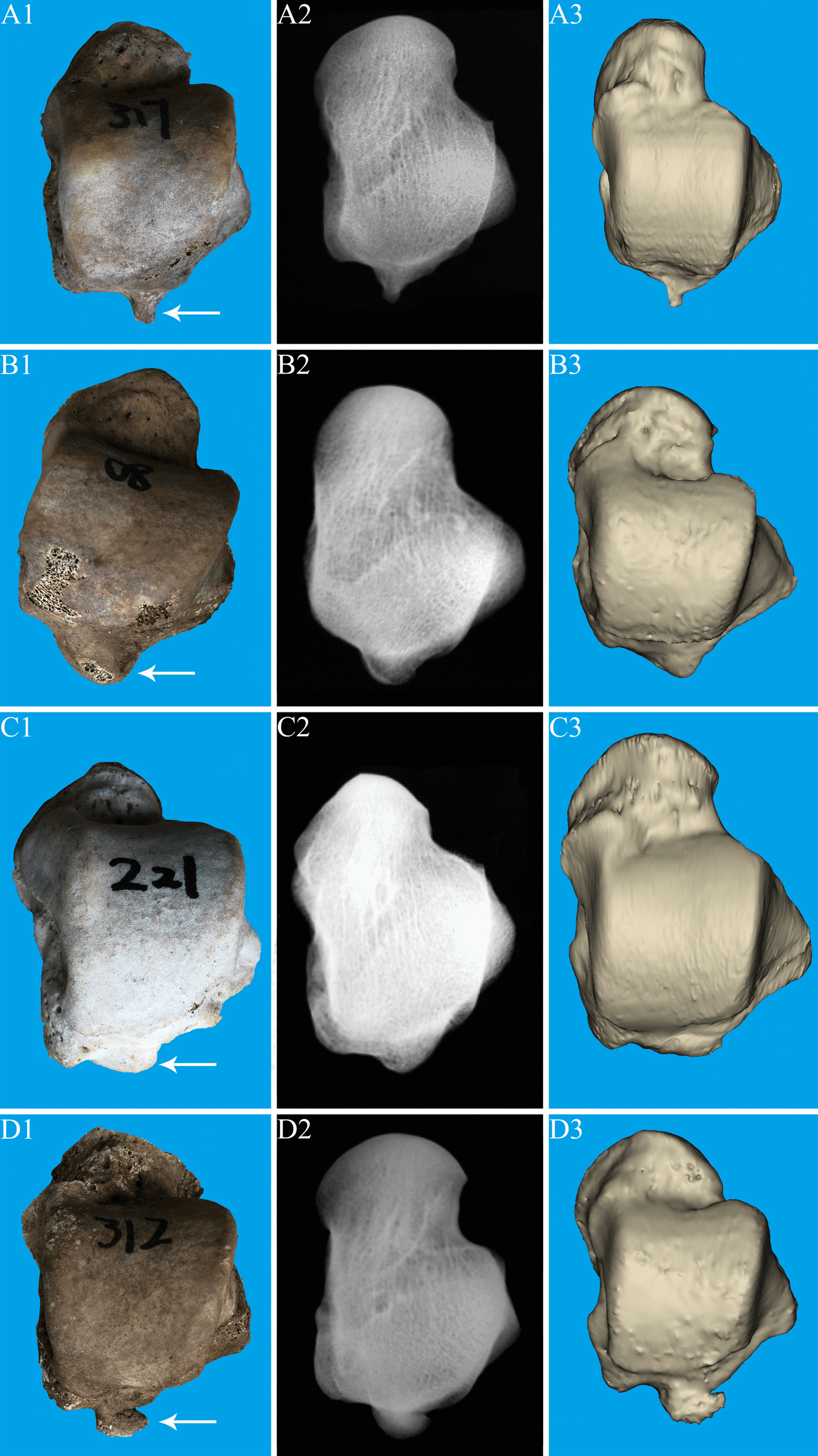
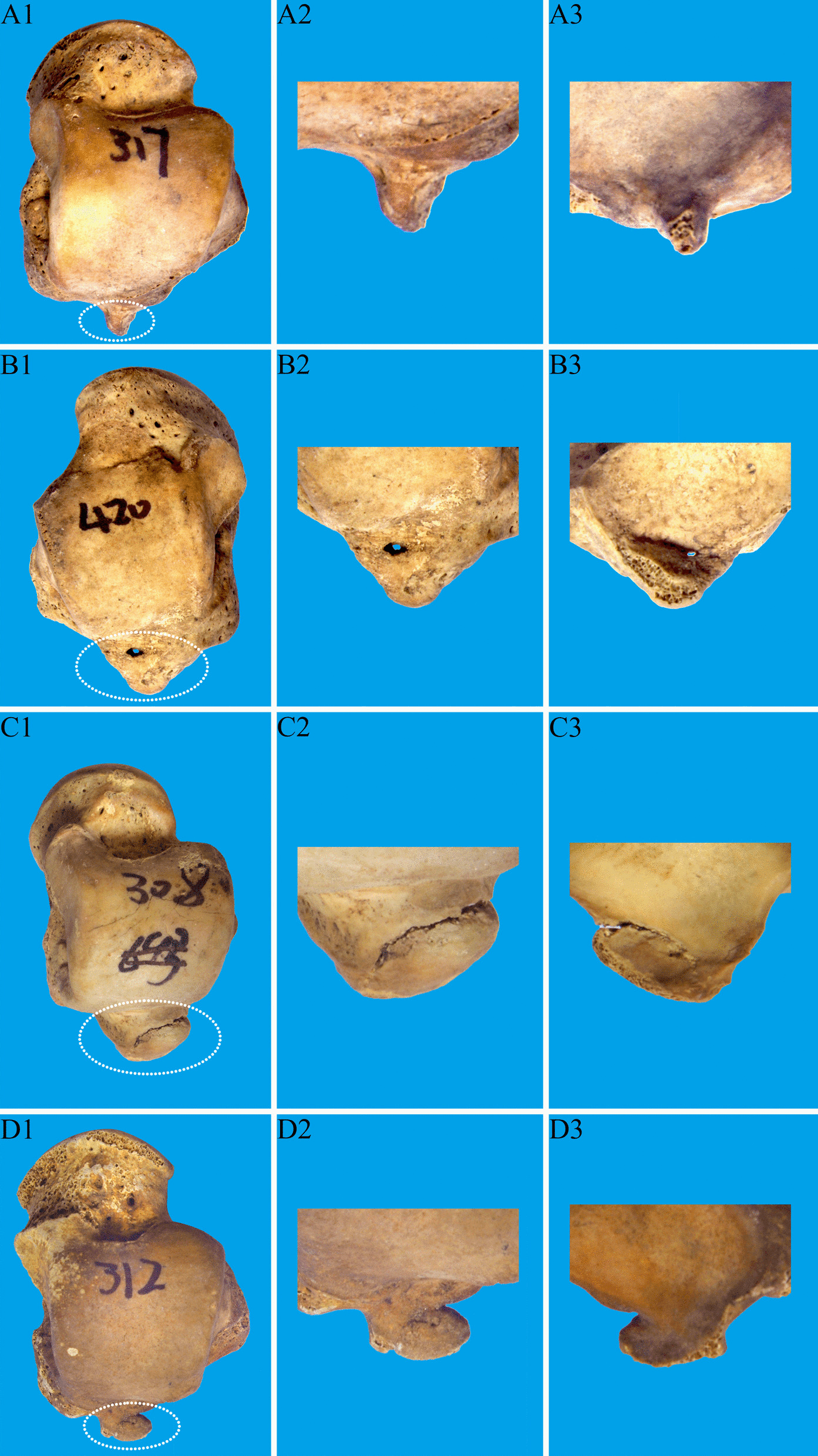
Morphology. The posterior process itself varies in shape, and one label can conceal different fragment geometry.
Clinical Assessment
History. Posterior ankle pain after forced plantarflexion, direct trauma to the posterior ankle or a sports injury. The swelling is localised to the posterior ankle and weight bearing is painful, so walking is difficult. A lateral tubercle fracture impinging on FHL adds pain on great toe flexion.
Examination. There is swelling in the posterior ankle and ecchymosis, which may be delayed; deformity is rare. The tenderness is over the posterior process, and the ankle joint itself is usually not tender. Ankle range of motion may be limited, and so may plantarflexion.
The FHL signs. The FHL impingement test is pain with active great toe flexion, and it is the key clinical test. Pain on passive hallux movement, which stretches FHL, is the other clue, and in the reported Cedell case it was a key clinical clue.
Pain with active great toe flexion indicates FHL impingement from a lateral tubercle fracture. This is pathognomonic for posterior process fracture with impingement.
Differential diagnosis. The single most important distinction is an acute fracture versus a symptomatic os trigonum, because the treatment, prognosis and medicolegal consequences of a missed fracture differ greatly.
- Key clinical clue
- Acute trauma, point tenderness, pain on passive FHL stretch
- Imaging discriminator
- Sharp irregular fracture line, marrow oedema on MRI, sclerotic acutely-displaced fragment on CT
- Management contrast
- May need ORIF or excision if displaced
- Key clinical clue
- Insidious, repetitive plantarflexion (dancers, footballers)
- Imaging discriminator
- Smooth corticated ossicle with rounded margins, no marrow oedema
- Management contrast
- Rest, then excision/hindfoot endoscopy if refractory
- Key clinical clue
- Pain at end-range plantarflexion, positive forced-plantarflexion test
- Imaging discriminator
- May show os trigonum or Stieda process, soft-tissue oedema
- Management contrast
- Activity modification, injection, endoscopic decompression
- Key clinical clue
- Pain along FHL behind medial malleolus, crepitus, triggering
- Imaging discriminator
- Fluid in FHL sheath on MRI, no bony fragment
- Management contrast
- Conservative, FHL release if refractory
- Key clinical clue
- Dorsiflexion/inversion injury, lateral tenderness below fibula
- Imaging discriminator
- Fragment at lateral process, not posterior, on mortise/CT
- Management contrast
- ORIF or excision per fragment size
- Key clinical clue
- Inversion injury, ATFL tenderness, negative bony signs
- Imaging discriminator
- Normal bone, soft-tissue swelling only
- Management contrast
- Functional rehabilitation
Investigations
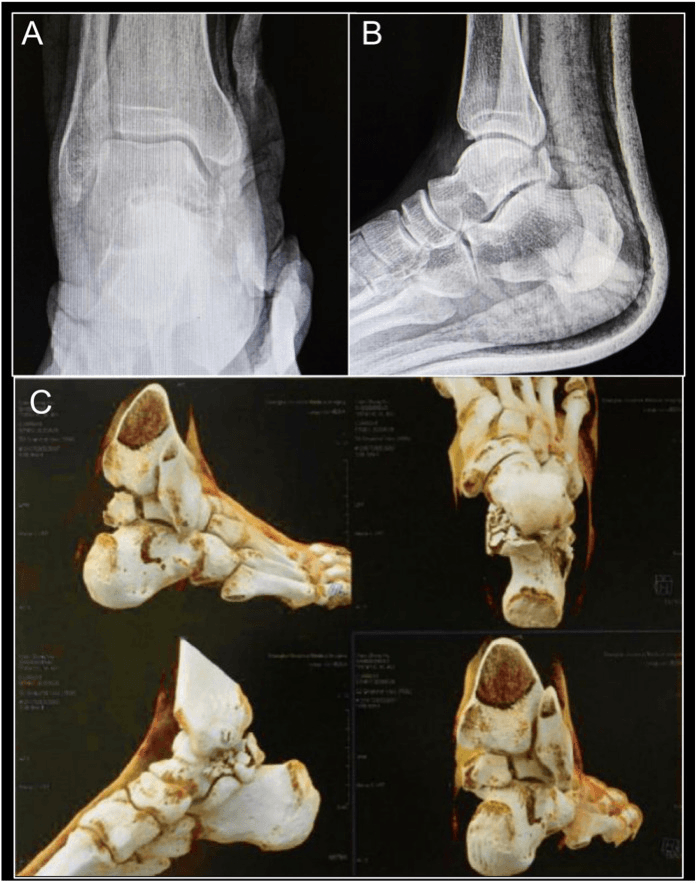
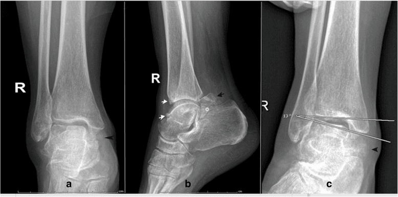
Why it is missed. Plain films detect only around three-quarters of talar fractures, a radiographic sensitivity of roughly 74% against CT. The posterior process is among the hardest regions to see, because the fibula, normal ossification variants and overlapping tarsal bones obscure it on routine projections, and the fracture is misdiagnosed as an os trigonum or an ankle sprain. A high index of suspicion is needed, and CT is often what makes the diagnosis.
Radiographs. A few tailored views materially improve detection before cross-sectional imaging:
- Lateral - the best plain view. It profiles the posterior process, but the fracture is often subtle and is easily mistaken for an os trigonum synchondrosis or overlooked entirely
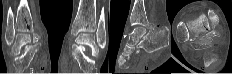
- External-rotation lateral - taken with the foot in approximately 30 degrees of external rotation, it rotates the posteromedial (Cedell) tubercle clear of overlapping bone. In the reported Cedell case it revealed a hypolucent shadow posterior to the talus that routine views had missed, and CT then confirmed the fracture
- AP - may show the fracture, but is less reliable than the lateral
- AP/mortise and oblique - help localise the tubercle and exclude a lateral process fracture or a malleolar injury
- Contralateral comparison films - help distinguish a smooth, corticated, often-bilateral os trigonum from a sharp, non-corticated acute fracture line
The os trigonum and Stieda process as normal variants are covered in the os trigonum and posterior ankle impingement topics; here the point is how to make the fracture visible.
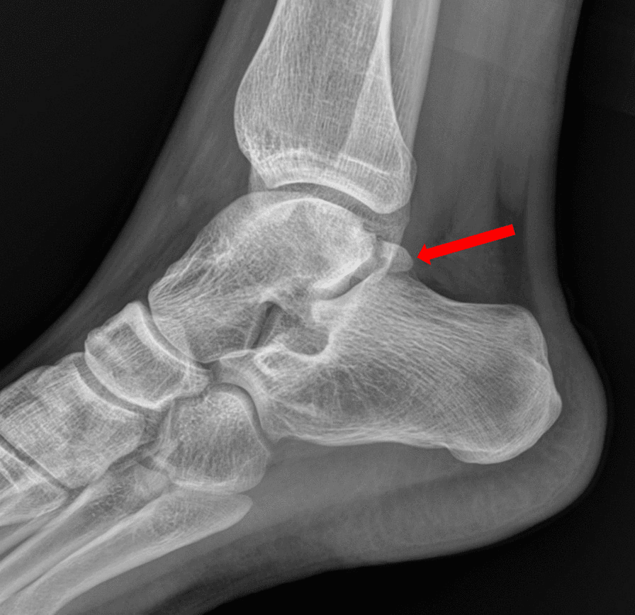
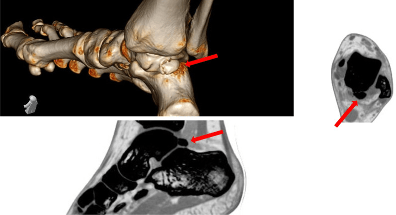
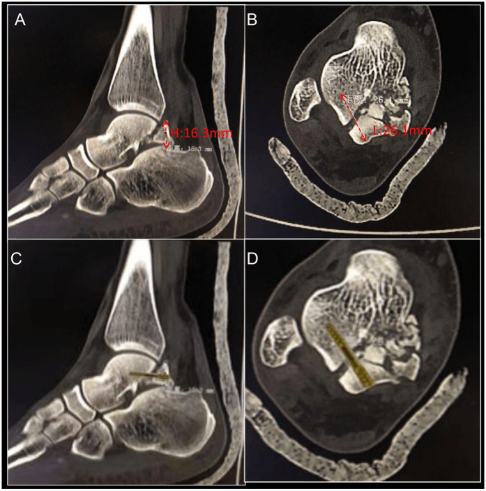
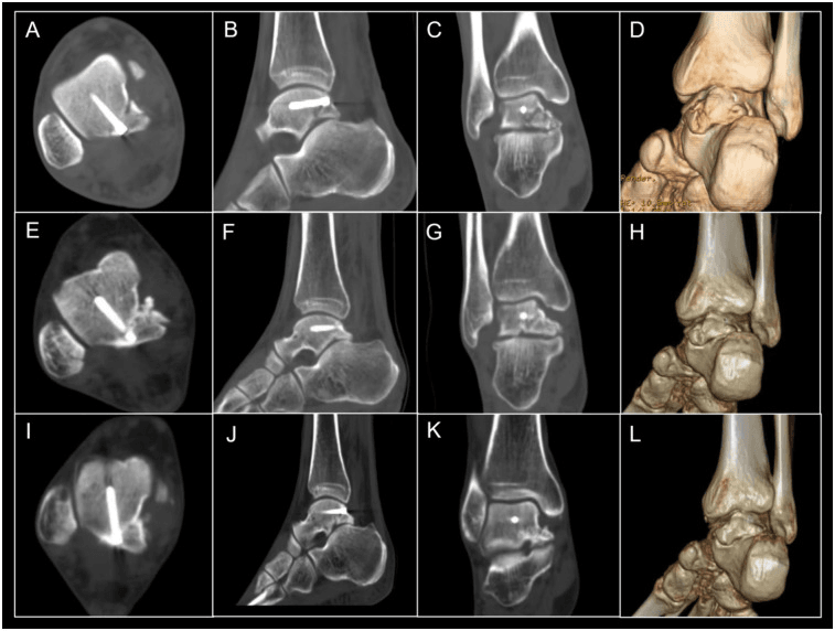
CT. CT is the reference standard, and it added information in 93% of talar fractures in trauma-centre data. It is recommended when the fracture is suspected clinically but the radiographs are negative, when displacement is unclear on the radiographs, and when planning surgery. It shows the fracture pattern, the displacement (the step-off is measured), the fragment size and the fragment's relationship to FHL.
If the history (forced plantarflexion) and focal posterior tenderness fit, a "normal" routine ankle series is not reassuring. Add an external-rotation lateral and compare with the contralateral foot, and if still equivocal obtain a CT rather than repeating the same view.
Associated Injuries and the Hindfoot Survey
A sentinel injury. A posterior process fracture is frequently a marker of a higher-energy hindfoot injury rather than an isolated event. In a CT-based trauma-centre series, talar fractures were accompanied by adjacent joint subluxation or dislocation in more than 66% of cases and by an adjacent fracture in more than 72%. Fall from height was the commonest mechanism (44.8%) in the largest posterior-process outcome series, and the visible fragment may be the least of the patient's problems.
What to look for. Deliberately exclude each of these, and route to the dedicated topics:
- Subtalar (peritalar) dislocation - the posterior process can avulse during a subtalar dislocation, so reduction and stability of the subtalar joint take priority (see subtalar-dislocations)
- Other talus fractures - lateral process (snowboarder's), talar neck and talar body fractures frequently coexist and change the whole management plan (see lateral-process-talus-fractures, talus-fractures, talar-body-fractures)
- Calcaneal and malleolar fractures - high-energy plantarflexion and inversion loads the entire hindfoot, so scrutinise the calcaneus and malleoli
- Chondral and subtalar facet damage - posterior process fragments involve the posterior subtalar facet, and articular involvement drives both the fixation decision and the later risk of post-traumatic subtalar arthritis (see subtalar-arthritis)
In practice. Keep a low threshold for whole-foot CT in any suspected posterior process fracture, and document the neurovascular and soft-tissue assessment: the posteromedial neurovascular bundle and the skin over a displaced fragment. Never plan fixation of the visible fragment in isolation; exclude subtalar dislocation and lateral process, neck and body fractures first. Counsel that prognosis is often set by the associated injury rather than by the process fragment, and in the delayed or high-energy case it usually is.
Management Algorithm
The decision. Suspicion comes first, because the fracture is often missed. Once it is confirmed, displacement, FHL impingement and fragment size set the treatment:
- Non-displaced (less than 2mm step-off) with no FHL impingement - conservative treatment
- Displaced (greater than 2mm) with a large fragment - ORIF is preferred. It relieves FHL impingement and prevents nonunion
- Displaced with a small fragment, or causing FHL impingement - excision is acceptable. It relieves the impingement and is better than malunion
Non-operative treatment. The indications are a non-displaced fracture (less than 2mm step-off), no FHL impingement, and patient preference. The protocol is a short leg cast, non-weight bearing, for 6-8 weeks, with serial radiographs to monitor healing.
Surgical indications. Operate within 2 weeks if possible.
- Absolute: displacement greater than 2mm step-off, FHL impingement (pain with great toe flexion), a large fragment
- Relative: a small fragment that is symptomatic, failed conservative treatment
Surgical Technique
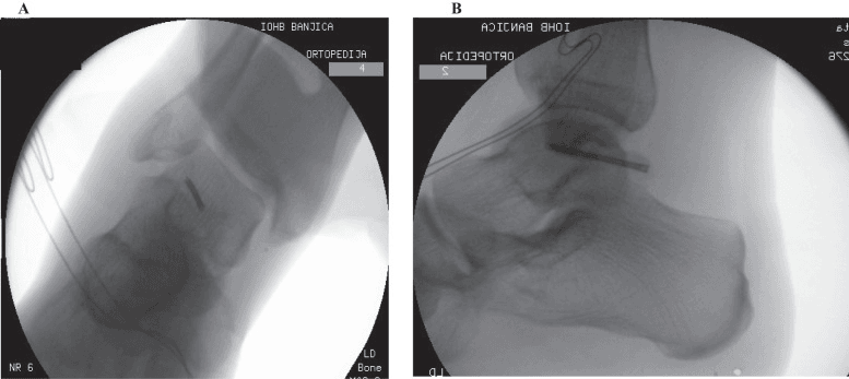
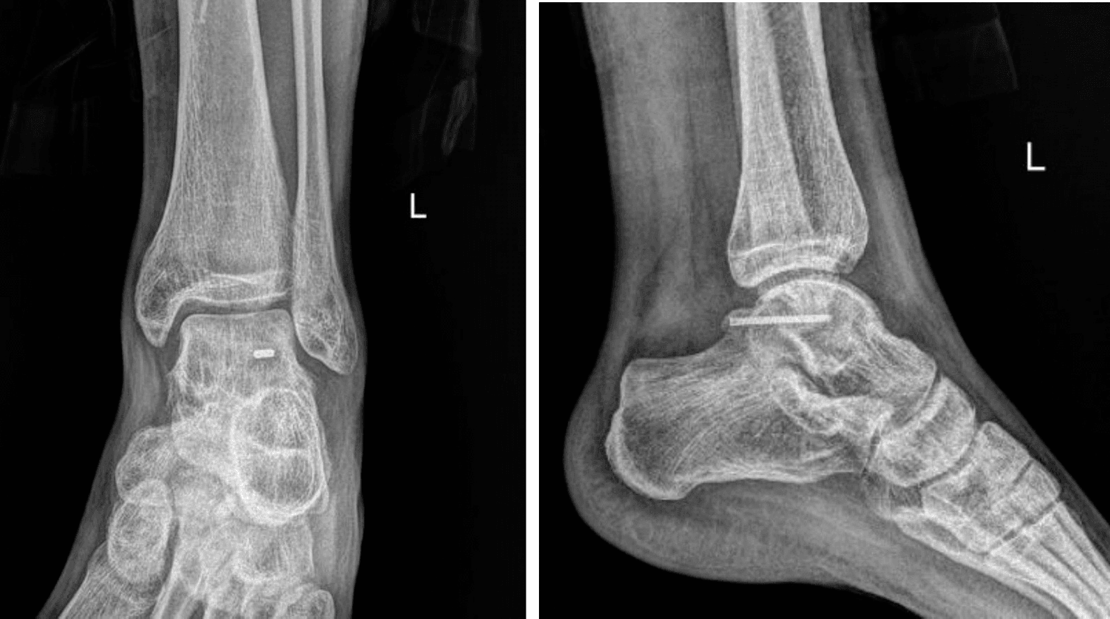
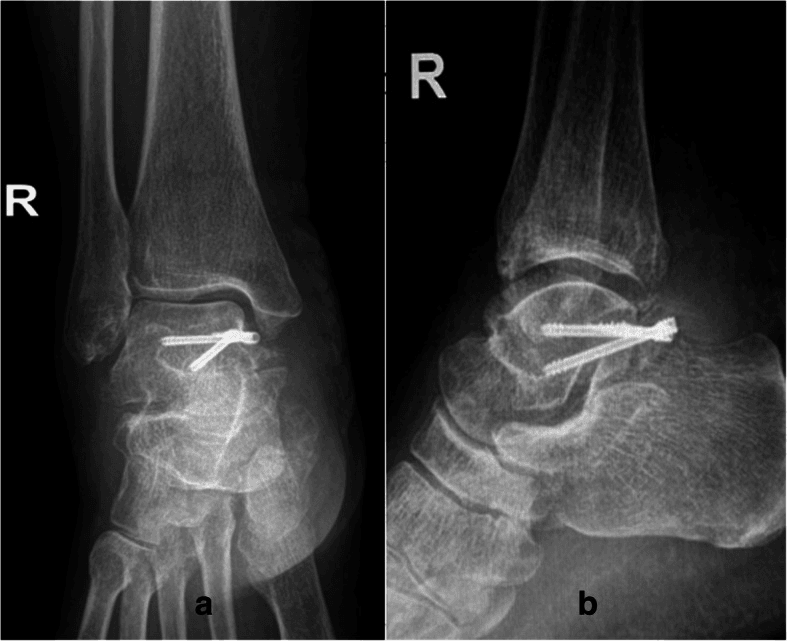
ORIF for the large fragment. The indications are a large, reconstructible fragment, displacement greater than 2mm and FHL impingement. Fixation relieves the impingement, prevents nonunion and gives better outcomes than excision for large fragments. The screw can be planned on CT beforehand, measuring the fragment and the screw length, with a virtual screw fit used to avoid articular penetration.
- Exposure through a posteromedial or posterolateral approach to the posterior process
- Protection - identify and protect the FHL tendon
- Reduction - anatomical reduction of the fragment
- Fixation - lag screws (2.0-2.7mm) or mini-fragment screws
- Verification - confirm the reduction and hardware position fluoroscopically
Complications
The incidence figures below are conventional teaching ranges, not rates measured in any cited series.
- Incidence
- 20-30% if untreated
- Risk Factors
- Lateral tubercle fracture
- Prevention/Management
- ORIF or excision relieves
- Incidence
- 10-15%
- Risk Factors
- Displacement, inadequate fixation
- Prevention/Management
- Rigid fixation, bone graft if needed; revision fixation with bone graft
- Incidence
- 30-40%
- Risk Factors
- Subtle on X-ray
- Prevention/Management
- High index of suspicion, CT
- Incidence
- 5-10%
- Risk Factors
- Posterior approach
- Prevention/Management
- Careful technique
Postoperative Care
Immobilisation. A short leg cast or boot, with the foot non-weight bearing for 6-8 weeks. Ankle range-of-motion exercises start in the first six weeks only if stable; otherwise ankle movement waits for cast removal. Physiotherapy covers FHL stretching and strengthening.
Protocol. Rehabilitation is staged by week:
- Weeks 0-6 - short leg cast, non-weight bearing, elevation to reduce swelling, ankle range-of-motion exercises if stable
- Weeks 6-8 - cast removal, transition to a walking boot, progressive weight bearing
- Weeks 8-12 - full weight bearing and progressive activity, with return to sport at 3-4 months
Outcomes and Prognosis
The percentages below are conventional teaching ranges; the cited literature (case reports and series of 29-33 patients) does not validate any numeric rate. For displaced fractures as a group, treated by ORIF or excision, the conventional figure is 75-85% good results.
- Success rate
- 80-90% (union, pain relief)
- Return to pre-injury level
- 75-85%
- FHL impingement
- Relieved in 90-95%
- Success rate
- 75-85% (pain relief)
- Return to pre-injury level
- 70-80%
- FHL impingement
- Relieved in 85-90%
- Success rate
- 85-90% (union, pain relief)
- Return to pre-injury level
- 80-85%
- FHL impingement
- Rare if non-displaced
Long-term prognosis. Displacement and delayed treatment are the risk factors for chronic FHL impingement.
Guidelines, Registries & Global Practice
These are rare injuries with no dedicated society guideline and no registry capture (talar process fractures are not separately tracked by arthroplasty registries such as the NJR, AJRR or AOANJRR, which record joint replacements rather than fixation of small process fractures). Practice is therefore guided by trauma principles and the small published literature, summarised below for a global readership.
- Position on posterior process fractures
- Anatomic reduction and stable fixation of displaced intra-articular talar fragments; protect FHL and neurovascular bundle via posteromedial/posterolateral approaches
- Practical implication
- Reconstruct large displaced fragments; lag-screw or mini-fragment fixation
- Position on posterior process fractures
- No consensus on optimal treatment; decision driven by size, location, displacement, cartilage damage and subtalar instability
- Practical implication
- Individualise — non-op for non-displaced, ORIF or excision for displaced
- Position on posterior process fractures
- Endorses two-portal hindfoot endoscopy for excision in athletes; supports operative management of extended fractures
- Practical implication
- Consider minimally invasive excision for small fragments in active patients
- Position on posterior process fractures
- High index of suspicion; CT for any suspected but radiographically occult posterior ankle fracture
- Practical implication
- Plain-film sensitivity is only ~74% — image early to avoid missed injury
Global epidemiology and practice variation
- Rarity and demographics: posterior process fractures are uncommon (the posterior tubercle accounts for around 21% of talar body fractures in CT-based trauma-centre data, but isolated process fractures are far rarer). They cluster in young, active adults — sport (especially football, dance, running and climbing) and falls from height are the dominant mechanisms worldwide.
- High-resource settings: ready CT/MRI access, fluoroscopy and hindfoot endoscopy support early diagnosis and minimally invasive excision or fixation; missed-diagnosis rates fall when CT is used liberally for unresolving posterior ankle pain.
- Limited-resource settings: reliance on plain radiographs increases missed and delayed diagnoses; pragmatic management leans toward immobilisation for presumed minor injuries and open excision/ORIF when surgery is indicated, since advanced imaging and endoscopic equipment may be unavailable.
Posterior process talus fractures are a common viva topic. Know that Shepherd fracture = lateral tubercle (FHL impingement), Cedell fracture = medial tubercle (rare), FHL impingement test = pain with great toe flexion, ORIF is preferred for large reconstructible fragments and excision is acceptable for small ones, and that these injuries are frequently missed because plain-film sensitivity is only around 74% — request CT when suspected. On the displacement threshold, say what is true: 2mm is a conventional working figure carried over from intra-articular fracture practice generally, not a validated cut-off for this fracture — the best available review states there is no consensus on management and lists fragment size, displacement, cartilage damage and subtalar instability as the criteria, without numbers. Be prepared to discuss FHL impingement, the differential against os trigonum, and the ORIF-versus-excision decision.
Related pages: Talus Fractures is the umbrella covering neck, body and dome injuries, and the Hawkins classification that does NOT apply here; Lateral Process Talus Fractures for the snowboarder's fracture at the other corner of the talus - the same story of a missed "sprain", a different mechanism and a different approach; Os Trigonum and Posterior Ankle Impingement for the single most important differential: a smooth, corticated, rounded ossicle is an os trigonum, a sharp irregular non-corticated edge is a Shepherd fracture, and CT is what separates them; FHL Tendinitis for the tendon running in the groove between the two tubercles, whose pain on passive hallux movement is the clinical clue here; Subtalar Arthritis for the endpoint of an intra-articular fragment left unreduced, and Subtalar Dislocations for the injury these fractures accompany; Osteochondral Lesion of the Talus for the other cause of persistent post-sprain ankle pain with normal radiographs; and Lateral Ankle Instability for the diagnosis these are routinely mislabelled as.
Controversies and Areas of Uncertainty
The evidence base for posterior process talus fractures is limited to small retrospective series and case reports — there are no randomised trials. The following points are genuine areas of debate worth raising in a viva.
There is no agreed fragment-size cut-off for fixation versus excision. The decision rests on whether the fragment is reconstructible, the degree of subtalar articular involvement and surgeon experience rather than a validated number. Excision (open or endoscopic) gives reliable symptom relief for small fragments.
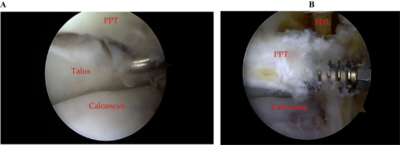
Two-portal hindfoot endoscopy is increasingly used for excision and even fixation, with good return-to-sport data in athletes, but comparative evidence against open techniques is absent and the learning curve and neurovascular proximity are real.
The widely quoted "greater than 2mm" surgical threshold is pragmatic and extrapolated from intra-articular fracture principles, not derived from outcome studies specific to this fracture.
Whether medial tubercle (Cedell) fractures are deltoid avulsions, FHL-retinaculum avulsions or direct compression injuries remains unresolved, with case evidence supporting more than one mechanism.
MCQ Practice Points
Q: What is the difference between Shepherd and Cedell fractures? A: Shepherd fracture = lateral tubercle of posterior process (more common, causes FHL impingement) - Cedell fracture = medial tubercle of posterior process (rare). Both are parts of posterior process of talus.
Q: What causes FHL impingement in posterior process talus fractures? A: Lateral tubercle fracture impinges on FHL tendon - FHL (Flexor Hallucis Longus) tendon runs between lateral and medial tubercles. Lateral tubercle fracture causes pain with great toe flexion. ORIF or excision relieves impingement.
Q: How do you test for FHL impingement? A: Active great toe flexion test - Pain with active great toe flexion indicates FHL impingement from lateral tubercle fracture. This is pathognomonic for posterior process fracture with impingement.
Q: When is ORIF preferred over excision for posterior process talus fractures? A: Large fragments (reconstructible) with displacement greater than 2mm - ORIF preserves anatomy and relieves FHL impingement (good results in most reported cases). Excision is acceptable for small comminuted fragments causing impingement (good results in most reported cases).
Q: Why are posterior process talus fractures often missed initially? A: Subtle on X-ray, often misdiagnosed as os trigonum or ankle sprain - 30-40% are missed initially. High index of suspicion needed, especially with FHL impingement symptoms. CT recommended if suspected clinically but X-ray negative.
Exam Viva Scenarios
Practise clinical reasoning and management decisions out loud
“A 28-year-old athlete presents with posterior ankle pain after forced plantarflexion injury. Clinical examination shows pain with active great toe flexion (FHL impingement test positive). Lateral X-ray shows fracture of lateral tubercle of posterior process. CT shows 3mm displacement.”
“A 25-year-old dancer presents with posterior ankle pain and pain with great toe flexion. Clinical examination shows positive FHL impingement test. CT shows small comminuted fragment of lateral tubercle (under 25% of process) with 4mm displacement. The fragment is not reconstructible.”
“A 34-year-old recreational footballer is referred with 14 months of persistent posteromedial ankle pain after an inversion injury initially diagnosed and treated as a sprain. Plain films were reported as normal. He has posteromedial tenderness and pain on passive great toe extension. How do you approach this?”
Key Concepts
- Posterior process has 2 parts: lateral tubercle (Shepherd) and medial tubercle (Cedell)
- FHL tendon runs between lateral and medial tubercles
- Lateral tubercle fracture causes FHL impingement
- Often missed initially (30-40%) - high index of suspicion needed
Classification
- Shepherd fracture: Lateral tubercle (more common, FHL impingement)
- Cedell fracture: Medial tubercle (rare)
- Non-displaced: Less than 2mm - conservative (good results typical)
- Displaced: Greater than 2mm - ORIF or excision (good results typical)
- Tubercle Location: Lateral (Shepherd) vs Medial (Cedell)
Treatment
- Non-displaced, no impingement: Conservative (cast, NWB 6-8 weeks)
- Large fragment, displaced: ORIF (relieves impingement; good results typical)
- Small fragment, impingement: Excision (relieves impingement; good results typical)
- FHL impingement: Requires surgical treatment (ORIF or excision)
Surgical Technique
- ORIF: Posteromedial or posterolateral approach, protect FHL, lag screws (2.0-2.7mm)
- Excision: Same approach, remove fragment, smooth edges
- Protect FHL tendon throughout
- Verify impingement relieved (test great toe flexion)
Complications
- FHL impingement: conventionally quoted 20-30% if untreated; surgery relieves most
- Nonunion: conventionally quoted 10-15% (prevent with rigid fixation)
- Missed diagnosis: 30-40% initially missed
- Wound complications: conventionally quoted 5-10% (posterior approach)
Evidence Base
Talar Process Fractures — Anatomy, Mechanism and Management
- Posterior process = medial + lateral tubercle, FHL groove between them
- Mechanism: forced hyperplantarflexion/inversion or PTFL avulsion
- Missed diagnosis risks malunion, nonunion and subtalar arthritis
Outcomes After Operative vs Nonoperative Treatment
- 29 patients, mean 6-year follow-up, mean AOFAS 78.7
- Operative management gave good function in extended fractures
- Non-operative care justified only in selected minor injuries
Medial Tubercle (Cedell) Fractures — Systematic Review
- 33 pooled Cedell cases; 58% sport, 73% misdiagnosed as sprain
- Diagnosis often delayed beyond one year
- Immobilisation, ORIF or excision all give reasonable-to-good results
Talar Fracture Patterns and Radiographic Sensitivity (CT)
- Posterior tubercle = 21% of talar body fractures
- Radiographic sensitivity only 74% vs CT
- High rate of associated dislocation and adjacent fracture
Safe Zone for Percutaneous Posterior Screw Fixation (3D CT)
- Lateral tubercle is optimal posterior-to-anterior screw entry
- Safe-zone geometry defined on 100 CT scans
- 3D planning may shorten surgery and reduce complications
Cedell Fracture — Diagnostic Dilemma and Fixation
- Painful passive FHL movement is a diagnostic clue
- 30-degree external-rotation lateral view aids detection
- Mini-open ORIF achieved union and full ROM at 6 months