Reverse Bankart | McLaughlin | Capsulorrhaphy | Bone Loss
- 2-5% of dislocations but frequently MISSED
- FAIR position of risk (flexion, adduction, internal rotation)
- Reverse Bankart = posterior labral tear
- McLaughlin lesion = reverse Hill-Sachs on anterior humeral head
- Posterior bone loss rare but important
- “Jerk test and Kim test specific for posterior labral lesions
- “Lightbulb sign on AP X-ray = locked posterior dislocation
- “Seizure patients have high rate of bilateral posterior dislocations
- “Avoid surgery for voluntary dislocators
Overview and Epidemiology
Posterior instability accounts for 2-5% of shoulder dislocations, and it matters out of proportion to that because most acute posterior dislocations are missed at first presentation. The figure usually quoted, 50-79%, is a range drawn from older case series rather than a single prospective study or one validated number, so the safe exam answer is "the majority", with the range offered if pressed.
Why it is missed. The AP film can look almost normal, because the humeral head is still roughly concentric in that projection. Suspect a posterior dislocation after a seizure or electrocution, or whenever the shoulder will not externally rotate, and always get an axillary or modified axial view.
Who. The rate is higher in patients with epilepsy, and after a seizure the dislocation is often bilateral. Athletes are the other group: overhead athletes, backhanders and linemen. Males are affected more often than females.
Mechanism. Four routes lead to a posterior shoulder:
- Seizure or electrocution - massive muscle contraction, in which the internal rotators (subscapularis, pectoralis, latissimus dorsi) overpower the external rotators
- Trauma - a posterior force with the arm in flexion, adduction and internal rotation, the FAIR position of risk
- Sport - repetitive microtrauma in the overhead athlete
- Voluntary - with a psychological component
SEEPPosterior Dislocation Causes
Hook:SEEP causes arm to SEEP backward!
The mirror of anterior instability. Each feature of posterior instability has an anterior counterpart.
- Posterior
- 2-5%
- Anterior
- 95%+
- Posterior
- FAIR (flex, add, IR)
- Anterior
- ABER (abd, ext rot)
- Posterior
- Reverse Bankart (posterior)
- Anterior
- Bankart (anterior)
- Posterior
- McLaughlin (anterior)
- Anterior
- Hill-Sachs (posterior)
- Posterior
- Posterior glenoid (reverse bony Bankart)
- Anterior
- Anterior glenoid
Pathophysiology and Mechanisms
The posterior restraints. The posterior glenoid rim gives bony stability, and the fibrocartilaginous posterior labrum deepens the socket. The capsular restraints are the posterior capsule and the posterior band of the IGHL, with infraspinatus and teres minor, the external rotators, as the muscular stabilisers. The posterior capsule is thinner than the anterior, so its stabilisation is less robust.
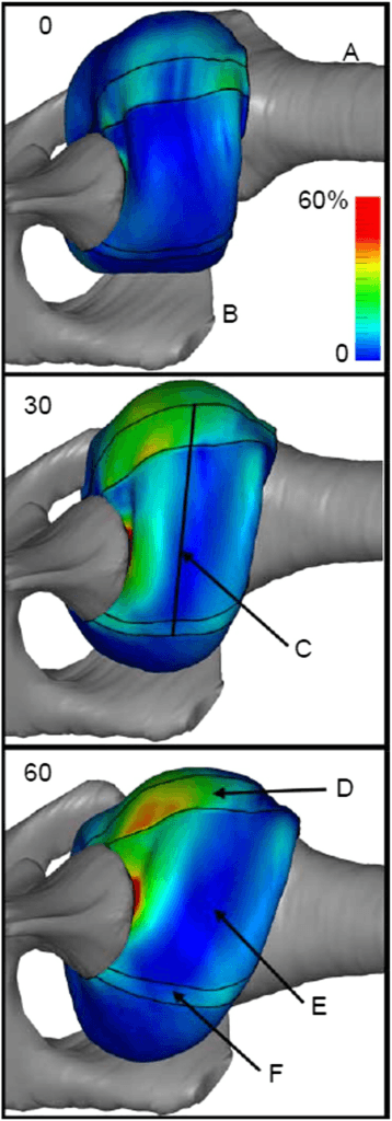
Why the FAIR position is the position of risk. Flexion, adduction and internal rotation place the humeral head against the posterior capsule and labrum.
The lesions. The posterior labral tear, the reverse Bankart, is the key lesion for surgical stabilisation. On the humeral side the McLaughlin lesion, or reverse Hill-Sachs, is an impaction fracture of the anteromedial humeral head, and it can engage on the posterior glenoid. A reverse bony Bankart adds posterior glenoid bone loss, and recurrent instability stretches the posterior capsule into laxity.
Locked posterior dislocation = humeral head trapped behind glenoid with McLaughlin engaging posteriorly. Requires specific reduction technique or open reduction.
The Posterior Capsulolabral Lesion Spectrum
Not every posterior instability is a reverse Bankart. Recognising the full spectrum matters because the site of the lesion dictates where the repair must be placed: a glenoid-sided labral repair will fail if the true lesion is on the humeral side.
- What it is
- Avulsion of the posterior labrum from the glenoid
- Why it matters
- Commonest lesion; glenoid-sided suture-anchor repair
- What it is
- Incomplete, concealed avulsion of the deep posteroinferior labrum beneath an intact superficial labrum
- Why it matters
- Looks normal at arthroscopy - must be probed, converted to a complete tear and repaired
- What it is
- Posterior labral avulsion with a glenoid rim fracture / bone loss
- Why it matters
- Bone loss must be quantified; large defects need a bone block
- What it is
- Posterior labrocapsular periosteal sleeve avulsion - labrum and capsuloperiosteum stripped as an intact sleeve (the posterior analogue of an ALPSA)
- Why it matters
- A medialised, malunited sleeve must be mobilised and re-tensioned to the rim
- What it is
- Humeral-sided avulsion of the posterior band of the IGHL from the humeral neck
- Why it matters
- A glenoid-sided labral repair will NOT address it - it needs humeral-sided repair; a classic cause of 'failed' stabilisation
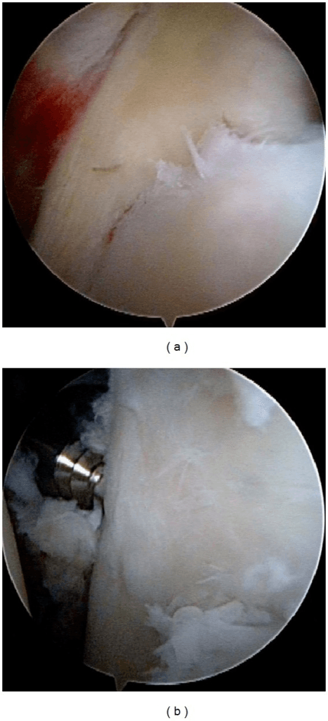
If posterior instability recurs after an apparently sound labral repair, think of a missed reverse HAGL or a concealed Kim lesion. On MR arthrogram, look for capsular extravasation or a J-sign of the posterior capsule.
Classification Systems
Posterior instability is classified three ways: by the type of instability, by the size of the humeral head defect, and by how long the shoulder has been out.
By type. The type sets the treatment approach.
- Mechanism
- Seizure, electrocution
- Features
- Often locked, bilateral
- Treatment Approach
- Closed reduction if possible
- Mechanism
- Previous dislocation
- Features
- Apprehension, recurrence
- Treatment Approach
- Surgical stabilisation
- Mechanism
- Microtrauma, laxity
- Features
- Athletes, overhead sports
- Treatment Approach
- Rehab first, surgery if fails
- Mechanism
- Patient induced
- Features
- Psychological aspect
- Treatment Approach
- Avoid surgery
The Stanmore polar framework (structural / muscular-patterning / atraumatic) increasingly guides whether to operate, replacing the rigid traumatic-versus-voluntary dichotomy.
By size of the McLaughlin lesion. Defect size dictates treatment strategy:
- Small, under 20% - stable after reduction, with a good prognosis; may do well without specific treatment
- Moderate, 20-40% - may engage
- Large, over 40% - high risk of engaging and re-dislocation
The threshold for engagement is also quoted as greater than 25-40% of the articular arc, at which the defect may engage and lock on the posterior glenoid.
By chronicity. Chronic dislocations have worse outcomes.
- Acute - within 6 weeks; closed reduction may be attempted
- Chronic locked - more than 6 weeks; usually needs open surgery
- Recurrent - repeated episodes; surgical stabilisation, after a failed rehabilitation trial if the instability is atraumatic
Clinical Assessment
History. Ask about the mechanism: a seizure, an electrocution, or trauma in the FAIR position. The pain is posterior, the patient cannot externally rotate, and there may have been previous episodes. A voluntary dislocator can demonstrate the instability.
Examination. The arm is held adducted and in internal rotation. Blocked external rotation is the key finding, and inability to externally rotate should raise suspicion of a locked posterior dislocation. The jerk and Kim tests are specific for posterior labral lesions:
- Jerk test - patient supine, arm at 90 degrees of flexion and internal rotation; the examiner applies an axial load and adducts the arm across the body. Posterior subluxation followed by a clunk as it reduces when the arm is returned to neutral is positive for a posterior labral tear
- Kim test - patient seated, arm at 90 degrees of abduction; a posterior force is applied with elevation, and posterior subluxation is positive
- Posterior apprehension - loading in the FAIR position
- Key discriminator
- Pain/apprehension in FAIR; positive jerk and Kim tests
- Best test
- MR arthrogram + dynamic exam
- Key discriminator
- Apprehension in ABER (abduction-external rotation)
- Best test
- Anterior apprehension/relocation
- Key discriminator
- Global laxity, sulcus sign, often bilateral, atraumatic
- Best test
- Sulcus sign, generalised laxity (Beighton)
- Key discriminator
- Fixed internal rotation, blocked external rotation
- Best test
- Axillary radiograph (humeral head behind glenoid)
- Key discriminator
- Pain on overhead throwing, positive O'Brien
- Best test
- MR arthrogram, dynamic labral tests
- Key discriminator
- Weakness, scapular winging, no true subluxation
- Best test
- Scapular assistance test, cuff strength
Investigations
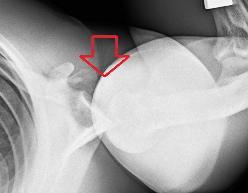
Radiographs. Take an AP, an axillary and a scapular Y view. The axillary view is essential and diagnostic, showing the posterior position of the head that the AP can hide.
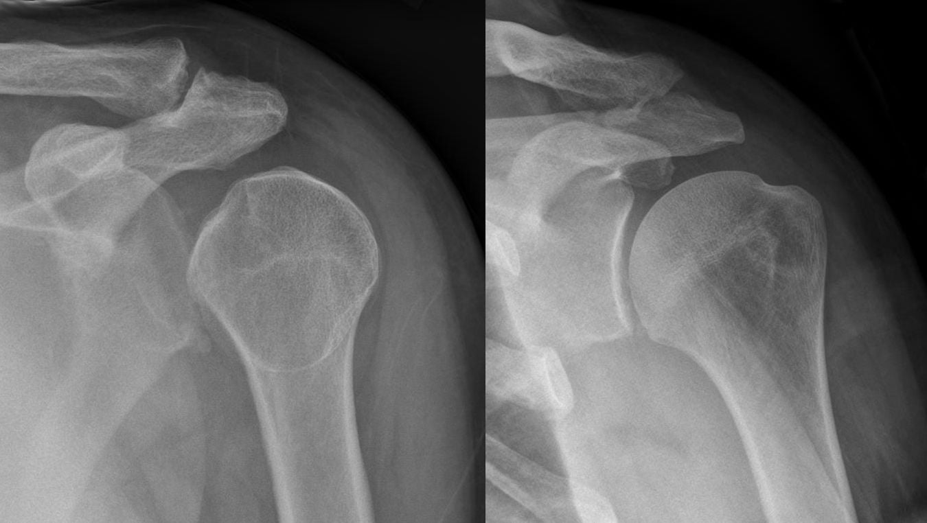
Lightbulb sign = internally rotated humerus on AP view looks like a lightbulb. Indicates locked posterior dislocation due to inability to externally rotate. DO NOT MISS.
Two further signs:
- Rim sign - increased space between the humeral head and the glenoid
- Through sign - overlap of head and glenoid on the Y view
CT. CT characterises bone loss and quantifies the McLaughlin lesion, and it is essential for planning bone grafting or arthroplasty. It gives three things:
- Posterior glenoid bone loss as a percentage
- McLaughlin lesion size as a percentage of the arc
- 3D reconstructions for surgical planning
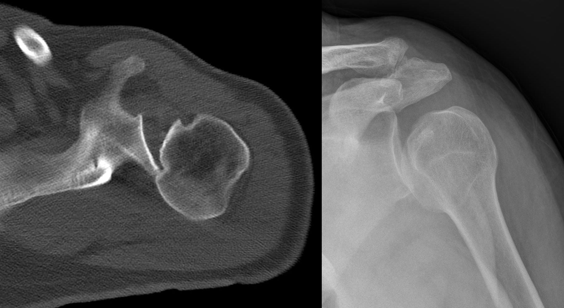
MRI. MRI shows the posterior labral tear (reverse Bankart), posterior capsular stretching or disruption, and the McLaughlin lesion as marrow oedema or a defect. It also picks up associated rotator cuff injuries, especially in older patients. MR arthrography enhances labral visualisation.
Management Algorithm
The acute dislocation. Four steps, in order:
- Diagnose - high suspicion with a seizure or electrocution; get the axillary view and check whether the dislocation is locked
- Size the defect - CT to assess the humeral head defect
- Closed reduction - traction and gentle external rotation, which may succeed if the dislocation is less than 6 weeks old
- Immobilise - brace in neutral or external rotation, avoiding internal rotation
Neutral versus external-rotation bracing after reduction or repair lacks robust evidence; the fixed point is keeping the arm out of internal rotation.
Recurrent instability. Atraumatic instability is treated conservatively first, with physical therapy, rotator cuff and scapular strengthening, and activity modification. If that fails, surgery addresses each lesion present: posterior labral (reverse Bankart) repair, posterior capsulorrhaphy, and any bone loss. Voluntary dislocators are the exception: surgical outcomes are poor, so surgery is avoided and psychological counselling is part of their care.
The McLaughlin lesion. The key is to fill or address the engaging defect, and its size decides how:
- Under 20% - conservative, or soft-tissue treatment only
- 20-40% - the McLaughlin procedure, which transfers subscapularis into the defect, or an osteochondral allograft
- Over 40% - osteochondral allograft or hemiarthroplasty
Surgical Technique
Arthroscopic posterior labral repair. The patient is in the beach chair or lateral decubitus position, with a standard posterior viewing portal, an anterior portal and a posterolateral working portal. Capsular plication is added for laxity.
- Mobilise the labrum from the glenoid
- Prepare a bleeding bone bed on the glenoid rim
- Place 2-4 suture anchors on the glenoid face
- Shuttle the sutures through the labrum
- Tie the knots to reduce the labrum to the glenoid
Posterior capsulorrhaphy. Indicated for capsular laxity or a patulous capsule, and often combined with labral repair. It can be done arthroscopically or open, shifting the redundant capsule superiorly with plication stitches through the capsule. Thermal capsular shrinkage has been abandoned because of chondrolysis and capsular necrosis, and is mentioned in a viva only to be rejected.
Avoid overtightening posterior capsule which leads to loss of internal rotation and anterior subluxation. Balance is important.
Posterior glenoid bone loss. Loss greater than 25-30% requires bone grafting. The exact loss at which soft-tissue repair fails is less certain: it is often quoted as 20-25%, is not well defined, and remains debated. Posterior bone grafting is less common than anterior bone loss surgery, and the graft options are:
- Iliac crest bone graft
- Distal clavicle autograft
- Fresh frozen allograft
The graft is placed through an open posterior approach and fixed to the glenoid with screws. Posterior bone-block techniques are less standardised than the anterior Latarjet, with no high-level comparative data on open versus arthroscopic blocks.
Glenoid Version, Dysplasia and Posterior Glenoid Osteotomy
Posterior bone loss (a reverse bony Bankart) is addressed by a bone block. A separate structural problem is excessive glenoid retroversion or posteroinferior dysplasia / hypoplasia, which biomechanically directs the head posteriorly and is a recognised cause of soft-tissue repair failure.
Measuring version. Version is measured on axial CT, commonly by the Friedman method, which uses a line from the medial scapular border to the glenoid centre as the reference axis. The normal glenoid is slightly retroverted, around 2 to 7 degrees. Pathological retroversion is generally taken as more than about 10 degrees, and it both causes instability and predicts failure of an isolated labral repair.
Dysplasia. Posteroinferior glenoid dysplasia, a deficient, rounded or hypoplastic ("lazy-J") posteroinferior glenoid, is a developmental contributor identified on CT or MRI.
Posterior glenoid (opening-wedge) osteotomy. It is reserved for instability driven by excessive retroversion, often quoted as more than about 10 to 15 degrees, where soft-tissue work alone is likely to fail. It is technically demanding with a historically high complication rate, so it is used selectively and in specialist centres:
- Graft-site fracture
- Avascular necrosis
- Intra-articular penetration
- Over-correction producing anterior subluxation
Complications
Recurrence after posterior stabilisation runs at 5-15% in experienced hands. It is worse with voluntary instability, bone loss and unrecognised pathology.
- Cause
- Missed pathology, undertightening
- Prevention
- Address all lesions
- Management
- Revision surgery
- Cause
- Overtightening, immobilisation
- Prevention
- Balanced repair
- Management
- Physical therapy
- Cause
- Overtightening posterior
- Prevention
- Avoid overtightening
- Management
- Rare, may need revision
- Cause
- Portal placement
- Prevention
- Safe portal placement
- Management
- Observation usually
Postoperative Care
The FAIR position stresses the posterior repair, so it is avoided during early rehabilitation and range of motion is progressed to full gradually.
Posterior Stabilisation Rehabilitation
Sling in neutral rotation (not internal rotation). Pendulums, elbow and wrist ROM.
Progressive ROM. Avoid combined flexion, adduction, internal rotation. Start external rotation.
Progressive strengthening. Rotator cuff and scapular stabilisers. Pool exercises.
Sport-specific training. Full return at 6-9 months if strength and stability adequate.
Outcomes and Prognosis
By type. Traumatic recurrent instability has good outcomes with surgery, with 85-90% stability. Atraumatic instability may respond to physiotherapy, with surgery if it fails. A large McLaughlin lesion carries a worse prognosis and may need arthroplasty if it engages.
Throwing athletes. They consistently return to their pre-injury level less often, so counselling and graded return-to-throw protocols are emphasised.
Guidelines, Registries & Global Practice
- Posterior instability is around 2-5% of all glenohumeral instability
- Acute locked posterior fracture-dislocation incidence around 0.6 per 100,000 per year (Robinson, Edinburgh population)
- Peak in middle-aged men (seizure, electrocution, high-energy trauma)
- A second peak in young overhead/collision athletes (atraumatic, repetitive microtrauma)
- Up to 50-79% of acute posterior dislocations are missed at first presentation
- No single dedicated international guideline exists - practice is consensus and registry-informed
- AAOS / ASES (US), BESS-BOA (UK), AO Foundation, and EFORT/SECEC (Europe) converge on the same principles
- Axillary or modified axial radiograph is mandatory in every suspected dislocation
- Atraumatic instability: structured physiotherapy first; surgery only after a failed dedicated rehab trial
- Voluntary/positional (muscular-pattern) instability: non-operative, with psychological input where relevant
- AAOS / ASES (US)
- Axillary lateral mandatory; CT for bone loss
- BESS-BOA (UK)
- Trauma series + axial/Velpeau view; CT if locked
- AO Foundation / EFORT (Europe)
- AO: axial view essential; CT to template head defect
- AAOS / ASES (US)
- Supervised rehab, scapular/cuff control
- BESS-BOA (UK)
- Physiotherapy-led muscle-patterning programme first
- AO Foundation / EFORT (Europe)
- Conservative rehab; surgery reserved for structural lesions
- AAOS / ASES (US)
- Arthroscopic capsulolabral repair with anchors
- BESS-BOA (UK)
- Arthroscopic labral repair / capsular plication
- AO Foundation / EFORT (Europe)
- Arthroscopic repair; posterior bone block (autograft) for bone loss
- AAOS / ASES (US)
- Subscap or lesser-tuberosity transfer (20-40%)
- BESS-BOA (UK)
- Modified McLaughlin; arthroplasty if greater than 40-50%
- AO Foundation / EFORT (Europe)
- AO: ORIF + grafting acutely; transfer/arthroplasty if neglected
- AAOS / ASES (US)
- Avoid surgery; psychological assessment
- BESS-BOA (UK)
- Non-operative; biofeedback rehab (Stanmore type 3)
- AO Foundation / EFORT (Europe)
- Conservative; surgery only if proven structural lesion
- Registry data for posterior instability are limited compared with arthroplasty (no large dedicated instability registry). Most evidence is single-centre cohorts and the DeLong/Bradley meta-analysis. National arthroplasty registries (NJR, AJRR, AOANJRR) capture only the small subset progressing to reverse shoulder arthroplasty for chronic locked fracture-dislocation.
- High-resource settings: MR arthrography and CT bone-loss templating are routine; arthroscopic suture-anchor repair is the default.
- Limited-resource settings: diagnosis rests on a careful axillary/Velpeau radiograph and clinical exam; open posterior repair, open McLaughlin/lesser-tuberosity transfer, and ORIF remain valid where arthroscopy or advanced imaging is unavailable. The priority everywhere is recognising the missed dislocation early.
Posterior shoulder instability is high-yield because it is commonly missed. Know the FAIR position of risk, the lightbulb sign and mandatory axillary view, why voluntary/muscular-pattern dislocators should not have surgery, and the size-based treatment of the reverse Hill-Sachs (McLaughlin) lesion. Examiners worldwide expect the same principles regardless of country.
MCQ Practice Points
Q: What position causes posterior shoulder instability? A: FAIR - Flexion, Adduction, Internal Rotation. This is opposite to ABER for anterior instability.
Q: Most common cause of acute posterior dislocation? A: Seizure (grand mal). Also electrocution. Internal rotators overpower external rotators during convulsion.
Q: What is the lightbulb sign? A: Internally rotated humeral head on AP X-ray with locked posterior dislocation. Looks like a lightbulb.
Q: What is a McLaughlin lesion? A: Reverse Hill-Sachs. Impaction fracture of anteromedial humeral head. May engage on posterior glenoid.
Q: What X-ray view is diagnostic for posterior dislocation? A: Axillary view (or a modified axial if the arm cannot be abducted). It shows the humeral head sitting posterior to the glenoid. The AP misses most of these - the quoted range is 50-79% - because the head remains roughly concentric in that projection.
Q: Should you operate on voluntary dislocators? A: NO. Poor surgical outcomes, high recurrence. Physiotherapy and psychological assessment indicated.
Exam Viva Scenarios
Practise clinical reasoning and management decisions out loud
“A 35-year-old epileptic presents after a grand mal seizure with bilateral shoulder pain. He cannot externally rotate either shoulder. AP X-rays appear normal. How would you manage this?”
“A 25-year-old tennis player has recurrent posterior shoulder subluxation episodes during serving. He has failed 6 months of physiotherapy. MRI shows a posterior labral tear. What would you recommend?”
“A 20-year-old can voluntarily dislocate her shoulders by positioning them. She has some pain and asks for surgical stabilization to stop the dislocations. What is your approach?”
Key Differences from Anterior
- 2-5% vs 95% of dislocations
- FAIR position (flex, add, IR) vs ABER
- Reverse Bankart (posterior labrum)
- McLaughlin lesion (anterior head)
Causes (SEEP)
- Seizure (most common)
- Electrocution
- Ethanol (withdrawal seizures)
- Posterior trauma
Imaging
- Lightbulb sign on AP = locked posterior
- Axillary view is DIAGNOSTIC
- CT for McLaughlin size
- MRI for labral tear
Key Clinical Tests
- Jerk test: axial load and adduction
- Kim test: posterior force with elevation
- Blocked external rotation = locked
- Posterior apprehension in FAIR
McLaughlin Lesion Treatment
- Less than 20%: conservative/soft tissue
- 20-40%: McLaughlin procedure
- Greater than 40%: allograft or arthroplasty
- Fill the engaging defect
Critical Pearls
- Missed at first presentation in most cases - quoted range 50-79%
- Always get axillary view
- Avoid surgery for voluntary
- Immobilize in neutral (not IR)
Evidence Base and Key Studies
Arthroscopic Posterior Capsulolabral Reconstruction (100 shoulders)
- 91 athletes (100 shoulders) with unidirectional recurrent posterior instability, mean 27-month follow-up
- Mean ASES score improved from 50.4 to 85.7 (p less than 0.001)
- 89% returned to sport; 67% returned at the same level
- Posterior-instability shoulders had significantly greater chondrolabral and osseous retroversion than controls
Arthroscopic Posterior Capsulolabral Reconstruction (200 shoulders)
- 183 athletes (200 shoulders), mean 36-month follow-up - the largest single-series cohort
- Mean ASES score improved from 45.9 to 85.1 (p less than 0.001)
- 90% returned to sport; suture-anchor plication gave significantly higher ASES and return-to-play rates than anchorless repair
- Contact athletes did as well as the overall cohort