Congenital Posteromedial Bowing | Angular Remodelling | Residual Shortening | Calcaneovalgus and Ankle Sequelae
- Two trajectories: angular components usually remodel substantially, while percentage shortening may partly improve but a clinically important absolute discrepancy can persist
- Do not promise a number: maturity discrepancies vary widely across small referral cohorts and projections are not measurements
- Assess the ankle separately: motion limitation, calf asymmetry and distal tibial/fibular morphology may persist
- Direction is essential but not sufficient: distinguish posteromedial bow from the anterolateral/CPT spectrum using bone architecture, associated features and serial imaging
- Most visible neonatal foot deformity improves, but surveillance follows length, alignment and function
- “Trap: angular remodelling does not prove that length, ankle alignment or function have normalised
- “Direction is the first discriminator; canal/cortex, fibula, fracture history and NF1 phenotype complete the differential
- “Repeat prediction as the child grows because multiplier estimates are model-dependent, especially in infancy
- “Surgery treats a projected functional length/alignment problem, not the diagnosis alone
Overview and Epidemiology
Congenital posteromedial bowing of the tibia (CPMBT) presents at birth with posterior and medial tibial angulation, tibiofibular shortening and usually a calcaneovalgus foot. The angular components usually remodel substantially and percentage shortening may partly improve, but an absolute limb-length discrepancy can persist or increase as the child grows. The long-term task is serial measurement of length, alignment, ankle motion and function, not automatic reassurance or automatic reconstruction.
Who. CPMBT is rare, reported in older series as roughly 1 per 1,000 to 1 per several thousand live births; the exact incidence is uncertain because most of the evidence comes from small single-centre cohorts. It is usually unilateral, and the left side is more frequently affected in collated case-series data. Pooled literature reports a slight male predominance, though individual series vary.
Inheritance. The condition is sporadic, with no Mendelian inheritance pattern and no reproducible familial clustering. An association with firstborn status has been noted, which supports an intrauterine-packaging mechanism.
Why it matters. Small historical cohorts found substantial angular remodelling but persistent shortening, and worse early deformity often accompanied greater later discrepancy. The widely quoted maturity ranges come from selected, incompletely mature cohorts, so they should frame the uncertainty in counselling rather than predict an individual child.
Pathophysiology and Mechanisms
The normal tibia. The tibia is normally straight in both the sagittal and coronal planes, with a triangular cross-section. Bone remodels throughout childhood in response to physeal growth and mechanical (Hueter-Volkmann) forces, which is why a flexible neonatal angular deformity can correct.
The deformity. The diaphysis is angulated posteriorly and medially, apex posteromedial and concavity anteromedial, and the initial bow at birth ranges from roughly 25 to 70 degrees. The bone architecture is normal: there is no narrowed or sclerotic canal, no cystic lesion and no hamartomatous fibrous tissue, which are features of the CPT spectrum rather than of posteromedial bow. The tibia and fibula are short from birth, and the shortening is progressive in absolute terms through growth.
How the angle corrects. Shah, Doddabasappa and Joseph (2009) described two mechanisms: rapid early physeal realignment and slower diaphyseal remodelling. Most correction occurs in the first year and slows markedly thereafter, and residual deformity may remain beyond 4 years of age, so it is incorrect to promise complete resolution by 2-3 years (PMID 19339901).
Aetiology. The cause is still uncertain. The leading hypothesis is intrauterine moulding or packaging, fetal constraint of a dorsiflexed, externally rotated foot pressing the leg posteromedially, supported by the firstborn association, the almost universal calcaneovalgus foot and prenatal ultrasound detection. It is not a positional deformity in the trivial sense, because the shortening is a true growth disturbance, not just a postural curve.
What comes with it. The calcaneovalgus foot is almost universal at birth and usually resolves. Reduced ankle dorsiflexion, sometimes with calf or peroneal weakness, may persist into later childhood and has its own section below. The progressive limb-length discrepancy is the dominant long-term issue.
Classification Systems
There is no formal eponymous classification for CPMBT. The clinically useful grading is by the initial angular bow because, per Hofmann & Wenger and Pappas, the greater the initial bow, the greater the eventual limb-length discrepancy. Severity therefore predicts the length problem, not whether the angle will correct, since the angle corrects in nearly all cases.
- Angular outcome
- Often remodels substantially
- Anticipated LLD risk
- Usually lower observed shortening
- Implication
- Observe and measure serially
- Angular outcome
- Variable residual component
- Anticipated LLD risk
- Prediction evolves with growth
- Implication
- Monitor length, alignment and ankle
- Angular outcome
- Residual deformity more plausible
- Anticipated LLD risk
- Greater discrepancy possible
- Implication
- Early limb-reconstruction input; no automatic procedure
Clinical Assessment
History. The bowing is noted at birth or in early infancy, sometimes with a calcaneovalgus foot. There is usually no pain or functional limitation and no history of trauma, and a family history is rare but may be present.
Look and feel. The tibia is bowed posteriorly and medially, and the bow is palpable. Look for the calcaneovalgus foot and for other deformities. Unlike CPT, there is no pseudarthrosis site and the bone structure is normal.
Movement. At birth the ankle sits in the characteristic calcaneovalgus posture, the foot dorsiflexed against the shin; later, reduced ankle dorsiflexion may persist. Test active plantarflexion and toe-walking, because calf and peroneal weakness can be present. The knee and hip are usually normal.
Measurements. The limb-length discrepancy is the key longitudinal measurement.
- Angular bow, clinically and by radiographic interphyseal angles on AP and lateral views
- Limb-length discrepancy, measured clinically (block test, Galeazzi) and confirmed radiographically
- Foot position (calcaneovalgus assessment)
Investigations
Tibial radiographs. AP and lateral films of the tibia confirm the posteromedial bow, measure it, and show whether the bone is normal, as it should be: normal cortical thickness and medullary canal, with no canal narrowing or sclerosis, no cyst and no pseudarthrosis. The tibia and fibula are short, often with distal fibular hypoplasia, and the distal tibial changes described under Classification may appear over time.
Measuring the bow. The angle is monitored on AP and lateral films as interphyseal or diaphyseal angles, and the length is tracked as percentage tibial shortening (affected/normal length).
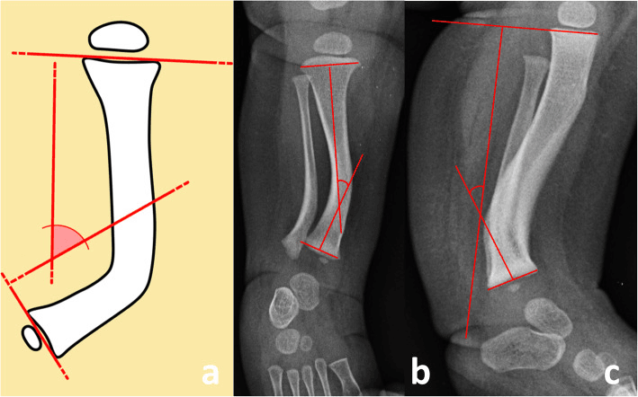
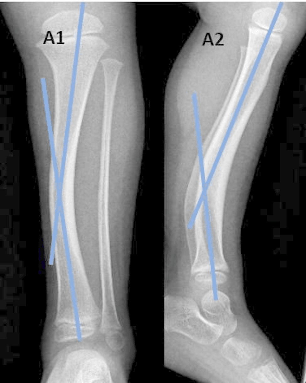
Full-length standing radiographs. These measure the bowing angle accurately and show overall alignment, the leg-length discrepancy and the position of the foot.
Other tests. CT and MRI are not routinely indicated and are reserved for surgical planning or atypical features. Genetic or NF1 testing is needed only if NF1 stigmata raise the possibility of an anterolateral (CPT) lesion instead.
Prenatal Diagnosis and Antenatal Counselling
An isolated tibial bow may be detected antenatally, but direction, mineralisation, the other bones, fractures and associated anomalies determine the differential and the counselling.
The ultrasound appearance. The features that point to CPMBT:
- An isolated, usually unilateral long-bone bow angulated posteriorly and medially, with the concavity anteromedial
- Normal bone echogenicity and mineralisation: the shaft is not thin, wide, fractured, sclerotic or cystic
- Normal length and morphology of the other long bones, a normal chest circumference and no other skeletal anomaly
- A commonly associated dorsiflexed (calcaneovalgus) foot pressed against the shin, a supportive clue consistent with the intrauterine-moulding hypothesis
- Mild-to-moderate tibial shortening, which may already be measurable relative to the contralateral side
The fetal differential. The mimics of a bent or short fetal long bone are the reason the read matters:
- Discriminating features
- Multiple bones affected, bilateral, short/broad or under-mineralised bones, in-utero fractures, small chest, other anomalies
- Discriminating features
- Bow apex anterolateral (opposite direction), may show cortical narrowing; associated NF1 family history
- Discriminating features
- Callus or a discrete fracture line, angulation at a focal point rather than a smooth diaphyseal curve
- Discriminating features
- Circumferential soft-tissue groove, distal lymphoedema, digital amputation
An isolated, unilateral, smoothly curved posteromedial tibial bow with normal mineralisation, no fractures and normal other bones is characteristic of CPMBT rather than a lethal or generalised skeletal condition. The single most useful discriminators are direction (posteromedial, not anterolateral), whether one bone or many are involved, and the presence of fractures or abnormal mineralisation (present in dysplasias, absent here).
Counselling. Antenatal counselling mirrors the postnatal message and avoids two opposite errors: needless alarm, mistaking a benign bow for a dysplasia, and false reassurance that "it will simply straighten". Parents should be told that the angular deformity is expected to remodel after birth, that the leg is nonetheless likely to remain somewhat short with a progressive discrepancy requiring surveillance and possibly later equalisation, and that the condition does not fracture or form a pseudarthrosis. Postnatal radiographs and examination, including a review of the newborn for the calcaneovalgus foot and reduced ankle dorsiflexion, complete the assessment.
Differential Diagnosis
The diagnosis is usually clinical, but the direction of the bow and the surrounding features separate it from dangerous mimics. The single most important distinction is from anterolateral bowing (the congenital pseudarthrosis spectrum), which is managed completely differently.
Making the distinction. Direction is essential but not sufficient. Confirm the posterior and medial components separately on orthogonal films, then look at the architecture: cortical or canal dysplasia, or a fracture, redirects the child to a CPT pathway. When CPT is suspected, the skin, the family history and other NF1 features matter.
- Posteromedial Bow
- Posterior + medial (concave anteromedial)
- Anterolateral Bow (CPT spectrum)
- Anterior + lateral (anterolateral)
- Posteromedial Bow
- Preserved
- Anterolateral Bow (CPT spectrum)
- Canal/cortical abnormality; possible fibular involvement
- Posteromedial Bow
- Not characteristic; assess if direction/architecture is atypical
- Anterolateral Bow (CPT spectrum)
- Common association in the CPT spectrum, but not universal
- Posteromedial Bow
- Not a defining natural-history feature
- Anterolateral Bow (CPT spectrum)
- Recognised risk in the CPT spectrum
- Posteromedial Bow
- Usually remodels substantially
- Anterolateral Bow (CPT spectrum)
- May progress or fracture
- Posteromedial Bow
- Calcaneovalgus is common
- Anterolateral Bow (CPT spectrum)
- No single obligatory posture
- Posteromedial Bow
- Residual shortening, ankle motion/alignment
- Anterolateral Bow (CPT spectrum)
- Fracture and pseudarthrosis risk
- Posteromedial Bow
- Observe remodelling; measure length and alignment
- Anterolateral Bow (CPT spectrum)
- Specialist CPT/NF1 pathway
When direction or bone architecture suggests the CPT spectrum, stop treating the case as benign positional bowing. Assess NF1 features, obtain specialist review and avoid an unplanned biopsy or corrective osteotomy through dysplastic bone because iatrogenic fracture or nonunion can be catastrophic.
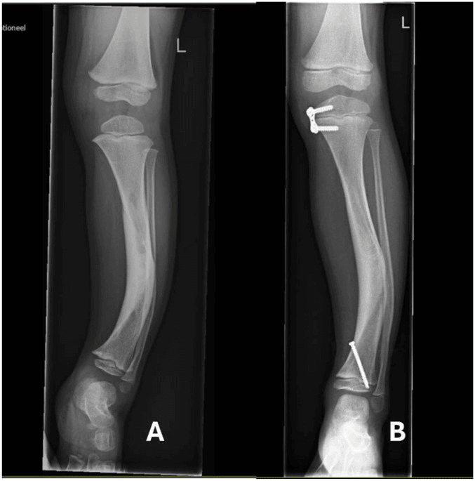
The other causes of a bowed infant or toddler tibia are separated by direction, symmetry and the rest of the skeleton:
- Direction of bow
- Coronal varus
- Key associations
- Usually bilateral and symmetric
- Natural history
- Age-dependent spontaneous evolution
- Distinguishing point
- No focal posterior component or true segmental shortening
- Direction of bow
- Proximal tibial varus/internal rotation
- Key associations
- Progressive proximal metaphyseal phenotype
- Natural history
- Variable progression
- Distinguishing point
- Apex and metaphyseal changes differ
- Direction of bow
- Variable
- Key associations
- Fibular/tibial and foot-ray deficiency or instability
- Natural history
- Fixed structural deficiency
- Distinguishing point
- Map every bone and ray
- Direction of bow
- Often anterolateral or varus, bilateral
- Key associations
- Metaphyseal cupping/fraying, biochemical changes
- Natural history
- Corrects with medical treatment
- Distinguishing point
- Bilateral, biochemistry abnormal, metaphyseal flaring
Management Algorithm
Management separates several related but non-identical problems: angular remodelling, residual length inequality, ankle and foot function, and distal alignment. Published operative proportions come from referral cohorts and must not be presented as the probability that an individual child will need surgery.
The goals:
- Confirm the posteromedial phenotype and exclude a high-risk mimic.
- Observe expected angular remodelling while measuring what actually persists.
- Treat foot or ankle limitations only when they remain clinically relevant.
- Recalculate the projected discrepancy and discuss equalisation only when its expected benefit exceeds treatment morbidity.
The options, and when each is used:
- Observation and serial measurement, for most young children
- Therapy, orthotic or casting support, selected from foot flexibility and function
- Contralateral growth modulation or epiphysiodesis, when projected discrepancy, remaining growth, expected stature and family priorities make shortening the longer limb acceptable
- Tibial lengthening with or without deformity correction, when the discrepancy, residual deformity, stature and patient goals justify reconstruction morbidity, as when preserving height or correcting complex deformity
- Targeted osteotomy or guided growth, for a symptomatic residual angular or ankle deformity rather than for a remodelling infant bow
Surgical Techniques
Indication. A measured or projected discrepancy, or a residual deformity, whose functional burden justifies reconstruction after shared decision-making.
Principles.
- Plan length and multiplanar alignment from calibrated standing imaging, rotational assessment and ankle and knee status.
- Select frame, rail, lengthening-over-nail or internal device from patient and bone size, physis, canal, deformity and centre experience.
- Place the corticotomy and fixation to control both length and alignment without creating a stress riser or injuring the physis.
- Titrate distraction to regenerate, pain, neurovascular status and joint motion rather than applying an immutable daily rate.
- Protect knee and ankle motion throughout; add foot control only when the construct or soft tissues require it.
Timing. Timing is individual. Early procedures may require later equalisation, while waiting may prolong functional inequality, and referral-series comparisons do not establish a universal age. Outcomes and complications vary by age, target, device, deformity and centre.
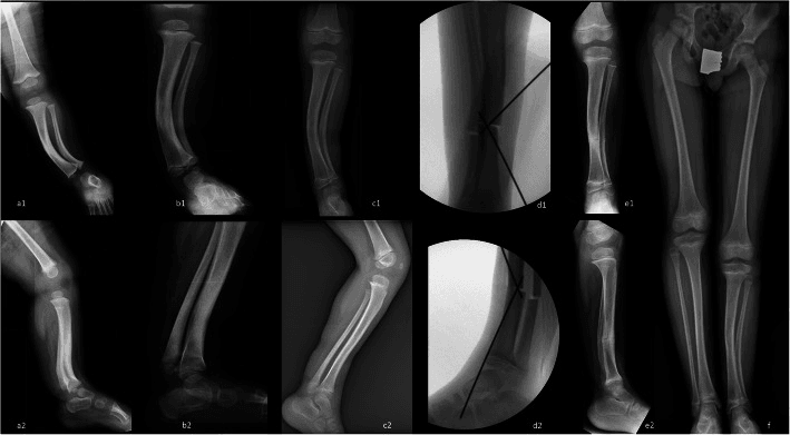
Complications
Natural-history sequelae. These are the things that actually cause morbidity:
- Residual limb-length discrepancy, which may remain clinically important even as percentage shortening partly improves
- Residual angular or rotational deformity, judged by function and mechanical axis rather than age alone
- Ankle motion limitation and calf asymmetry
- Ankle valgus or other distal alignment change from distal tibial and fibular morphology
Complications of lengthening or osteotomy. During treatment and early:
- Pin-tract infection and other pin or wire problems
- Premature or delayed consolidation and regenerate problems
- Neurovascular irritation or injury, and joint subluxation during distraction
Late:
- Regenerate fracture, axis drift and tibial valgus malalignment
- Ankle stiffness and equinus
- Repeat discrepancy after early lengthening as growth continues
- Need for staged or repeat equalisation when the original target or growth trajectory changes
Prevention. Accurate prediction (multiplier method) and correct timing of equalisation, circular or hexapod fixation or internal lengthening with adequate stability, and proactive ankle protection and physiotherapy during distraction.
The distinction to draw. Untreated, the complication that matters is the progressive discrepancy, not fracture, because this condition does not pseudarthrose. The treatment-related complications are those of limb reconstruction.
Follow-up and Postoperative Care
After reconstruction. Progress loading and frame or device removal from the evidence of regenerate and fixation, not a universal healing index. Protect knee and ankle motion throughout lengthening and consolidation, and monitor for axis drift, joint contracture, regenerate fracture and recurrent discrepancy.
After epiphysiodesis. Recovery is brief and mobilisation early. Follow the child to confirm that the predicted equalisation is achieved at maturity.
Outcomes and Prognosis
The angle. The prognosis is excellent: the posteromedial bow remodels spontaneously and substantially, with most correction in the first year, and the majority of children need no angular surgery. A mild residual medial component may remain.
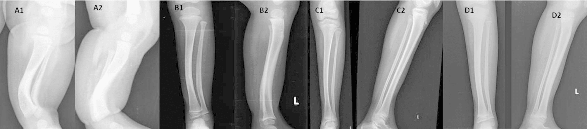
The length. The prognosis is guarded without planning, because the discrepancy is progressive and proportional to the initial bow.
- Hofmann & Wenger (1981): mean LLD 3.1 cm at follow-up, with 9 of 13 children over 2.5 cm and usually progressing
- Pappas (1984): absolute tibial discrepancy 3.3-6.9 cm at maturity, greater with greater initial bow
- Modern series confirm progressive shortening and a frequent need for equalisation by epiphysiodesis or lengthening
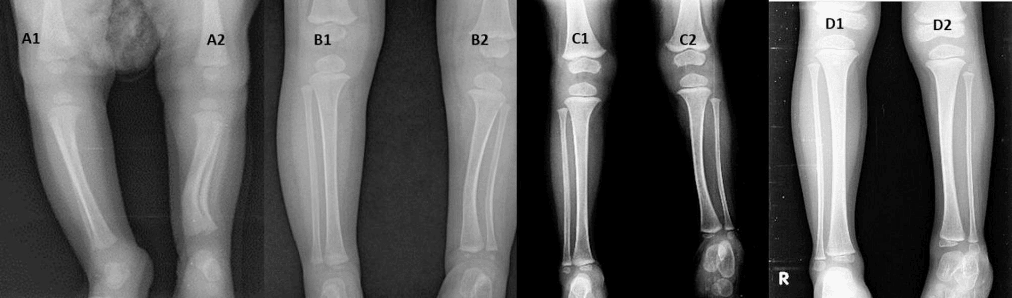
The ankle and foot. The calcaneovalgus foot resolves, but reduced ankle dorsiflexion may persist and ankle valgus can develop from the distal tibial changes.
Predictors. A greater initial angular bow is the single best predictor of a worse length outcome, and greater early percentage shortening also predicts it. Distal tibial epiphyseal wedging and fibular hypoplasia predict ankle valgus.
Quality of life. It is generally good. With appropriate prediction and timely, well-executed equalisation, most patients reach maturity with near-equal leg lengths, neutral alignment and good function, and outcomes after lengthening are good in modern series, though at the cost of the morbidity of reconstruction. Good outcomes depend on surveillance and planning, not on assuming the condition disappears on its own.
The Posterior Soft-Tissue Mass, Calf Atrophy and the Persistent Ankle Dorsiflexion Deficit
Natural history. Pappas (1984) described enlargement of the posterior soft tissues of the affected leg in early life that evolves into a relative muscle atrophy in later years, together with an initial calcaneovalgus foot and a decrease in ankle motion that does not improve with age (PMID 6490868). Shah, Doddabasappa and Joseph (2009) likewise recommended following all children to maturity specifically to detect residual muscle weakness alongside limb-length inequality and ankle deformity (DOI).
The deficit. The neonatal calcaneovalgus posture resolves, but the underlying posterior-compartment weakness and a fixed reduction in ankle dorsiflexion frequently persist and do not fully normalise. The functional consequence is weak plantarflexion and push-off: the child may have difficulty with toe-walking and heel-rise, and a subtly weaker calf on the affected side. The calcaneovalgus foot itself is a flexible neonatal posture covered as its own entity; the concern here is the leg's posterior musculature and the ankle's motion arc, which are CPMBT-specific and long-lasting.
Assessment. At every review, compare calf girth side to side and document active and passive ankle dorsiflexion and plantarflexion, adding single-leg heel-rise as the child matures. Reduced dorsiflexion here is a motion and strength deficit, separate from the later ankle valgus produced by distal tibial wedging and fibular hypoplasia; the two can coexist and should be recorded separately.
Management. Physiotherapy and stretching for the ankle motion arc and calf strength are the mainstay, and surgery is rarely needed. A fixed equinus or limited-dorsiflexion contribution should be recognised before any lengthening, because distraction tends to aggravate equinus and valgus, so the ankle must be protected during reconstruction with physiotherapy and sometimes a temporary foot-frame extension. Operative soft-tissue correction (tendo-Achilles or posterior release) is reserved for the rare rigid or functionally limiting deformity.
Guidelines, Registries & Global Practice
There is no dedicated AAOS, BOA, EFORT or AO guideline and no population registry for CPMBT. Evidence consists mainly of small single-centre cohorts and referral reconstruction series.
- Principle
- Confirm direction and architecture on orthogonal radiographs
- Resource adaptation
- Plain calibrated imaging is sufficient for most baseline assessment
- Principle
- Track angle, absolute/percentage shortening, ankle and function
- Resource adaptation
- Clinical block testing plus serial radiographs can define trajectory
- Principle
- Use more than one maturity input and repeat over time
- Resource adaptation
- Refer before the growth-modulation window closes
- Principle
- Match device and timing to deformity, size and expertise
- Resource adaptation
- Circular fixation remains versatile where internal devices are unsuitable
High-resource systems may offer low-dose biplanar imaging, hexapod planning and internal lengthening devices. Other settings may rely on block testing, conventional standing films, epiphysiodesis and circular fixation. The invariant is not a device: it is accurate phenotype, serial measurement, honest prediction uncertainty and timely referral.
Related pages: Congenital Pseudarthrosis of the Tibia, Neurofibromatosis, Blount Disease, Fibular Hemimelia, Causes of Limb Length Discrepancy, and Limb Length Discrepancy and Epiphysiodesis.
Controversies and Areas of Uncertainty
Earlier lengthening may reduce years lived with inequality but can require later equalisation; waiting improves size/maturity but prolongs discrepancy. Referral cohorts are confounded by severity and do not define one best age.
Growth modulation is lower burden but reduces final stature and is timing-sensitive. Lengthening preserves height and can correct deformity but carries regenerate, joint and device morbidity. No universal discrepancy boundary separates them.
Contemporary serial data show partial improvement in percentage shortening, yet absolute discrepancy may persist or grow. Percentage and centimetres must be reported separately.
Small cohorts suggest multiplier estimates stabilise with age, but most children were not observed to maturity. Treat prediction as a range checked against serial growth.
MCQ Practice Points
Q: What is the natural history of congenital posteromedial tibial bowing? A: Posterior and medial angular components usually remodel substantially. Percentage shortening may partly improve, while an absolute discrepancy can persist or increase during growth. Follow length, alignment, ankle motion and function; do not substitute a quoted maturity range for serial measurement.
Q: What distinguishes posteromedial bow from the anterolateral/CPT spectrum? A: Direction on orthogonal views, preserved versus dysplastic cortex/canal, fibular findings, fracture history and associated NF1 phenotype. Suspected CPT requires specialist planning and protection from unplanned biopsy or osteotomy.
Q: How is maturity discrepancy estimated? A: Use multiplier or growth-remaining methods together with chronological age, skeletal maturity and serial observed growth. Repeat the estimate; stability of a model output does not prove individual accuracy.
Q: When is surgery considered? A: For a measured/projected discrepancy, residual deformity or ankle problem whose expected functional benefit justifies growth-modulation or reconstruction morbidity. The procedure follows the whole deformity and child, not the diagnosis or one centimetre threshold.
Exam Viva Scenarios
Practise clinical reasoning and management decisions out loud
“An infant has unilateral posteromedial tibial bowing and a calcaneovalgus foot. How do you assess and counsel the family?”
“A toddler has a bowed tibia and possible NF1 features. How do you distinguish posteromedial bow from an anterolateral/CPT lesion?”
“A child whose bow has remodelled has a growing clinical discrepancy and a multiplier projection. How do you plan?”
Phenotype
- Posterior and medial tibial bow present at birth
- Tibiofibular shortening and calcaneovalgus are common
- Confirm each component on orthogonal radiographs
- Document foot flexibility, ankle motion and calf bulk
Natural History
- Angular components usually remodel substantially
- Percentage shortening may partly improve
- Absolute discrepancy can remain or increase
- Distal alignment and ankle function need separate follow-up
CPT Differential
- Anterolateral direction raises CPT concern
- Inspect cortex, canal, fibula, fracture and NF1 phenotype
- Direction alone is insufficient
- Avoid unplanned biopsy or osteotomy through dysplastic bone
Planning
- Measure angle plus absolute and percentage shortening
- Repeat growth prediction with skeletal maturity and serial data
- Growth modulation trades discrepancy for reduced stature
- Lengthening trades preserved height for reconstruction morbidity
Surgical Safety
- Plan mechanical axis, rotation and ankle/knee status
- Choose device from size, physis, canal and deformity
- Titrate distraction to regenerate and joint tolerance
- Monitor axis drift, contracture, regenerate failure and recurrent inequality
Evidence Base
Progression of Leg-Length Discrepancy (Landmark)
- 13 children followed a mean of 4 years from diagnosis
- Discrepancy at follow-up 1.9-5.6 cm (mean 3.1 cm), usually progressive
- 9 of 13 had a discrepancy greater than 2.5 cm
- Direct relationship between initial tibial bow and eventual leg-length discrepancy
Growth Pattern and Predictability (Landmark)
- 33 patients with congenital posteromedial bowing of tibia and fibula
- Bow 25-70 degrees at birth; absolute tibial discrepancy 3.3-6.9 cm at maturity
- Proportional length difference stable after 12 months, allowing prediction at maturity
- Initial calcaneovalgus foot and reduced ankle motion that does not improve with age
Mechanisms of Correction and Distal Tibial Changes
- 20 children; two mechanisms of correction - physeal realignment and diaphyseal remodelling
- Rapid angular resolution in year one, then markedly slower; residual deformity can persist beyond age 4
- Shortening proportional to severity (up to 40% in one case)
- Distal tibial epiphyseal wedging and fibular hypoplasia with ankle valgus in some
A Benign Condition or a Case for Limb Reconstruction?
- 38 patients; greatest deformity correction in the first year, limited remodelling after age 4
- Absolute LLD increased throughout growth (mean 14.3% tibial discrepancy)
- 20 of 38 (53%) required limb reconstruction for residual deformity and/or worsening LLD
- Higher LLD recurrence when lengthening performed under age 10
Deformity Progression in a Large Series
- 44 children - one of the largest single-institution series
- Progressive spontaneous correction of angular deformity but progressive increase in LLD
- 26 of 44 underwent surgery (mostly limb lengthening; also epiphysiodesis, osteotomy, distal tibial hemiepiphysiodesis)
- Final lateral distal tibial angle ankle valgus correlated with residual lateral interphyseal angle
Timing of Lengthening - Early vs Late
- 28 tibial lengthenings in 23 patients with circular fixators
- Mean initial LLD 3.4-4.1 cm; projected LLD at maturity often greater than previously reported (up to about 9.5 cm)
- Younger children (5 years or under) had larger deformities but shorter fixator duration/index and fewer complications
- Good/excellent bone results in both age groups
Triplanar Remodelling and Resolution of Shortening (Recent)
- 51 children prospectively followed (median 48 months)
- Posterior diaphyseal bow remodels fastest; remodelling minimal after 4-6 years of age
- Shortening partially resolves (20% at presentation to 13% at age 6 years) but persists
- Multiplier-method prediction of shortening at maturity is reliable after age 2 years