Fragility Fracture | Assess for Ring Involvement
- Key question: is there occult posterior-ring injury, displacement or inability to mobilise?
- Population: distinguish older low-energy fragility fracture from young high-energy pelvic trauma
- Imaging: CT defines sacral, SI, acetabular and anterior-ring morphology; MRI detects occult marrow injury when CT is unrevealing
- Treatment: start with analgesia and mobilisation for stable fragility patterns; consider fixation when pain, displacement or instability defeats mobilisation
- Goal: restore safe mobility while treating frailty, osteoporosis risk and immobility complications
- “Pubic rami fractures are common fragility fractures in the elderly.
- “Posterior sacral injury is common and easily missed on AP radiographs; lower the threshold for CT in fragility fractures, high-energy injury or failure to mobilise.
- “LC-1 is a Young-Burgess high-energy morphology; FFP/OFP classifications describe fragility patterns and should not be used as synonyms.
- “Focus on early mobilization to prevent immobility complications.
Overview and Epidemiology
A pubic rami fracture involves the superior pubic ramus, the inferior ramus or both. It may be isolated, with the ring intact, or associated with a posterior ring injury: a sacral fracture or an SI joint injury.
Two populations. The most common presentation is the elderly fragility fracture after a low-energy fall. It is a very common fragility fracture and an often under-recognised one. The other patient is young and has had high-energy trauma, a motor vehicle accident or a fall from height, and in them the rami fracture is often part of a major pelvic ring injury.
Anatomy and Pathophysiology
The rami. The superior pubic ramus connects the pubic body to the acetabulum and forms the anterior column. The inferior pubic ramus connects the pubic body to the ischium. The two pubic bones meet at the symphysis, a fibrocartilaginous joint in the midline.
The ring. The pelvis is a closed loop of sacrum, SI joints, innominate bones (ilium, ischium and pubis) and symphysis. A complete ring cannot break in only one place, so fractured rami are a reason to look posteriorly.
Where the rule bends. Osteoporotic bone can have incomplete or subtle posterior fractures, the sacral insufficiency fractures. Rami fractures frequently coexist with a sacral or SI injury, but the ring aphorism is not a law. Do not assume the fracture is isolated, and do not claim a second break automatically either: CT and the patient's function define the actual pattern.
Classification
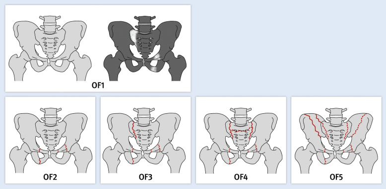
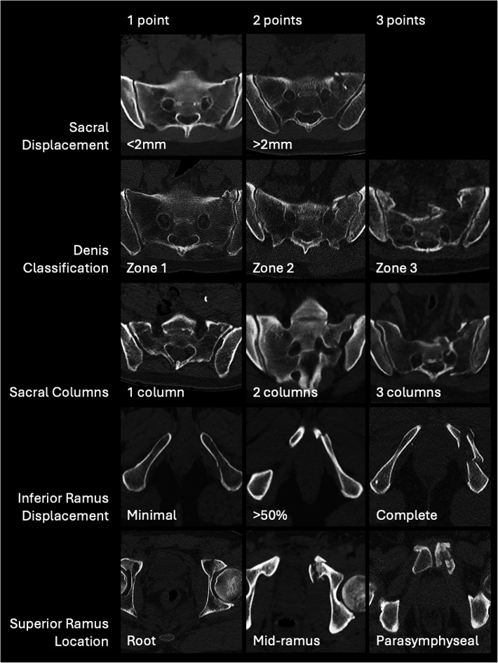
Isolated or ring injury. The first division is whether the posterior ring is involved, and CT is essential to determine it.
- Isolated rami: superior and/or inferior rami only, with the posterior ring intact on CT
- Ring involvement: rami plus a sacral fracture (LC-1), an SI injury or contralateral rami fractures
Young-Burgess, when the ring is involved. Lateral compression is a side-impact mechanism that compresses the pelvis, and the higher the LC type, the more unstable the ring.
- LC-1: ipsilateral rami fractures anteriorly and ipsilateral sacral compression posteriorly, which may be subtle. Usually stable; the most stable LC type
- LC-2: more severe lateral compression, with rami fractures anteriorly and a crescent (iliac wing) fracture posteriorly. More unstable
Two languages. LC-1 is a Young-Burgess high-energy mechanism; FFP and OFP are fragility classifications. Do not call FFP II and LC-1 interchangeable.
Clinical Assessment
History. The mechanism sorts the patient: a low-energy fall in the elderly, high-energy trauma in the young. The pain is in the groin and anterior pelvis. Ask whether they can walk and bear weight.
Examination. Tenderness is over the pubic rami, in the groin and anterior pelvis. An isolated stable fracture usually has no deformity; pelvic compression or distraction may elicit pain, and the gait is antalgic.
- Genitourinary: blood at the meatus, for urethral injury in high-energy trauma, especially in males
- PR/PV: for associated injury
Lower Urinary Tract Injury with Anterior Pelvic Fractures
Who is at risk. High-energy rami fractures may carry a bladder or urethral injury, the limb-and-life-relevant association of the anterior ring and the one that changes the very first action at the bedside, so check for haematuria. It is a young, high-energy problem, from straddle injuries, falls astride and anteroposterior-compression patterns, not a feature of the elderly low-energy fragility fracture.
Urethral injury. Predominantly male: the longer male urethra is the one at risk. The membranous urethra at the urogenital diaphragm is the classic site in pelvic-fracture (anteroposterior) injuries, and the bulbar urethra is torn in straddle mechanisms. The signs:
- Blood at the external meatus, the cardinal sign
- Inability to pass urine
- A high-riding, boggy or impalpable prostate on rectal examination
- A perineal or scrotal "butterfly" haematoma
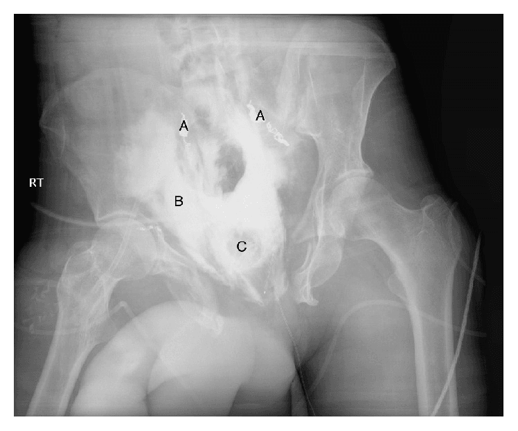
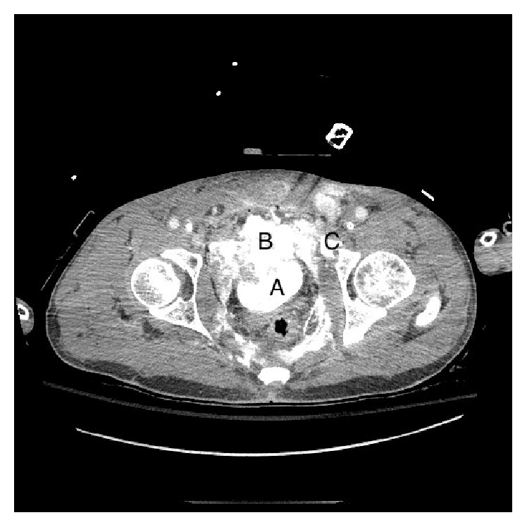
If a urethral injury is suspected, do NOT pass a urethral catheter. Perform a retrograde urethrogram first: blind catheterisation can convert a partial tear into a complete disruption and worsen later stricture. Involve urology early.
Bladder injury. Gross haematuria is the cardinal sign and mandates a CT cystogram, with retrograde gravity filling rather than just a passively clamped catheter. The pattern of rupture decides the treatment:
- Extraperitoneal rupture, the common pattern with pelvic-ring fractures: usually catheter drainage alone
- Intraperitoneal rupture, a free leak into the peritoneal cavity, often from a dome tear: operative repair
Investigations
The sequence.
- AP pelvis radiograph: the rami fractures are usually visible, but the posterior injury may be missed
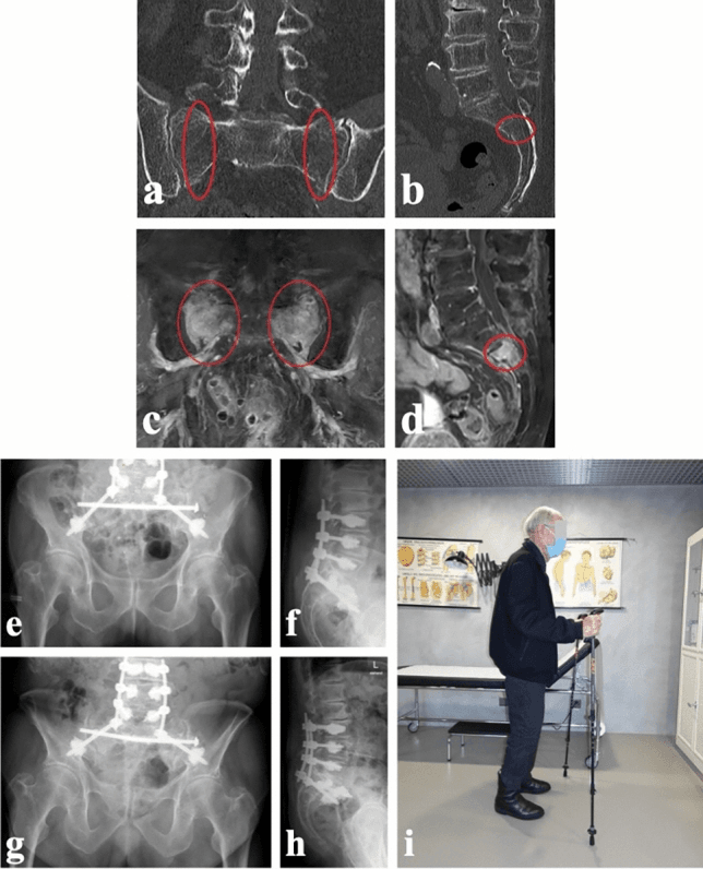
- CT pelvis: essential; assesses the posterior ring, the sacrum and SI joints
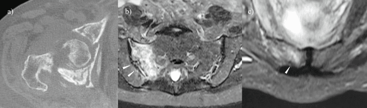
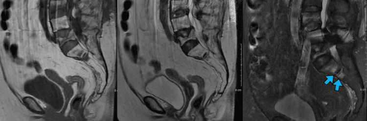
- MRI: when CT is negative but clinical suspicion is high, for the occult sacral insufficiency fracture
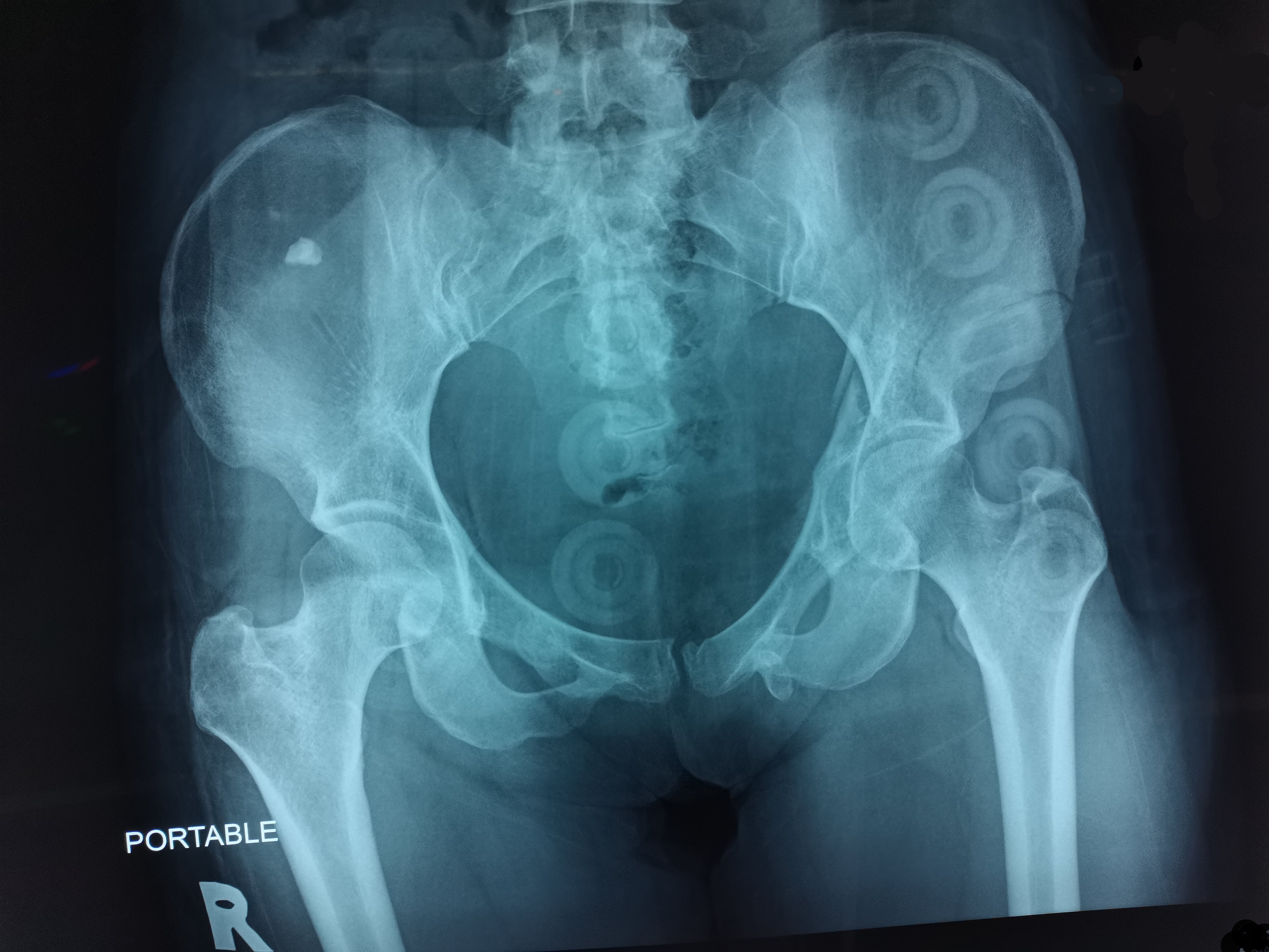
Why the CT. Posterior sacral injury is common and easily missed on AP radiographs.
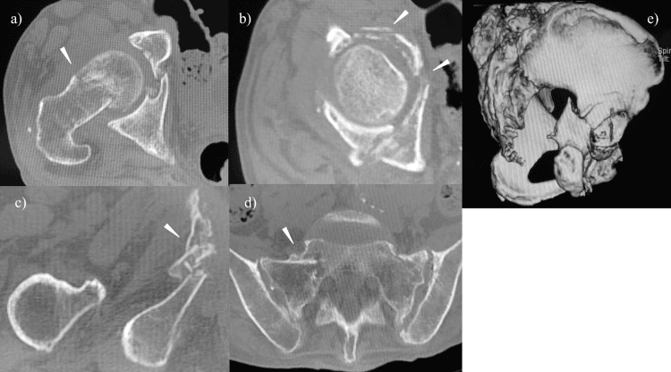
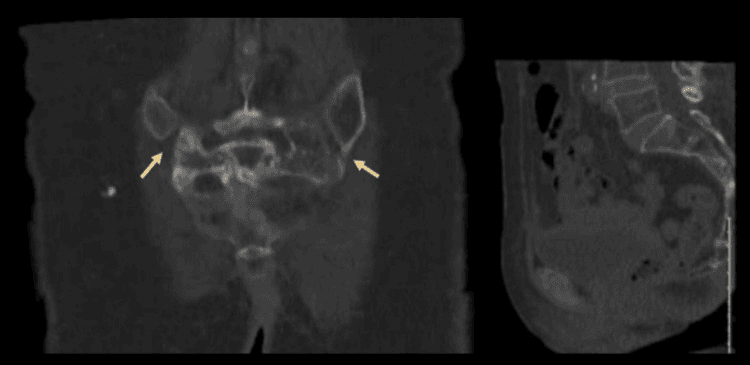
What to look for posteriorly.
- Sacral compression (LC-1) may be subtle on CT: look for buckling of the sacral ala
- Sacral insufficiency fracture: the classic H-sign (Honda sign) on bone scan or MRI, from bilateral sacral ala and transverse fractures
When the pain does not fit. Persistent immobilising pain after an apparently limited rami injury should prompt posterior-ring reassessment.
RINGPosterior-Ring Check
Hook:A rami fracture prompts a posterior-ring check, not an automatic second-break claim.
Differential Diagnosis
The elderly patient with groin or buttock pain who cannot weight-bear does not always have a rami fracture.
- Discriminating Feature
- Anterior pelvic / groin tenderness, ring tenderness; often able to lie still
- Key Test
- AP pelvis then CT for posterior ring
- Discriminating Feature
- Pain on log-roll, shortened/externally rotated leg, axial load pain
- Key Test
- MRI hip if radiograph negative
- Discriminating Feature
- Low back / buttock pain, sacral tenderness, may have no rami fracture
- Key Test
- MRI / bone scan: Honda (H) sign
- Discriminating Feature
- Atraumatic or trivial-trauma pain, lytic lesion, weight loss, raised markers
- Key Test
- CT, bloods (Ca, ESR, electrophoresis), bone scan
- Discriminating Feature
- Pain on axial loading through the femur, present in ~29% with rami fractures on CT
- Key Test
- CT pelvis
- Discriminating Feature
- No bony tenderness, normal imaging, mechanical pain
- Key Test
- Clinical, exclude fracture first
An elderly faller who cannot weight-bear with a normal pelvic radiograph may have an occult femoral neck fracture, not just a rami fracture. If pain persists or examination points to the hip, obtain MRI (or CT) before attributing everything to the rami.
Management Algorithm
The decision. Treatment follows what CT shows posteriorly and whether the patient can mobilise. Isolated pubic rami fractures rarely need operative fixation.
- Anterior-Only on CT
- No fracture detected
- Posterior Ring Injury Present
- Sacral fracture or SI injury
- Anterior-Only on CT
- Usually stable
- Posterior Ring Injury Present
- Ranges from nondisplaced to unstable
- Anterior-Only on CT
- Analgesia and mobilisation
- Posterior Ring Injury Present
- Trial mobilisation or selective fixation
- Anterior-Only on CT
- CT confirms anterior-only pattern
- Posterior Ring Injury Present
- CT defines displacement; MRI for occult insufficiency injury
- Anterior-Only on CT
- Unexpected pain or failure to mobilise
- Posterior Ring Injury Present
- Persistent immobilising pain, progression or instability
The older adult with a low-energy rami fracture. Inability to mobilise is a trigger to reassess both the diagnosis and the treatment.
- Confirm anterior and posterior morphology with CT when clinically indicated.
- Give prompt renal-, gastrointestinal- and delirium-conscious multimodal analgesia, opioid-sparing and with consideration of regional blocks, so that the patient can actually move. Pain that prevents mobilisation is itself a driver of mortality.
- Mobilise from day 1 with physiotherapy, weight-bearing as tolerated and an aid (frame or walker), as pain and stability permit. The explicit aim is to break the immobility cascade.
- Assess venous-thromboembolism risk and prescribe prophylaxis individually.
- Start the falls, frailty and bone-health pathway (below).
- Reassess persistent or worsening pain for occult or progressive posterior injury.
Surgical Technique
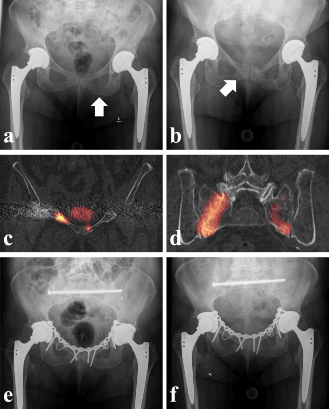
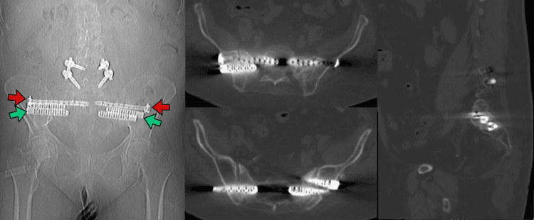
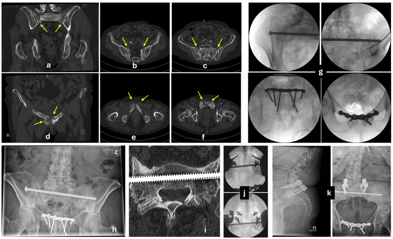
Selection. Sacroplasty is considered for a selected sacral insufficiency fracture when:
- Sacral pain remains immobilising despite optimised analgesia and rehabilitation
- Imaging confirms a morphology with a safe cement corridor
- Neurological compromise, infection and tumour requiring another strategy have been excluded
Technique. Cement leakage and neural injury must be considered.
- Select position and imaging guidance to visualise the sacral ala, foramina and canal.
- Place the trocar within the planned osseous corridor.
- Inject high-viscosity PMMA incrementally under continuous imaging, and stop for leakage.
- Reassess pain, neurology and mobilisation after the procedure.
Complications
Immobility is the biggest risk. Prolonged immobility leads to DVT, PE, pneumonia, deconditioning and death. Mobilise early.
- Risk Factor
- Immobility
- Prevention/Management
- Early mobilization + Prophylaxis
- Risk Factor
- Bed rest
- Prevention/Management
- Early mobilization + Resp physio
- Risk Factor
- Bed rest
- Prevention/Management
- Early mobilization
- Risk Factor
- Immobility
- Prevention/Management
- Pressure care
- Risk Factor
- Sacral involvement
- Prevention/Management
- Analgesia / Sacroplasty
- Risk Factor
- Frailty, Immobility
- Prevention/Management
- Orthogeriatric care
Mortality. One-year mortality after an elderly pelvic fragility fracture is around 10-20%, and the best single figure is Studer's 18.5% in 132 consecutive patients of mean age 84. That is in the same range as hip fracture, which is the comparison that justifies the pathway. It is crude mortality in a very frail population, though, and no study has shown that the fracture causes the deaths rather than marking the frailty that does. An orthogeriatric model of care improves outcomes.
The Orthogeriatric / Fragility-Fracture Care Pathway
Co-management. Beyond the analgesia, mobilisation and VTE risk assessment in the management protocol, orthogeriatric co-management adds:
- Delirium prevention and screening (for example the 4AT), with reorientation, sensory aids, hydration, sleep hygiene and avoidance of precipitating drugs
- Pressure-area, continence, nutrition and hydration care
- Review and deprescribing of falls-risk and sedating medications
Bone health. This fragility fracture is a sentinel event signalling high future fracture risk; in the elderly a rami fracture means osteoporosis, and it is treated. Enrol the patient in a fracture liaison service, arrange bone-density assessment and start the osteoporosis work-up. Correct calcium and vitamin-D deficiency, and select osteoporosis medication after contraindication and renal review; the choice between anti-resorptive and anabolic agents is developed in the osteoporosis topic.
Falls. Undertake a multifactorial falls assessment (gait and balance, vision, postural blood pressure, home hazards) to prevent the next fracture.
The hip-fracture parallel. National hip-fracture programmes and their best-practice standards (early surgery where relevant, orthogeriatric review, bone-health and falls assessment) increasingly bring pelvic fragility fractures onto the same audited pathway, recognising them as an equivalent frailty event rather than a benign isolated injury.
A fragility pelvic or rami fracture carries hip-fracture-level mortality and loss of independence, so it earns hip-fracture-level care. The pathway, not the fracture line, decides the outcome.
Postoperative Care
Conservative care. Mobilisation starts on day 1 with a walker or frame and daily physiotherapy. Discharge, home or to rehabilitation, comes when it is safe.
After sacroplasty. Mobilise the same day or the next, weight-bearing as tolerated, with a follow-up radiograph.
Outcomes
An isolated rami fracture has a good outcome if mobilised early. With ring or sacral involvement there may be chronic pain, and sacroplasty can help. Frail elderly patients have high morbidity and mortality from immobility.
Guidelines, Registries & Global Practice
Global epidemiology:
- Fragility fractures of the pelvis are rising rapidly with population ageing; the typical patient is a woman in her 80s after a low-energy fall.
- Women are affected roughly 5-6 times more often than men, reflecting postmenopausal bone loss.
- One-year mortality (around 15-20%) and loss of independence approach those of hip fracture, making this a sentinel frailty event rather than a benign injury.
Side-by-side guidance:
- Core Recommendation
- Classify by posterior instability (FFP I-IV); image the whole ring with CT; reserve surgery for higher grades or failed mobilization.
- Core Recommendation
- Manage as a fragility fracture on an orthogeriatric pathway: early mobilization, bone-health assessment, falls prevention.
- Core Recommendation
- Treat the underlying osteoporosis after any fragility fracture; close the post-fracture care gap with anti-resorptive or anabolic therapy.
- Core Recommendation
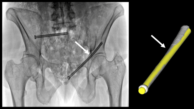
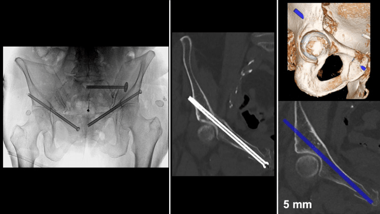
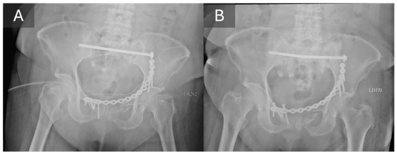
- Increasing use of minimally invasive posterior fixation (sacroiliac/transsacral screws) for unstable FFP III-IV in fit elderly patients.
- No dedicated pelvic-fragility registry exists; data come from national hip-fracture registries and trauma databases, which increasingly capture pelvic fragility fractures under the same frailty pathways.
- Bone-health drugs (bisphosphonates, denosumab, and anabolic agents such as teriparatide/romosozumab) are standard secondary prevention after a fragility fracture; agent choice and access vary by region and fracture risk.
- High-resource settings: routine CT, orthogeriatric co-management, fracture liaison services, and percutaneous fixation/sacroplasty for refractory pain.
- Limited-resource settings: diagnosis often rests on plain radiographs (risking missed posterior injury), with conservative analgesia and mobilization the mainstay and surgery reserved for clear instability.
Related pages: Pelvic Ring Injuries is the parent topic and the one to read for the high-energy patterns and for posterior fixation technique - the rule that a ring rarely breaks in one place comes from there and is why the CT is mandatory here; Sacral Insufficiency Fracture is the posterior half of this injury and, in most of these patients, the half that actually hurts - the sacroplasty evidence carded above properly belongs to it; Sacral Fractures for the traumatic counterpart and the Denis zones; Lateral Compression Injuries for the mechanism that produces the FFP II pattern in a younger patient; Sacropelvic Fixation for the constructs used when a fragility pattern does need stabilising; Pelvic Discontinuity for the arthroplasty problem that shares this anatomy in the same age group; and Pelvic Avulsion Fractures for the adolescent injury at the other end of life that must not be confused with it radiographically.
Controversies & Areas of Uncertainty
Operative or conservative for FFP II? The dominant pattern, rami plus a non-displaced unilateral sacral fracture, sits in a grey zone. A randomised pilot found no functional, pain or mortality advantage for surgery at 1 year, but it was small and underpowered. Most centres start conservatively and fix only those who fail to mobilise.
CT for every rami fracture? CT uncovers a posterior lesion in the majority, from 54% of Studer's consecutive cohort to 96.8% of Scheyerer's selected one, yet most are still treated conservatively. The debate is whether universal CT changes management enough to justify its dose and cost, or whether it should be reserved for persistent pain or inability to mobilise.
How much sacroplasty adds. Cohort data show rapid, early pain relief, but selection bias and limited comparative data prevent a universal success percentage. There is no high-quality randomised comparison against optimised conservative care, so its true added benefit and patient selection remain debated.
Reliability of the FFP grade. Interobserver agreement on the FFP grade is only moderate, and worst exactly where it matters most: complete versus incomplete sacral fractures, the distinction that swings the operative decision.
Viva Scenarios
Clinical Decision Scenarios
Practise clinical reasoning and management decisions out loud
“What is your assessment and management?”
“Explain the 'Ring Rule'.”
“Discuss the mortality risk.”
MCQ Practice Points
Q: Why must you image the posterior pelvis if pubic rami are fractured? A: A ring cannot break in only one place. If the anterior ring (rami) is broken, there is likely a posterior injury (sacral fracture, SI injury).
Q: What is the classic LC-1 injury pattern? A: Ipsilateral pubic rami fractures (anterior) + Ipsilateral sacral compression (posterior).
Q: What is the treatment for isolated pubic rami fractures? A: Conservative - Analgesia, Early Mobilization (WBAT), DVT Prophylaxis, Osteoporosis Treatment.
Q: What is sacroplasty? A: Percutaneous cement injection into a symptomatic sacral insufficiency fracture to provide pain relief and allow mobilization.
Q: What is the 1-year mortality for elderly patients with pelvic fragility fractures? A: 10-20%, similar to hip fractures. Immobility is the main risk factor.
Key Points
- Ring breaks in 2 places
- CT to assess posterior
- LC-1 = Rami + Sacrum
- Mobilize early
Treatment
- Conservative most cases
- Analgesia + Mobilize
- DVT prophylaxis
- Osteoporosis treatment
Sacroplasty
- For painful sacral insufficiency fractures
- Cement injection (PMMA)
- Pain relief + Earlier Mobility
- Consider for non-union sacral fractures
Mortality
- 10-20% at 1 year (elderly)
- Similar mortality to hip fractures
- Immobility is the killer
- Fragility fracture = fall risk protocol
Evidence Base
FFP Classification (Landmark)
- Comprehensive classification of Fragility Fractures of the Pelvis (FFP) from 245 consecutive patients.
- Graded I to IV by increasing instability (anterior only, non-displaced posterior, displaced unilateral posterior, bilateral/displaced posterior).
- Most FFP are minimally displaced and managed conservatively; higher grades and progressive displacement favour surgical stabilization.
Occult Posterior Ring Injury on CT
- 177 patients with pubic rami fractures and a CT at admission.
- A posterior pelvic ring injury was present on CT in 96.8% of patients who had no obvious posterior injury on the AP radiograph.
- A coexisting acetabular fracture was found in 28.8%; 30% of those with dorsal injury ultimately required operative treatment.
Mortality & Loss of Independence
- 132 patients aged over 65 with low-energy pubic rami fractures; mean age 84 years, women affected 6x more often.
- One-year mortality was 18.5% and almost 30% permanently lost their prior independence.
- A concomitant posterior ring lesion was found on CT in 54%; only 4% required secondary fixation.
Operative vs Conservative FFP II (RCT)
- Randomized pilot of 39 patients over 60 with FFP II (rami + non-displaced unilateral sacral fracture): 17 operative vs 22 conservative.
- No significant difference at 12 months in Barthel index, pain (VAS), quality of life (EQ-5D) or Tinetti gait, and no mortality difference.
- Two conservative patients crossed over to surgery for persistent pain/immobility.
Sacroplasty for Sacral Insufficiency Fractures
- Prospective multicentre cohort of 52 osteoporotic patients undergoing percutaneous sacroplasty.
- Mean VAS pain fell from 8.1 at baseline to 3.4 within 30 minutes and 0.8 at 52 weeks.
- One transient S1 radiculitis was the only significant complication.
FFP: From Eminence to Evidence (Review)
- Synthesis of the evidence accumulated since the 2013 FFP classification.
- Mortality exceeds the reference population and is lower after operative treatment, at the cost of surgical complications.
- Mobility, independence and quality of life remain worse than pre-injury regardless of FFP grade or treatment type.
FFP Classification Reliability
- 100 CT scans of low-energy pelvic ring injuries classified by 4 observers.
- Interobserver reliability was only moderate (kappa 0.42-0.59); intraobserver substantial (0.68-0.72).
- Reliability was poorest for the FFP IIc/IIIc/IVb subtypes involving a complete sacral fracture, where interobserver kappa fell as low as 0.10 - effectively chance agreement.