Direct Impact Injury | Flexion Key Prognostic Factor | Myositis Ossificans Prevention Critical
- Knee flexion at 24 hours is the key prognostic indicator
- Immediate 120 degree flexion position reduces hematoma and speeds recovery
- Myositis ossificans develops in 9-17%, more common with severe grades
- Avoid early aggressive ROM and massage - increases MO risk
- NSAIDs and aspiration have role in preventing myositis ossificans
- “Jackson and Feagin showed military cadets in flexion splints recovered 3x faster
- “Never massage a quadriceps contusion - promotes MO development
- “Heterotopic ossification appears on X-ray at 3-4 weeks post-injury
- “Compartment syndrome rare but possible - monitor tight thighs
Flexion at 24 hours determines grade: greater than 90 degrees = mild, 45-90 degrees = moderate, less than 45 degrees = severe. This predicts recovery time and MO risk.
Immediate 120 degree flexion (Jackson-Feagin protocol) shown to reduce recovery time from 88 to 13 days in military study. Compress hematoma between muscle bellies.
Avoid heat, massage, aggressive stretching in first 2-3 weeks. Consider indomethacin prophylaxis in severe grades. Aspiration of large hematomas may help.
Anterior thigh compartment syndrome rare but described. High index of suspicion with severe contusions, anticoagulated patients, or coagulopathy.
- ROM at 24h
- Greater than 90 degrees flexion
- Expected Recovery
- 2-3 weeks
- Key Management
- RICE, active ROM, progress when pain-free
- ROM at 24h
- 45-90 degrees flexion
- Expected Recovery
- 3-6 weeks
- Key Management
- Flexion splinting, protected rehab, monitor MO
- ROM at 24h
- Less than 45 degrees flexion
- Expected Recovery
- Greater than 6 weeks
- Key Management
- Flexion splinting, consider NSAIDs prophylaxis, MRI
- ROM at 24h
- Persistent pain, palpable mass
- Expected Recovery
- 3-6 months natural history
- Key Management
- Wait for maturation, excise if symptomatic
RICERICE Plus - First 48 Hours
Hook:RICE is not enough for severe contusions - add NSAIDs for protection
MOMO RISK - Myositis Ossificans Risk Factors
Hook:These factors put you at MO RISK for heterotopic bone formation
Overview and Epidemiology
Quadriceps contusions are the second most common injury in contact sports after muscle strains. They result from direct blunt trauma to the anterior thigh, causing intramuscular hemorrhage and tissue damage.
- Direct blow - knee or helmet strike to anterior thigh (most common)
- Contact sports - rugby, Australian football, martial arts, football
- Falls - direct impact onto hard surface
- Motor vehicle accidents - dashboard injury
- Rugby and Australian Rules Football - highest incidence
- American football - common in receivers/running backs
- Hockey - puck or stick trauma
- Combat sports - knee strikes
The vastus intermedius is most commonly injured as it lies directly on the femur with no muscle posterior to provide cushioning. The rectus femoris is protected by the intermedius layer.
Pathophysiology and Mechanisms
- Rectus femoris - only biarticular muscle, hip flexor
- Vastus lateralis - largest, lateral aspect
- Vastus medialis - medial aspect, VMO critical for patella tracking
- Vastus intermedius - deepest, directly on femur
- Branches of profunda femoris (lateral and medial circumflex femoral)
- Rich vascular supply explains significant hematoma formation
- Intramuscular bleeding contained by fascia
Contusion Evolution
Acute hemorrhage into muscle belly. Vasospasm initially limits bleeding. Hematoma begins forming in the intramuscular space.
Inflammatory response peaks. Edema develops. ROM rapidly decreases. Critical window for classification by flexion.
Hematoma organization begins. Fibroblast migration starts. Continued inflammation. Key period for flexion positioning.
Granulation tissue formation. Early scar tissue. Risk period for myositis ossificans if tissue is reinjured or aggressively mobilized.
Muscle regeneration phase. Satellite cell activation. Progressive strengthening safe to begin if pain-free.
Heterotopic bone forms when pluripotent mesenchymal cells differentiate into osteoblasts instead of myoblasts. Risk factors include re-injury, massage, aggressive passive stretching, and large hematoma size.
120 degree flexion compresses the hematoma between the rectus femoris and vastus intermedius, limiting expansion. It also maintains muscle length, preventing adaptive shortening. Jackson and Feagin demonstrated recovery in 13 days vs 88 days with extension splinting.
Classification Systems
Ryan Classification (by ROM at 24 hours - most commonly used)
- Knee Flexion at 24h
- Greater than 90 degrees
- Gait
- Normal
- Swelling
- Minimal
- Recovery Time
- 2-3 weeks
- Knee Flexion at 24h
- 45-90 degrees
- Gait
- Antalgic
- Swelling
- Moderate
- Recovery Time
- 3-6 weeks
- Knee Flexion at 24h
- Less than 45 degrees
- Gait
- Unable to walk
- Swelling
- Severe
- Recovery Time
- Greater than 6 weeks
The Ryan classification is the most practical as it uses a single objective measure (ROM) taken at a standardized time point (24 hours) to predict prognosis and guide treatment intensity.
This classification informs treatment intensity and helps you counsel athletes and coaches appropriately.
- Distinguishing Features
- Direct blunt blow, diffuse intramuscular swelling, ROM loss tracks severity
- Key Investigation
- Clinical grading by 24h flexion; ultrasound for haematoma
- Distinguishing Features
- Indirect eccentric overload (sprinting), pain at musculotendinous junction, no impact history
- Key Investigation
- Ultrasound/MRI showing fibre disruption at junction
- Distinguishing Features
- Persistent firm mass and ROM plateau weeks after contusion
- Key Investigation
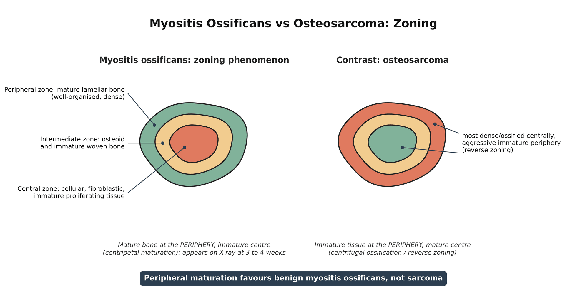
- Radiograph at 3-4 weeks (peripheral zoning); CT if uncertain
- Distinguishing Features
- Pain out of proportion, tense compartment, pain on passive stretch, saphenous paraesthesia
- Key Investigation
- Compartment pressure; urgent fasciotomy
- Distinguishing Features
- High-energy mechanism, deformity, inability to bear weight
- Key Investigation
- Plain radiograph
- Distinguishing Features
- Painless enlarging mass, no clear trauma, deep to fascia, more than 5 cm
- Key Investigation
- MRI then biopsy of periphery; do NOT assume myositis ossificans
Clinical Assessment
History
- Mechanism - direct blow, contact event, speed of impact
- Immediate response - able to continue playing? (poor prognostic sign if yes - suggests ongoing bleeding)
- Time since injury - critical for classification at 24 hours
- Previous quad contusions - risk factor for recurrence
- Anticoagulation status - increased bleeding risk
- Severe pain out of proportion
- Numbness in leg
- Progressive swelling despite rest
- Inability to weight bear at all
Documenting the history carefully guides prognosis and identifies high-risk patients.
Examination
Physical examination
- Swelling location and extent
- Ecchymosis (may appear 24-48h later)
- Muscle contour - bulge or defect
- Point of maximal tenderness
- Palpable hematoma or mass
- Warmth
- Active and passive knee flexion
- Document angle at 24 hours for classification
- Compare to uninjured side
- Resisted knee extension
- Straight leg raise ability
- Document any quad lag
Pain with passive stretch, tense anterior compartment, paresthesias in saphenous nerve distribution, weak knee extension. Rare but requires urgent fasciotomy.
Investigations
- Not routinely required for typical contusion
- Indicated if fracture suspected (significant trauma)
- Baseline for monitoring myositis ossificans
- MO visible at 3-4 weeks post-injury
- First-line imaging for soft tissue assessment
- Quantifies hematoma size
- Differentiates intramuscular vs intermuscular
- Can guide aspiration
- Dynamic assessment of muscle integrity
- Reserved for severe grades or diagnostic uncertainty
- Quantifies extent of muscle damage
- Identifies associated injuries
- Useful for return-to-play decisions in elite athletes
Acute hematoma appears hypoechoic or anechoic. Organized hematoma becomes more heterogeneous. Early myositis ossificans shows increased echogenicity before calcification is visible on X-ray.

Management Algorithm
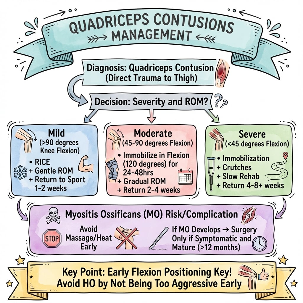
- 1Acute Injury
Immediate RICE plus 120 degree flexion positioning
Compress hematoma, limit bleeding
- 224-Hour Assessment
Measure passive knee flexion
Classify as mild/moderate/severe
- 3Mild Grade (greater than 90 degrees)
Active ROM, crutches PRN, progress as tolerated
RTS 2-3 weeks
- 4Moderate Grade (45-90 degrees)
Flexion splint 24h, protected rehab, consider NSAIDs
RTS 3-6 weeks
- 5Severe Grade (less than 45 degrees)
Continuous flexion splint, indomethacin, consider aspiration
RTS greater than 6 weeks, monitor for MO
- 6Rehabilitation Phase
Progressive ROM then strengthening when 90 degrees achieved
Sport-specific training when criteria met
- 7Return to Sport
Full ROM, strength greater than 90 percent, functional testing passed
Graduated return to contact
- Remove from play - do not continue with injury
- RICE protocol immediately
- 120 degree flexion position - wrap knee flexed
- Crutches - non-weight bearing to partial
- Ice - 20 min every 2 hours
- Knee wrapped in maximal comfortable flexion
- Elastic bandage over ice pack
- Maintained for 24 hours initially
- Shown to reduce recovery time significantly
- Analgesia as needed (paracetamol preferred)
- NSAIDs controversial early - may increase bleeding
- After 48-72h, indomethacin for MO prophylaxis in severe grades
This acute phase management is critical for optimal outcomes.
The reason NSAIDs are delayed and used selectively in quadriceps contusion is a genuine trade-off, not just bleeding risk. By inhibiting cyclo-oxygenase and the prostaglandin-driven inflammatory phase, NSAIDs (especially indomethacin) suppress the heterotopic-ossification pathway - hence their value for myositis ossificans prophylaxis in severe grades. But that same early inflammatory phase drives satellite-cell activation and muscle regeneration, and experimental work shows NSAIDs can blunt myoblast proliferation and impair the quality and strength of muscle repair. So the acute strategy is deliberate: avoid NSAIDs in the first 48-72 hours (bleeding risk, and they are not yet needed), favour paracetamol for early analgesia, and reserve an NSAID course for MO prophylaxis in moderate-to-severe contusions where the heterotopic-bone risk outweighs the small regenerative cost. This is why the protocol times the NSAID rather than giving it from minute one.
FLEXFLEX - Acute Management
Hook:Keep it FLEXED to compress the hematoma between muscle bellies
Surgical Technique
Surgical excision of myositis ossificans:
Surgery is rarely required for quadriceps contusions. The primary surgical indication is symptomatic mature myositis ossificans that fails conservative management.
- Persistent pain limiting function after MO maturation
- Mechanical symptoms (limited ROM, catching)
- Large MO causing cosmetic concern
- Failed conservative management for 6+ months
- Immature MO (less than 6 months, hot bone scan)
- Asymptomatic MO
- Active infection
Careful patient selection is essential for successful surgical outcomes.
Complications
Myositis Ossificans Traumatica (MOT)
9-17% of quadriceps contusions
- Severe grade (ROM less than 45 degrees)
- Delay in treatment greater than 72 hours
- Early massage or aggressive stretching
- Re-injury before full healing
- Large intramuscular hematoma
- Previous MO
- Persistent pain beyond expected recovery
- Palpable firm mass in muscle
- Plateau or decrease in ROM
- Pain with activity
- Appears 3-4 weeks post-injury
- Zoning phenomenon - mature bone peripherally
- Matures over 3-6 months
- Initially conservative - wait for maturation
- NSAIDs may limit progression if caught early
- Surgical excision only if symptomatic AND mature (6-12 months)
- Pre-op bone scan to confirm maturity
Never excise immature myositis ossificans - high recurrence rate. Wait minimum 6 months, confirm maturity with bone scan (cold lesion), then excise with margin. Consider radiation prophylaxis post-excision in recurrent cases.
A blunt tangential or shearing blow to the thigh (the same rugby/AFL/road mechanism) can also produce a Morel-Lavallée lesion - a closed internal degloving in which the skin and subcutaneous fat are sheared off the underlying fascia, tearing perforating vessels and lymphatics and filling the new potential space with a haemolymphatic collection. Unlike a contusion's intramuscular haematoma it sits in the subcutaneous-fascial plane (classically over the greater trochanter or proximal thigh), presents as a fluctuant, sometimes mobile swelling with overlying skin hypoaesthesia and ecchymosis, and may enlarge or present late. It is its own diagnosis because management differs: small/acute lesions get compression, but persistent collections need percutaneous drainage and sclerodesis (e.g. doxycycline) or operative debridement, and a chronic encapsulated lesion can become infected or recur. MRI shows the characteristic fluid collection between fat and fascia, often with a capsule. Suspect it whenever a "contusion" is fluctuant, superficial and not resolving.
Rehabilitation and Return to Sport
Rehabilitation Phases
RICE protocol, 120 degree flexion positioning, crutches, ice every 2 hours. Goal is to minimize hematoma size and inflammation. No stretching or strengthening activities.
Begin active ROM exercises, continue ice, pain-free weight bearing progression. Stationary bike when 90 degrees flexion achieved. Avoid massage, heat, passive stretching.
Progressive resistance exercises when ROM greater than 90 degrees pain-free. Isometrics progressing to isotonics. Pool running and swimming permitted.
Sport-specific drills, agility training, plyometrics when strength greater than 80 percent. Non-contact training initially, then progress to contact with padding.
Full ROM, strength greater than 90 percent, functional testing passed. Protective thigh padding for first 2-4 weeks of competition. Monitor for any symptoms.
Return to Sport Criteria (all must be met):
- Requirement
- Equal to uninjured side
- Testing Method
- Goniometry
- Requirement
- Greater than 90 percent
- Testing Method
- Isokinetic or 1RM
- Requirement
- Less than 1cm difference
- Testing Method
- Tape measure
- Requirement
- None with activity
- Testing Method
- Functional testing
- Requirement
- Pass hop tests
- Testing Method
- Single leg hop for distance
In professional athletes, MRI may be used to confirm complete muscle healing before return to sport. Some teams use isokinetic testing with peak torque and total work comparisons to uninjured side, requiring greater than 90 percent symmetry.
RTSRTS Criteria - Return to Sport
Hook:RTS when ROM, Thigh circumference, and Strength are symmetric
Outcomes and Prognosis
Prognosis by grade:
- Expected Recovery
- 2-3 weeks
- Return to Sport
- Full recovery expected
- MO Risk
- Low (under 5%)
- Expected Recovery
- 3-6 weeks
- Return to Sport
- Full recovery expected
- MO Risk
- Moderate (10-15%)
- Expected Recovery
- 6+ weeks
- Return to Sport
- May have prolonged course
- MO Risk
- High (15-20%)
- Time to treatment initiation (earlier is better)
- Compliance with flexion positioning protocol
- Avoidance of re-injury during recovery
- Strict adherence to RTS criteria
- Most athletes return to pre-injury level
- Recurrent contusion possible if RTS too early
- Myositis ossificans may require delayed surgical excision
- Chronic pain rare if properly managed
Key prognostic indicator is ROM at 24 hours. Athletes who follow the Jackson-Feagin flexion protocol and meet all RTS criteria have excellent outcomes with low recurrence rates. Premature return is the main risk factor for complications.
Guidelines, Registries & Global Practice
Global epidemiology:
- Figure
- Contusion second only to strain as cause of sports-injury morbidity
- Source
- Beiner & Jokl (PMID 11476532)
- Figure
- Among most common injury categories over five seasons
- Source
- Larruskain et al (PMID 28207979)
- Figure
- 4.82x higher incidence in men vs women (contact exposure)
- Source
- Larruskain et al (PMID 28207979)
- Figure
- 9% of quadriceps contusions (up to 9-17% in series)
- Source
- Ryan et al (PMID 1867338)
The injury is fundamentally a contact-sport problem and is over-represented wherever collision codes dominate (the football codes, rugby, American football, ice hockey, martial arts and combat sports). The driver is exposure to direct blunt impact rather than any geographic or health-system factor.
Guidance and consensus, side by side:
There is no high-level RCT-based guideline body that issues a quadriceps-contusion-specific protocol; practice is built on the West Point/Naval Academy military series and sports-medicine consensus, which are remarkably consistent worldwide.
- Position
- Immediate sustained knee flexion (aim 120 degrees), grade by 24-hour ROM, avoid heat/massage
- Evidence level
- Level IV consensus from prospective military series
- Position
- RICE plus early protected flexion; criteria-based graded return
- Evidence level
- Expert consensus
- Position
- Same acute principles; emphasis on ultrasound/MRI grading and objective return-to-sport testing
- Evidence level
- Expert consensus
- Position
- Exclude fracture and compartment syndrome; conservative care for isolated contusion
- Evidence level
- Expert consensus
There is no joint or implant registry relevant to this soft-tissue injury, so registry-based survival or revision data do not apply.
Global practice variation:
- Typical approach
- Immediate pitch-side flexion bracing, early ultrasound/MRI, isokinetic-guided return
- Reason
- Resourced medical teams, financial stakes of recurrence
- Typical approach
- Clinical grading by ROM, RICE, physiotherapy-led rehabilitation, imaging only if severe or atypical
- Reason
- Cost-effective and adequate for most cases
- Typical approach
- Clinical diagnosis and graded rehabilitation without routine imaging
- Reason
- Imaging access constrained; clinical course usually sufficient
Across every region and resource setting the core message is identical: immediate sustained flexion, grade by 24-hour ROM, avoid heat and massage, and use objective criteria for return to sport. Premature return is the dominant modifiable risk factor for recurrence and myositis ossificans.
MCQ Practice Points
Q: What is the key prognostic indicator for recovery time in quadriceps contusions? A: Knee flexion at 24 hours. Recoveries vary from 2-3 weeks (greater than 90 deg) to greater than 6 weeks (less than 45 deg).
Q: What is the most important acute intervention? A: Immediate 120 degree knee flexion positioning. This compresses the hematoma and prevents stiffness.
Q: What radiographic sign distinguishes Myositis Ossificans from malignancy? A: Zoning Phenomenon. MO has mature cortical bone peripherally with a central lucency. Osteosarcoma is the reverse (central ossification, indistinct margin).
Q: What modalities should be absolutely avoided in the acute phase? A: Heat and Massage. Both increase blood flow and risk of Myositis Ossificans.
Q: What is a mandatory criterion for return to contact sports? A: Full ROM and greater than 90% Strength. Plus protective padding is essential.
Exam Viva Scenarios
Practise clinical reasoning and management decisions out loud
“A 22-year-old rugby player presents 6 hours after a direct knee strike to his anterior thigh. He has marked swelling and can only flex his knee to 30 degrees. How would you manage this player?”
“A footballer returns 4 weeks after a moderate quadriceps contusion with persistent pain and a firm palpable mass in the anterior thigh. X-ray shows early calcification. How would you manage this?”
“A professional AFL player sustained a moderate quadriceps contusion 5 weeks ago. He has regained 100 degrees of knee flexion and wants to return for finals. The coach is putting pressure on medical staff. How do you approach this?”
Classification (Ryan)
- Mild: Flexion greater than 90 degrees at 24h - RTS 2-3 weeks
- Moderate: Flexion 45-90 degrees - RTS 3-6 weeks
- Severe: Flexion less than 45 degrees - RTS greater than 6 weeks
- Prognosis: 24h ROM is key predictor
Acute Management
- IMMEDIATE 120 degree flexion positioning
- RICE protocol - ice 20 min every 2 hours
- Non-weight bearing with crutches
- NSAIDs AFTER 48-72h only (bleeding risk earlier)
Jackson-Feagin Protocol
- Wrap knee in maximal comfortable flexion
- Elastic bandage over ice pack
- Reduced recovery 88 days to 13 days
- Compresses hematoma between muscle bellies
Myositis Ossificans
- Develops in 9-17% of quad contusions
- Visible on X-ray at 3-4 weeks
- ZONING phenomenon = mature bone peripherally
- Surgery only after maturation (6-12 months)
Return to Sport Criteria
- Full ROM equal to uninjured side
- Strength greater than 90 percent (isokinetic)
- Thigh girth within 1cm
- Pain-free with sport-specific activity
Key Exam Points
- Never massage acute quad contusion
- Avoid heat and aggressive stretching early
- MO vs Osteosarcoma: zoning phenomenon key differentiator
- Compartment syndrome rare but possible - high suspicion
Evidence Base
Jackson-Feagin Study (1973) - Landmark
- Severity of injury correlates directly with disability and complication rate
- Heat, massage and premature activity were associated with worse outcomes and myositis ossificans
- Established graded rehabilitation and the principle of early protected motion
Ryan et al - West Point Update (1991) - Landmark Classification
- 24-hour knee ROM is the key prognostic indicator and basis of the grading system
- Mean disability 13 days (mild), 19 days (moderate), 21 days (severe)
- Myositis ossificans in 9%; five risk factors - ROM less than 120 degrees, football mechanism, previous quadriceps injury, treatment delay greater than 3 days, ipsilateral knee effusion
Aronen et al - Immediate 120 degree Flexion Immobilisation (2006)
- Immediate 120 degree flexion held for 24 hours dramatically shortened disability (mean 3.5 days)
- Only 1 case of myositis ossificans on 3- and 6-month radiographs of the first 23 patients
- Earlier intervention from the moment of injury improved results compared with prior series
Diaz et al - Severe Quadriceps Contusions (2003)
- Severe contusions produce large intramuscular haematoma and markedly prolonged recovery
- Imaging (ultrasound/MRI) defines haematoma size and guides rehabilitation
- Highlights need for protected, graded return to avoid re-injury and myositis ossificans
Beiner & Jokl - Contusion Injury and Myositis Ossificans (2002)
- Healing is a balance between muscle regeneration and scar/heterotopic bone formation
- Myositis ossificans is a major debilitating complication of contusion injury
- Reviews risk factors and prevention strategies including avoidance of re-injury and aggressive early mobilisation
Larruskain et al - Contusion Epidemiology in Elite Football (2018)
- Contusions are among the most common injuries in elite football
- Contusion incidence 4.82 times higher in male than female players (95% CI 2.30-10.08)
- Reflects greater contact exposure as the driver of contusion injury