AORI Classification | Constraint Selection | Bone Loss Management | Outcomes
- Aseptic loosening is the most common indication for revision TKA (50% of cases)
- AORI classification guides bone loss management: Type 1 (intact), Type 2 (damaged), Type 3 (deficient)
- Constraint progression: PS to CCK to hinged based on bone loss and soft tissue competence
- Infection must be ruled out in ALL cases before revision - aspirate, inflammatory markers, culture
- Two-stage revision is gold standard for chronic PJI with antibiotic spacer interval
- “AORI Type 3 requires structural support (metaphyseal sleeve, cone, or bulk allograft)
- “Constrained condylar knee (CCK) for MCL/LCL insufficiency but intact bone stock
- “Rotating hinge for massive bone loss or global instability
- “Modular stems for stability and load sharing, cemented distally in diaphysis
Type 1 = Intact metaphysis (standard components). Type 2A/B = Damaged (augments, cement). Type 3 = Deficient (structural graft/sleeve/cone). Know which reconstruction for which defect.
Aspirate ALL knees before revision. ESR, CRP, synovial WBC and differential. Alpha-defensin if clinical suspicion. Never proceed without excluding PJI.
PS to CCK to hinged based on:
- Bone loss (AORI grade)
- Collateral ligament integrity
- Flexion-extension gap balance
- Extensor mechanism function
Stage 1: Debridement, explant, antibiotic spacer. 6-8 weeks IV antibiotics then 2-week holiday. Stage 2: Aspirate negative, then reimplant. 90% infection control rate.
- AORI Grade
- Type 1 (intact)
- Constraint Level
- PS or CCK
- Key Technique
- Standard components, cement fixation
- AORI Grade
- Type 2A/B (damaged)
- Constraint Level
- CCK with augments
- Key Technique
- Metal augments or cement-fill, stems for stability
- AORI Grade
- Type 3 (deficient)
- Constraint Level
- Rotating hinge
- Key Technique
- Metaphyseal sleeve/cone, long cemented stems
- AORI Grade
- Variable (assess at stage 2)
- Constraint Level
- CCK or hinge
- Key Technique
- Two-stage with spacer, 6-8 weeks antibiotics
CLAMPConstraint Selection Principles
Hook:CLAMP down on instability - use increasing constraint as needed to stabilize the revision knee!
Overview and Epidemiology
With national registries recording over 100,000 primary TKAs annually in high-income settings and far greater volumes worldwide, the burden of revision surgery is substantial. Revision TKA is technically demanding, associated with higher complication rates (20-30% vs 5-10% for primary), longer operative times, and inferior functional outcomes. Understanding indications, bone loss classification (AORI), and constraint selection is critical for Orthopaedic exam success and clinical practice.
- Age: Mean 65-70 years at revision (younger than primary)
- Gender: F greater than M (2:1) - same as primary TKA
- Time to revision: Mean 7-10 years post-primary
- Re-revision risk: 15-20% within 5 years
- Complication rate: 20-30% (vs 5-10% primary)
- Operative time: 2-3 hours (vs 1-1.5 hours primary)
- Functional scores: 70-80% of primary TKA outcomes
- Survivorship: 85-90% at 10 years (vs 95% primary)
Indications for Revision TKA
- Frequency
- 50%
- Key Features
- Radiolucent lines over 2mm, progressive, painful
- Treatment Approach
- Revise components, address bone loss, stems
- Frequency
- 20%
- Key Features
- MCL/LCL insufficiency, flexion-extension gap imbalance
- Treatment Approach
- Increase constraint (CCK or hinge), soft tissue reconstruction
- Frequency
- 15%
- Key Features
- Chronic pain, effusion, elevated inflammatory markers
- Treatment Approach
- Two-stage revision with antibiotic spacer
- Frequency
- 10%
- Key Features
- Focal osteolysis, thin poly, no gross loosening
- Treatment Approach
- Poly exchange, curettage and grafting of lesions
- Frequency
- 3-5%
- Key Features
- Less than 90° flexion, failed manipulation
- Treatment Approach
- Open arthrolysis, poly exchange, consider hinge
Every revision knee must have infection excluded before proceeding with aseptic revision. Perform:
- Serum: ESR, CRP
- Synovial fluid: WBC (greater than 3000 cells/µL), PMN% (greater than 80%), culture
- Alpha-defensin or synovial CRP if high suspicion
- Consider aspiration biopsy if imaging shows loosening
Proceeding with aseptic revision in the setting of occult infection results in failure in over 90% of cases.
FAIL-TIndications for Revision TKA
Hook:The knee has FAILED T(otally) - these are the five main reasons for revision TKA!
Anatomy and Biomechanics
Understanding knee anatomy is critical for revision TKA. The metaphysis (wide flared bone at joint) provides primary component support but is often compromised in revision. The diaphysis (narrow shaft) is where stems gain fixation. The MCL and LCL (collateral ligaments) provide varus-valgus stability - their integrity determines constraint needs. The popliteal artery runs posteriorly and is at risk during exposure in revision cases.
- Metaphysis: Wide flared bone at joint - primary component support
- Compromised in revision: Bone loss (AORI classification) affects this region
- Diaphysis: Narrow shaft below metaphysis - stem fixation site
- Cemented stems: 14-16cm fixation length in diaphysis for stability
- MCL (medial collateral): Prevents valgus instability
- LCL (lateral collateral): Prevents varus instability
- Integrity assessment: Intraoperative stress testing determines constraint
- CCK vs hinge: One collateral deficient = CCK, both = hinge
The popliteal artery runs posterior to the knee joint, approximately 1cm from the posterior capsule. In revision TKA:
- Posterior capsule is attenuated from prior surgery, wear, loosening
- Anatomical planes are lost - difficult to identify neurovascular structures
- Posterior releases for stiff knees bring instruments close to artery
Protection strategies: Stay subperiosteal, use retractors carefully, avoid blind posterior releases, gentle tissue handling.
Classification Systems
The Anderson Orthopaedic Research Institute (AORI) classification is the gold standard for assessing bone loss in revision TKA. It guides selection of augments, stems, and structural grafts. Type 1 defects can be managed with standard components, Type 2 requires augmentation, and Type 3 demands structural support (metaphyseal sleeve, cone, or bulk allograft).
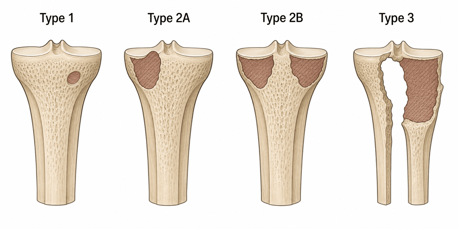
AORI Classification - Femoral Bone Loss
- Description
- Intact metaphyseal bone with cancellous defects only
- Bone Loss
- Minimal - contained defects
- Treatment
- Standard components, cement fill or morcellized graft
- Description
- Damaged metaphysis - one femoral condyle
- Bone Loss
- Moderate - unilateral condylar loss
- Treatment
- Metal augments (blocks), cement, or autograft
- Description
- Damaged metaphysis - both femoral condyles
- Bone Loss
- Moderate - bilateral condylar loss
- Treatment
- Bilateral metal augments or step-cut sleeves
- Description
- Deficient metaphysis - major loss affecting stability
- Bone Loss
- Severe - threatens component stability
- Treatment
- Metaphyseal sleeve, cone, or structural allograft
Femoral bone loss is common at the posterior condyles due to posterior capsular release and wear patterns.
I-D-D-DAORI Bone Defect Classification
Hook:I'm D-D-D (I'm devastated) - the progressive severity of bone loss in revision TKA!
Clinical Assessment
A painful TKA is not automatically a loose TKA. Before committing to revision, systematically exclude infection (always first), intrinsic and extrinsic causes. Up to a quarter of painful TKAs have no identifiable intra-articular cause - revising these blindly worsens outcomes. Localise pain, characterise it (start-up vs activity-related vs constant), and correlate with imaging and aspiration.
- Typical Features
- Rest/night pain, effusion, warmth, early failure, sinus
- Key Discriminating Test
- ESR/CRP, aspiration (WBC/PMN%, culture), alpha-defensin
- Action
- Exclude in EVERY case before any aseptic revision
- Typical Features
- Start-up pain, progressive, late onset
- Key Discriminating Test
- Progressive radiolucent lines, subsidence; bone scan if equivocal
- Action
- Revise components, address AORI bone loss
- Typical Features
- Giving way, recurrent effusion, gap imbalance
- Key Discriminating Test
- Stress radiographs, exam under anaesthesia
- Action
- Increase constraint (CCK/hinge), rebalance
- Typical Features
- Late, often painless until lysis advanced
- Key Discriminating Test
- Focal lysis on radiographs, thin poly, CT for lesions
- Action
- Poly exchange, debride and graft lysis
- Typical Features
- Anterior knee pain, patellar maltracking
- Key Discriminating Test
- Rotational CT (Berger protocol), skyline view
- Action
- Component revision if internal rotation excessive
- Typical Features
- Hip/spine pathology, neuropathic, vascular claudication
- Key Discriminating Test
- Hip/spine exam and imaging, vascular assessment
- Action
- Treat the true source - do NOT revise the knee
- Typical Features
- Patellar clunk, tendinitis, bursitis, CRPS
- Key Discriminating Test
- Clinical exam, ultrasound; CRPS is a clinical diagnosis
- Action
- Targeted non-operative management first
Constraint selection follows a ladder approach: start with the least constraint possible (PS if feasible), escalate to CCK if one collateral ligament is deficient or moderate bone loss exists, and use rotating hinge only for massive bone loss (AORI Type 3) or global instability. Over-constraining increases interface stress and loosening risk; under-constraining risks instability.
Posterior-Stabilized (PS) Revision
Indications:
- AORI Type 1 bone loss
- Intact MCL and LCL
- Balanced flexion-extension gaps
- Simple aseptic loosening or poly wear
Requirements:
- Adequate metaphyseal bone stock
- Competent collateral ligaments (MCL and LCL)
- No significant instability
- Good extensor mechanism
- Lowest constraint = least interface stress
- Better ROM preservation
- Lower revision rate compared to hinged
- Familiar implant for surgeons
- Requires good bone stock and ligaments
- Not suitable for moderate-severe bone loss
- Risk of instability if mis-selected
- May need stems for fixation
PS revision is rarely used in practice - most revisions require at least CCK constraint.
Investigations
Preoperative Workup
History: Onset of symptoms, pain character, mechanical symptoms (locking, catching), prior infections, previous surgeries. Examination: ROM, stability (varus-valgus, AP drawer), extensor lag, wound integrity, neurovascular status.
Serum: ESR, CRP (elevated suggests infection) Aspiration: Synovial WBC, PMN%, culture (aerobic, anaerobic, fungal) Alpha-defensin or synovial CRP if high clinical suspicion Aspiration biopsy: If imaging shows loosening
AP and lateral radiographs: Component position, alignment, radiolucent lines, osteolysis Long-leg alignment films: Assess mechanical axis CT scan: If severe bone loss suspected - quantify defects, plan reconstruction Nuclear medicine: If infection suspected but aspiration negative (Tc-99m, Indium-111 WBC)
AORI classification: Estimate bone loss from imaging Constraint level: Based on bone loss and ligament integrity Augments and stems: Plan for Type 2/3 defects Implant availability: Ensure CCK, hinge, sleeves, cones available
Management Algorithm
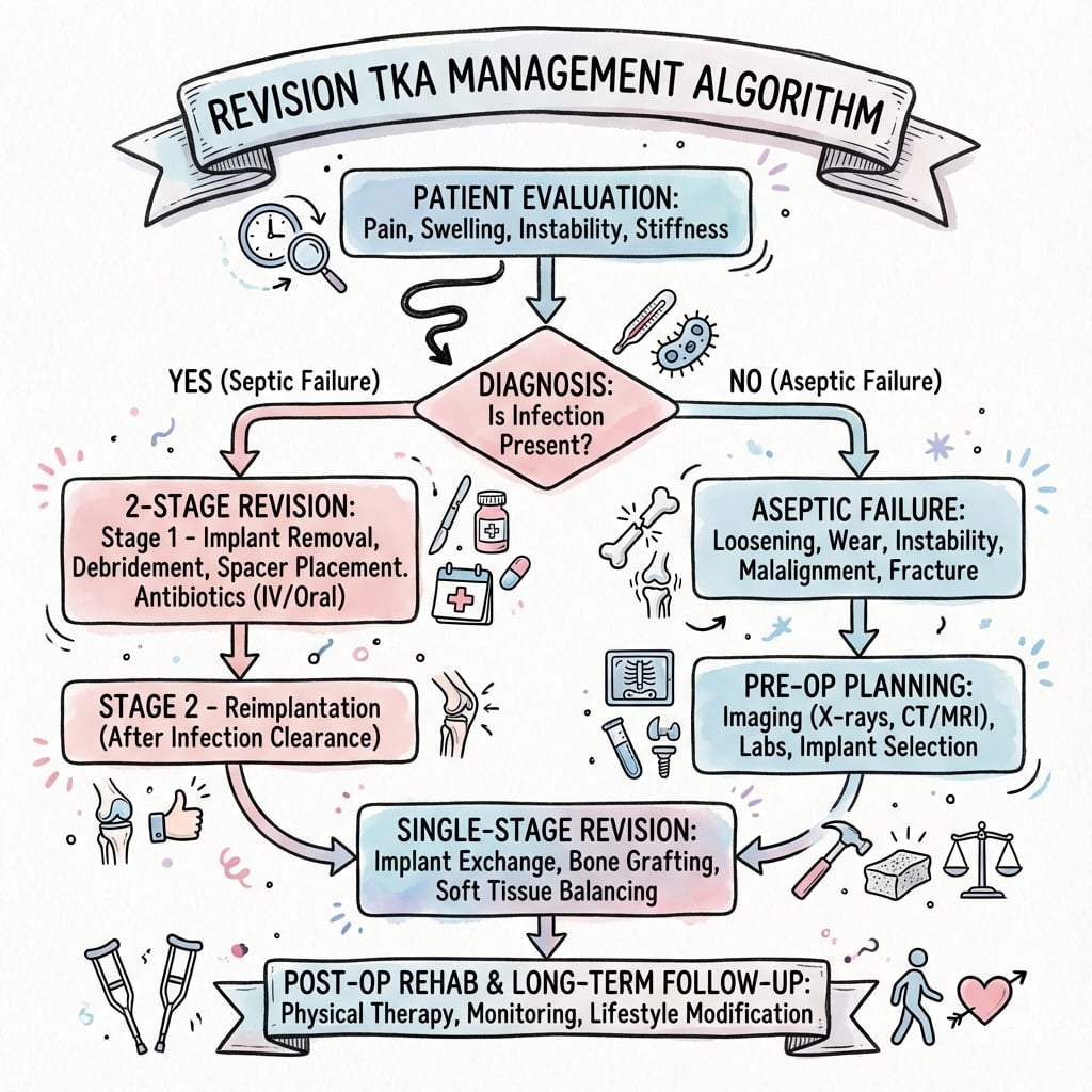
Revision TKA requires a systematic algorithm: (1) Rule out infection first (aspirate, ESR/CRP), (2) Assess bone loss (AORI classification from imaging and intraoperative findings), (3) Evaluate soft tissues (collateral ligament integrity, extensor mechanism), (4) Select constraint (PS to CCK to hinge based on bone loss and instability), (5) Plan reconstruction (augments for Type 2, sleeves/cones for Type 3, stems for stability). This stepwise approach ensures appropriate implant selection and technique.
Algorithm for Aseptic Loosening (50% of Revisions)
Management Steps
Radiographs: Radiolucent lines greater than 2mm, progressive, component subsidence. Rule out infection: Aspirate (WBC, PMN%, culture), ESR/CRP normal or mildly elevated. CT scan: If severe bone loss suspected - quantify AORI grade preoperatively.
AORI classification: Type 1 (intact), Type 2A/B (damaged), Type 3 (deficient). Constraint needs: Assess collateral ligament integrity from examination and prior radiographs. Implant availability: Ensure CCK, augments, stems, sleeves/cones available.
Remove loose components and cement - assess final bone loss intraoperatively. Type 1: Standard components, cement fill, short stems (50-75mm). Type 2: Metal augments, modular stems (75-100mm), CCK constraint. Type 3: Metaphyseal sleeve/cone, long cemented stems (100-150mm), hinge constraint.
Weight-bearing: WBAT for most cases (protected if TTO performed). ROM: Start immediately, goal 0-110° by 6 weeks. Surveillance: Annual radiographs and clinical exam for loosening.
The critical decision is constraint selection: PS if bone loss minimal and ligaments intact (rare in revision), CCK if Type 2 bone loss or one collateral deficient (most common), hinge if Type 3 bone loss or both collaterals deficient. Over-constraining increases loosening risk; under-constraining causes instability.
Surgical Technique
Pre-operative Planning
- Infection: 4-8% (higher than primary)
- Aseptic loosening: 5-10% at 10 years
- Periprosthetic fracture: 3-5% (higher with stems)
- Stiffness: 10-15% (less than 90° flexion)
- Neurovascular injury: Less than 1% (popliteal artery at risk)
- Re-revision: 15-20% at 5 years
- DVT/PE: Standard prophylaxis required
- Revision implant system: CCK and hinge options
- Augments: Metal blocks (5mm, 10mm, 15mm)
- Stems: Modular, various lengths (50-150mm)
- Metaphyseal sleeves/cones: For Type 3 defects
- Extraction tools: Component removal set, cement removal
- C-arm: Essential for alignment and stem placement
- Pulse lavage: High-volume irrigation
Surgical Approach and Exposure
Exposure Steps
Use prior incision if feasible - most lateral if multiple scars. Extend proximally and distally as needed for exposure (often 20-25cm). Skin flaps: Medial and lateral, full-thickness to preserve blood supply.
Medial parapatellar arthrotomy (standard approach). Extend proximally into vastus medialis obliquus if tight. Release adhesions carefully - dense scar tissue common.
Attempt gentle eversion - do NOT force if tight. If unable to evert: Consider quadriceps snip (45° oblique extension of VMO) or tibial tubercle osteotomy (TTO). TTO indications: Severe stiffness, patella baja, need for extensile exposure.
Release scar tissue from suprapatellar pouch, medial and lateral gutters. Protect popliteal artery - posterior capsule may be attenuated. Assess collateral ligaments - note MCL/LCL integrity for constraint decision.
The popliteal artery is at greater risk in revision TKA due to:
- Posterior capsule thinning from wear and loosening
- Loss of anatomical planes from prior surgery
- Need for posterior releases in stiff knees
Protection strategies: Stay subperiosteal, use retractors carefully, avoid blind releases posteriorly.
Modular stems provide rotational stability and load sharing, protecting the metaphyseal bone-implant interface. Use stems in:
- AORI Type 2: 75-100mm press-fit metaphysis, cemented diaphysis
- AORI Type 3: 100-150mm fully cemented diaphysis (14-16cm fixation length)
- Rotating hinge: Mandatory - long cemented stems always
Stems reduce stress at bone-component interface and improve survivorship.
Restoring the Joint Line
A stable, well-fixed revision can still function poorly if the joint line is not restored. In revision TKA the joint line is easily elevated (proximalised), because distal femoral bone is lost or resected and the temptation is to "build down" the tibia or under-augment the distal femur. Because the collaterals are only isometric about the native joint line, an elevated line produces mid-flexion instability and, by lowering the patella relative to the trochlea, patella baja (anterior knee pain, impingement, lost flexion). Restore the joint line to its native level using bony landmarks.
- Approximate relationship to the native joint line
- Joint line lies about 25-30 mm distal
- Approximate relationship to the native joint line
- Joint line lies about 25 mm distal
- Approximate relationship to the native joint line
- Joint line lies about 1-1.5 cm proximal to it
- Approximate relationship to the native joint line
- Sits about 1 cm above the joint line; a low-lying patella (baja) signals an elevated joint line
- Approximate relationship to the native joint line
- Secondary references when the primary landmarks are obscured
In revision TKA the joint line is easily ELEVATED, because distal femoral bone is lost or resected and it is tempting to 'build down' the tibia or under-augment the distal femur. An elevated joint line causes patella baja (anterior knee pain, impingement, lost flexion) and mid-flexion instability (the collaterals become non-isometric and lax in mid-flexion). Restore it using bony landmarks - roughly 25-30 mm distal to the epicondyles, about 1-1.5 cm proximal to the fibular head, and an inferior patellar pole sitting about 1 cm above the joint line in extension - and prefer DISTAL femoral augments (restoring distal femoral offset) over building up the tibia to bring the joint line back to its native level.
Diaphyseal Stem Fixation: Cemented vs Cementless
Stems offload the metaphyseal interface and add rotational stability, and the diaphyseal stem can be fixed cemented or cementless (press-fit) - a genuine, frequently examined controversy (the metaphyseal cone/sleeve hardware itself is covered in the dedicated bone-loss topic).
- Cementless (press-fit)
- Tight canal-filling diaphyseal cortical fit
- Cemented
- Cement mantle in the diaphysis - immediate fixation
- Cementless (press-fit)
- Needs good diaphyseal cortical bone
- Cemented
- Works in osteoporotic or wide/mismatched canals
- Cementless (press-fit)
- Stem can DICTATE position (bowed canal causes malalignment); use an offset coupler to recentre the component
- Cemented
- Component positioned independently of the canal axis (cement fills the offset)
- Cementless (press-fit)
- End-of-stem ('tip') pain in roughly 10-15%
- Cemented
- Cement burden; harder to remove at any future re-revision
- Cementless (press-fit)
- Easier to remove
- Cemented
- More difficult (cement removal)
A cementless press-fit stem relies on a canal-filling diaphyseal cortical fit, is easier to remove at future revision, but can DICTATE component position (a long straight stem in a bowed canal forces malalignment - use an offset coupler to recentre the baseplate) and causes end-of-stem pain in roughly 10-15%. A cemented stem gives immediate fixation, accommodates osteoporotic or wide/mismatched canals and lets you position the component independently of the canal axis, but adds cement burden and is harder to remove. Neither is universally superior; the contemporary compromise endorsed by current reviews is 'dual-zone' (hybrid) fixation - biological metaphyseal fixation with a cone or sleeve combined with a relatively short fully cemented stem.
Two-Stage Revision for Periprosthetic Joint Infection
Two-stage revision is the gold standard for chronic periprosthetic joint infection (PJI) in the knee, with infection control rates of 85-90%. Stage 1 involves component removal, debridement, and antibiotic spacer placement. After 6-8 weeks of IV antibiotics and a 2-week antibiotic holiday, Stage 2 reimplantation proceeds if infection markers have normalized and aspiration is negative.
Two-Stage Protocol
Remove all components: Femur, tibia, polyethylene, cement. Aggressive debridement: Synovectomy, remove all infected tissue, necrotic bone. Send cultures: Multiple samples (5-6) from different areas - aerobic, anaerobic, fungal. Antibiotic spacer: Static (block) or articulating (prefabricated or hand-made with mold). Antibiotics in cement: Vancomycin and tobramycin or gentamicin (organism-directed if known).
Organism-specific therapy: Based on intraoperative cultures (ID consult). Typical duration: 6 weeks IV antibiotics. Monitor: ESR, CRP weekly - should trend downward. Mobilization: Weight-bearing as tolerated with spacer in situ (articulating spacers allow ROM).
Stop all antibiotics for 2 weeks before reimplantation. Allows clearance of antibiotics from system for accurate aspiration. Repeat inflammatory markers: ESR, CRP should be normalizing (may not be fully normal).
Aspirate knee joint (through spacer) for culture and cell count. Criteria for reimplantation:
- Synovial WBC less than 3000 cells/µL
- PMN% less than 80%
- Negative culture (or low virulence organism with biofilm coverage)
- ESR and CRP trending down (not necessarily normal)
Remove spacer: Similar to primary revision technique. Debride again: Remove spacer cement, reassess bone loss (final AORI grading). Implant revision components: CCK or hinge based on bone loss and instability. Augments and stems: As needed for AORI Type 2/3 defects. Antibiotic-loaded cement: Consider organism-directed antibiotics in cement (controversial). Postoperative antibiotics: 6 weeks oral antibiotics (organism-directed, ID consult).
- Advantages
- Simple, inexpensive, high antibiotic elution
- Disadvantages
- No ROM, muscle atrophy, difficult Stage 2
- Use Case
- Severe bone loss, unable to achieve stability
- Advantages
- Maintains ROM, easier Stage 2, patient mobility
- Disadvantages
- More expensive, lower antibiotic concentration
- Use Case
- Moderate bone loss, able to achieve stability
- Advantages
- Customizable, high antibiotic dose, maintains ROM
- Disadvantages
- Technically demanding, spacer fracture risk
- Use Case
- When prefab spacer unavailable or special sizing needed
SPACETwo-Stage Revision Protocol
Hook:Create SPACE for the knee to heal - the two-stage revision protocol for chronic PJI!
Complications
- Incidence
- 5-10% at 10 years
- Risk Factors
- AORI Type 3, hinged implants, inadequate fixation
- Management
- Re-revision with longer stems, structural grafts, increase constraint
- Incidence
- 4-8%
- Risk Factors
- Diabetes, obesity, prior infection, prolonged surgery
- Management
- Two-stage revision with spacer, 6-8 weeks antibiotics
- Incidence
- 3-5%
- Risk Factors
- Osteoporosis, long stems, stress riser at stem tip
- Management
- ORIF if stem stable, revision if stem loose, consider strut allografts
- Incidence
- 5-10%
- Risk Factors
- Under-constraining, flexion-extension gap imbalance, poly wear
- Management
- Increase constraint (PS to CCK to hinge), thicker poly, revise components
- Incidence
- 10-15%
- Risk Factors
- Arthrofibrosis, oversized components, overstuffing joint
- Management
- Manipulation under anesthesia (within 12 weeks), open arthrolysis, poly exchange
- Incidence
- 2-5%
- Risk Factors
- Multiple prior surgeries, TTO, patellar fracture
- Management
- Primary repair if acute, allograft reconstruction if chronic, consider gastrocnemius flap
- Incidence
- Less than 1%
- Risk Factors
- Popliteal artery injury during exposure, common peroneal nerve palsy
- Management
- Vascular repair emergently if arterial, nerve exploration if palsy (often traction neurapraxia)
Periprosthetic fractures often occur at the stem tip - a stress riser in osteoporotic bone. Prevention strategies:
- Use cemented stems in osteoporotic bone (better load sharing)
- Bypass prior stress risers by 2 cortical diameters (extend stem past screw holes)
- Consider strut allografts prophylactically in very osteoporotic bone
- Weight-bearing precautions (TDWB) for 6-12 weeks if high risk
Management: ORIF if stem stable (plate and strut allograft), revision to longer stem if loose.
Postoperative Care and Rehabilitation
Standard Revision TKA Postoperative Protocol
Rehabilitation Timeline
DVT prophylaxis: Chemical (enoxaparin or rivaroxaban) and mechanical (SCDs). Pain control: Multimodal analgesia (acetaminophen, NSAIDs, opioids as needed). Mobilization: Out of bed to chair on Day 1, ambulate with walker Day 2. Weight-bearing: WBAT (weight-bearing as tolerated) for most revisions. ROM: CPM or bedside PT - goal 0-90° by discharge. Drain removal: 24-48 hours when output less than 30mL per 8 hours.
Weight-bearing: Progress to full weight-bearing with assistive device. ROM exercises: Active-assisted and passive, goal 0-110° by 6 weeks. Quadriceps strengthening: Straight leg raises, quad sets, terminal knee extension. Gait training: Progress from walker to cane to independent. Wound care: Staples removed at 2 weeks, monitor for infection. Radiographs: 6-week films to assess alignment, component position, no early loosening.
Discontinue assistive device when safe gait without limp. Resistance exercises: Progress resistance training for quads, hamstrings, hip abductors. Functional activities: Stairs, sit-to-stand, balance exercises. ROM maintenance: Continue ROM exercises, goal 0-120° flexion. Return to activities: Light ADLs, avoid high-impact (running, jumping).
Functional milestones: Independent ADLs, driving (8-12 weeks), return to work (12-16 weeks). Activity modification: Avoid high-impact sports - low-impact only (cycling, swimming, golf). Annual follow-up: Radiographs and clinical exam to monitor for loosening, wear, osteolysis. Expectations: 70-80% function of primary TKA, pain improvement in 80-85% of patients.
Outcomes and Prognosis
- Survivorship (10 yr)
- 90-95%
- Functional Outcome
- Good - similar to primary TKA
- Re-revision Risk
- 5-8%
- Survivorship (10 yr)
- 85-90%
- Functional Outcome
- Fair - 70-80% of primary
- Re-revision Risk
- 10-15%
- Survivorship (10 yr)
- 80-85%
- Functional Outcome
- Fair to poor - 60-70% of primary
- Re-revision Risk
- 15-20%
- Survivorship (10 yr)
- 80-85% (infection control 85-90%)
- Functional Outcome
- Fair - 60-70% of primary
- Re-revision Risk
- 20-25%
Factors associated with worse outcomes in revision TKA:
- AORI Type 3 bone loss (requires structural grafts - higher failure)
- Rotating hinge implant (higher loosening and infection rates)
- Multiple prior revisions (re-revision risk doubles with each revision)
- Chronic PJI (two-stage has lower function than aseptic revision)
- BMI over 35 (higher infection and complication rates)
- Extensor mechanism insufficiency (quadriceps dysfunction limits function)
Counseling patients realistically about expectations is critical - revision TKA does NOT achieve primary TKA outcomes.
Guidelines, Registries & Global Practice
Revision TKA is a globally rising burden driven by an ageing population and expanding primary TKA volumes. Across major national registries the leading reasons for revision are broadly consistent - infection, aseptic loosening and instability dominate - but exact proportions, implant choices and PJI protocols vary by region. Candidates for any board should know the common diagnostic framework (ICM/EBJIS for PJI, AORI for bone loss) and where major guidelines genuinely differ.
Global epidemiology
- Rising volume: registry data consistently report year-on-year growth in revision TKA tracking the global rise in primary TKA (de Steiger/AOANJRR, PMID 35271977)
- Leading failure modes: infection, aseptic loosening and instability predominate across registries
- Demographics: mean age 65-70 at revision; female predominance mirrors primary TKA
- Time to revision: bimodal - early failures (infection, instability, malposition) vs late (loosening, wear)
- AOANJRR distal femoral replacement for periprosthetic fracture: cumulative second-revision 12% at 6 years; infection (37%) and aseptic loosening (33%) the main re-revision causes (PMID 35271977)
- German EPRD: aseptic revision higher after constrained vs unconstrained TKA (3.3% vs 2.8% at 7 years); ligament instability the commonest aseptic cause in unconstrained TKA (PMID 39313693)
- Registries (NJR, AJRR, AOANJRR, SHAR, NZJR) are the primary source of real-world implant survival
MCQ Practice Points
Q: A revision TKA patient has bone loss affecting both tibial plateaus but the metaphyseal rim is intact. What AORI grade is this and what reconstruction is appropriate? A: AORI Type 2B (damaged metaphysis, both condyles/plateaus involved). Appropriate reconstruction includes bilateral metal augments (5-15mm blocks) cemented to the host bone, with a modular tibial baseplate and press-fit stem (75-100mm length, cemented in diaphysis) for rotational stability and load sharing. Type 2B requires augmentation but does NOT need structural grafts (sleeves/cones).
Q: What is the key difference in indications between a CCK and a rotating hinge implant in revision TKA? A: CCK (constrained condylar knee) is indicated for AORI Type 1-2 bone loss with MCL or LCL insufficiency (one collateral deficient), or moderate flexion-extension gap imbalance (3-5mm). Rotating hinge is reserved for AORI Type 3 bone loss (deficient metaphysis), MCL and LCL insufficiency (global instability), or massive flexion-extension gap imbalance. CCK provides varus-valgus constraint through a taller post (5-10° laxity) but still relies on collaterals; hinge is a mechanical hinge allowing only flexion-extension (0° varus-valgus laxity).
Q: What are the criteria for proceeding to Stage 2 reimplantation after a two-stage revision for chronic PJI? A: Criteria include: (1) Completed 6-8 weeks IV antibiotics organism-specific, (2) 2-week antibiotic holiday to clear antibiotics from system, (3) Aspiration of knee joint showing synovial WBC less than 3000 cells/µL and PMN less than 80%, (4) Negative culture (or low-virulence organism with biofilm antibiotic coverage planned), (5) ESR and CRP trending downward (may not normalize completely, but should be improving). All criteria must be met before reimplantation - proceeding with persistent infection results in failure in over 90% of cases.
Q: What is the recommended stem fixation technique in revision TKA for AORI Type 3 defects? A: Long cemented stems (100-150mm length) with 14-16cm cemented fixation in the diaphysis. The stem provides load sharing and bypasses the deficient metaphysis (Type 3). Cementation technique: place cement restrictors, pressurize cement in the diaphyseal canal, insert stem and allow polymerization. Press-fit fixation in the metaphysis is inadequate for Type 3 defects - must have diaphyseal fixation. Stem length should bypass any stress risers (prior screw holes) by 2 cortical diameters to prevent periprosthetic fracture.
Q: What are the advantages of metaphyseal sleeves over structural bulk allografts for AORI Type 3 defects? A: Metaphyseal sleeves have superior survivorship (92% at 5 years vs 78% for allografts), lower complication rates (10% vs 25%, particularly nonunion and resorption seen with allografts), biological fixation through osseointegration (porous-coated titanium), and faster rehabilitation (immediate press-fit stability vs waiting for graft incorporation). Allografts have risks of nonunion (10-15%), resorption over time, disease transmission (very low but non-zero), and technically demanding shaping/fixation. Sleeves are now first-line treatment for Type 3 defects.
Q: How do functional outcomes and survivorship of revision TKA compare to primary TKA? A: Revision TKA has inferior outcomes to primary TKA: (1) Functional scores 70-80% of primary TKA (worse with hinge or two-stage), (2) Survivorship 85-90% at 10 years vs 95% for primary TKA, (3) Re-revision rate 15-20% at 5 years vs 2-3% for primary, (4) Complication rate 20-30% vs 5-10% for primary. Patients must be counseled realistically - revision TKA does NOT restore primary TKA function and has higher risk of failure. Two-stage revisions for PJI have even worse outcomes (60-70% function, 80-85% survivorship).
Exam Viva Scenarios
Practise clinical reasoning and management decisions out loud
“A 68-year-old woman presents with progressive medial knee pain 8 years after primary TKA. Examination shows stable knee with full extension and 110° flexion, no effusion. Radiographs demonstrate 2mm radiolucent lines under the medial tibial baseplate and small focal osteolysis. ESR 18, CRP 8. How would you assess and manage this patient?”
“A 72-year-old diabetic man with prior TKA presents with chronic pain and recurrent effusions for 6 months. He has had two aspirations showing low-grade coagulase-negative staph. ESR 45, CRP 22. You have performed Stage 1 debridement and spacer placement. Walk me through your Stage 2 planning and technique.”
“Intraoperatively during revision TKA, after component removal you find severe tibial bone loss - the entire medial and lateral plateaus are gone down to the metaphyseal-diaphyseal junction (AORI Type 3). How do you reconstruct this defect?”
Key Indications
- Aseptic loosening: 50% of revisions - radiolucent lines, subsidence, pain
- Instability: 20% - MCL/LCL insufficiency, flexion-extension gap imbalance
- Infection (PJI): 15% - two-stage revision gold standard (85-90% control)
- Polyethylene wear/osteolysis: 10% - focal lysis, thin poly
- Stiffness: 3-5% - less than 90° flexion, failed manipulation
AORI Bone Defect Classification
- Type 1 (Intact): Metaphyseal bone intact - standard components, cement fill
- Type 2A (Damaged one): One condyle/plateau - metal augment, cement, stem
- Type 2B (Damaged both): Both condyles/plateaus - bilateral augments, stem
- Type 3 (Deficient): Metaphysis deficient - sleeve/cone/allograft, long stem (14-16cm)
Constraint Selection Algorithm
- PS: AORI Type 1, intact MCL and LCL, balanced gaps (rarely used in revision)
- CCK: Type 2, MCL OR LCL deficient, 3-5mm gap imbalance (most common)
- Rotating hinge: Type 3, MCL AND LCL deficient, massive bone loss, global instability
- Increase constraint as bone loss worsens and soft tissue insufficiency increases
Surgical Technique Pearls
- Rule out infection FIRST: ESR/CRP, aspirate (WBC, PMN%, culture) in ALL cases
- Component removal: Peripheral osteotomes, circumferential disruption, preserve bone
- Cement removal: Meticulous debridement - final AORI only visible after cement out
- Stems: Modular, 75-100mm Type 2, 100-150mm Type 3, cemented in diaphysis (14-16cm)
- Metaphyseal sleeves: Press-fit biological fixation for Type 3, 92% survivorship at 5 years
Two-Stage Revision Protocol
- Stage 1: Debridement, explant, antibiotic spacer (vancomycin + tobramycin in cement)
- 6-8 weeks IV antibiotics organism-specific (ID consult), then 2-week holiday
- Reimplantation criteria: WBC less than 3000, PMN less than 80%, negative culture, ESR/CRP trending down
- Stage 2: Remove spacer, debride again, revise with CCK/hinge, augments/stems as needed
- Infection control: 85-90%, but re-revision risk 20-25%, function 60-70% of primary
Complications and Outcomes
- Aseptic loosening: 5-10% at 10 years (higher with hinge, Type 3 defects)
- Infection: 4-8% (higher than primary 1-2%)
- Periprosthetic fracture: 3-5% (stem tip stress riser, osteoporotic bone)
- Survivorship: 85-90% at 10 years (vs 95% primary), re-revision 15-20% at 5 years
- Function: 70-80% of primary TKA (worse with hinge or two-stage)
Evidence Base and Key Trials
Tantalum Femoral Cones for Severe Femoral Bone Loss (AORI Type 3)
- Single-institution series: 159 tantalum metaphyseal femoral cones in 157 patients, mean follow-up 5 years
- 5-year survivorship 96% with cone aseptic loosening as the endpoint
- 5-year survivorship 84% for revision of the cone for any reason, 70% free of any reoperation
- All 134 unrevised cones were radiographically well-fixed without loosening
- Aseptic cone failure was associated with hinged TKA used for a Type 3 defect
Metaphyseal Sleeves for Bone Loss in Revision TKA
- Prospective series: 96 knees revised with metaphyseal sleeves, 83 with minimum 2-year follow-up (mean 2.4 years)
- Sleeves used predominantly for AORI Type 2B defects (femoral and tibial revisions)
- Mean Knee Society function score improved from 47.9 to 61.1 points
- No progressive radiolucent lines were seen around the metaphyseal sleeves
- Only 2 of 73 tibial components (2.7%) required revision for aseptic loosening at short-term follow-up