Muscle Breakdown | CK Elevation | Myoglobinuria | Acute Kidney Injury
- CK over 10,000 U/L is diagnostic threshold for rhabdomyolysis
- Myoglobinuria (dark tea-colored urine) indicates significant muscle breakdown
- Acute kidney injury from myoglobin tubular obstruction - prevent with aggressive fluids
- Hyperkalemia from muscle necrosis - cardiac risk, monitor ECG
- Urine alkalinization (pH over 6.5) reduces myoglobin precipitation in tubules
- “Target urine output over 200ml/hr with aggressive IV fluids (4-6L first 24h)
- “Sodium bicarbonate alkalinizes urine - prevents myoglobin precipitation
- “Compartment syndrome is most common orthopaedic cause
- “CK peaks at 24-48 hours, then declines if treated appropriately
Dark tea-colored or cola-colored urine indicates myoglobinuria - this is the clinical sign of significant rhabdomyolysis. Requires immediate aggressive fluid resuscitation to prevent acute kidney injury from myoglobin tubular obstruction.
CK over 10,000 U/L is diagnostic threshold. Levels can exceed 100,000 in severe cases. CK peaks at 24-48 hours after injury, then declines if treated. Monitor serial levels.
Aggressive IV fluids target urine output over 200ml/hr (4-6L first 24 hours). Alkalinize urine with sodium bicarbonate (target pH over 6.5) to prevent myoglobin precipitation in renal tubules. This is the key to preventing AKI.
Hyperkalemia from massive muscle necrosis can cause cardiac arrhythmias and arrest. Monitor potassium levels and ECG. May require calcium, insulin/glucose, or dialysis. Life-threatening complication.
- Myoglobinuria
- Absent
- Urine Output
- Normal
- Management
- IV fluids, monitor CK
- Myoglobinuria
- Present
- Urine Output
- Target 200ml/hr
- Management
- Aggressive fluids, alkalinize urine
- Myoglobinuria
- Severe
- Urine Output
- Over 200ml/hr
- Management
- ICU, consider dialysis
Overview and Epidemiology
Rhabdomyolysis is a syndrome of skeletal muscle breakdown with release of intracellular contents into the circulation, leading to myoglobinuria and potential acute kidney injury. It is a serious complication of compartment syndrome, crush injuries, and other muscle-damaging conditions.
Epidemiology:
- Common complication of compartment syndrome (especially if delayed over 6 hours)
- Crush injuries (earthquakes, building collapse)
- Prolonged immobility (unconscious patients)
- Exercise-induced (exertional rhabdomyolysis)
- Drug-induced (statins, alcohol)
- Heat-related illness
Mechanism of Muscle Breakdown:
Compartment syndrome is the most common orthopaedic cause of rhabdomyolysis. When compartment syndrome is delayed (over 6 hours), muscle necrosis occurs, releasing myoglobin. This is why post-fasciotomy monitoring for rhabdomyolysis is essential.
Anatomy and Pathophysiology
- Skeletal muscle contains high concentrations of CK and myoglobin
- Myoglobin is an oxygen-binding protein in muscle
- Muscle cell membrane disruption releases these into circulation
- Renal tubules filter myoglobin, which can precipitate
- Glomeruli filter myoglobin from blood
- Renal tubules are where myoglobin precipitates
- Acidic urine (pH under 5.6) promotes precipitation
- Tubular obstruction leads to decreased GFR and AKI
Whatever the trigger (direct trauma, ischaemia, metabolic, toxic), the end result is an uncontrolled rise in intracellular calcium. ATP depletion impairs the Na+/K+ and Ca2+ ATPase pumps, calcium floods the myocyte, activating proteases and phospholipases that destroy the sarcolemma. Membrane rupture releases creatine kinase, myoglobin, potassium, phosphate, urate and purines into the circulation, while extracellular fluid and calcium are sequestered into damaged muscle (third-spacing causing hypovolaemia and early hypocalcaemia).
Three mechanisms of myoglobinuric AKI (all favoured by hypovolaemia and aciduria):
- Renal vasoconstriction / ischaemia - hypovolaemia plus scavenging of nitric oxide by myoglobin reduces medullary blood flow
- Intratubular cast formation - myoglobin precipitates with Tamm-Horsfall protein, especially in acidic urine (pH under 5.6), obstructing distal tubules
- Direct heme-mediated tubular toxicity - iron-catalysed lipid peroxidation and free-radical injury of proximal tubular cells
- Process
- Sarcolemma rupture, calcium influx, protease activation
- Consequence
- Release of intracellular contents
- Process
- CK, myoglobin, K+, PO4, urate enter blood; fluid sequestered in muscle
- Consequence
- Rising serum markers + hypovolaemia
- Process
- Myoglobin filtered, casts form, heme toxicity, vasoconstriction
- Consequence
- Tubular obstruction + ischaemia
- Process
- Acute tubular necrosis, falling GFR
- Consequence
- Acute kidney injury, hyperkalaemia
Classification Systems
Rhabdomyolysis Severity
- CK Level
- 5,000-10,000 U/L
- Myoglobinuria
- Absent
- Management
- IV fluids, monitor
- CK Level
- 10,000-50,000 U/L
- Myoglobinuria
- Present
- Management
- Aggressive fluids, alkalinize urine
- CK Level
- Over 50,000 U/L
- Myoglobinuria
- Severe
- Management
- ICU, dialysis if needed
Usually resolves with simple hydration. Monitor CK levels.
Requires aggressive management to prevent AKI. Most common presentation.
Life-threatening, requires ICU care. High risk of AKI and hyperkalemia.
Clinical Assessment
History:
- Compartment syndrome (most common orthopaedic cause)
- Crush injury (prolonged compression)
- Prolonged immobility (unconscious patient)
- Exercise-induced (exertional)
- Drug history (statins, alcohol)
- Heat exposure
Physical Examination:
- Swollen, tender muscles
- Weakness
- Myalgia
- Compartment syndrome signs (if present)
- Dark tea-colored urine (myoglobinuria)
- Decreased urine output
- Signs of hyperkalemia (ECG changes)
- Signs of AKI (oliguria, fluid overload)
Urine Assessment:
- Color: Dark tea-colored or cola-colored = myoglobinuria
- Dipstick: Positive for blood (but no RBCs on microscopy) = myoglobin
- Output: Decreased output indicates AKI developing
Dark tea-colored or cola-colored urine is the clinical hallmark of myoglobinuria. Dipstick will be positive for blood, but microscopy shows no red blood cells (myoglobin, not hemoglobin). This requires immediate aggressive management.
Differential Diagnosis
The "blood-positive dipstick with no red cells" picture and dark urine have a short, high-yield differential. The key discriminator is serum CK and the urine microscopy.
- Distinguishing Feature
- Muscle pain/weakness, swelling, recent crush/exertion
- Serum CK
- Markedly raised (over 5x normal)
- Urine Microscopy / Dipstick
- Dipstick blood positive, NO red cells; pigmented granular casts
- Distinguishing Feature
- Anaemia, raised LDH/bilirubin, low haptoglobin; pink plasma
- Serum CK
- Normal
- Urine Microscopy / Dipstick
- Dipstick blood positive, no red cells; plasma pink (myoglobin clears, Hb stains plasma)
- Distinguishing Feature
- Renal/urological cause, clots
- Serum CK
- Normal
- Urine Microscopy / Dipstick
- Dipstick blood positive WITH red cells on microscopy
- Distinguishing Feature
- Chest pain, no myalgia
- Serum CK
- Raised but CK-MB fraction high, troponin positive
- Urine Microscopy / Dipstick
- Urine clear, dipstick negative
- Distinguishing Feature
- Subacute proximal weakness, autoantibodies
- Serum CK
- Moderately raised, fluctuating
- Urine Microscopy / Dipstick
- Usually clear urine
- Distinguishing Feature
- Dietary/drug history
- Serum CK
- Normal
- Urine Microscopy / Dipstick
- Dipstick blood NEGATIVE
Investigations
Laboratory Tests:
- Finding
- Over 10,000 U/L (diagnostic)
- Significance
- Peaks 24-48h, then declines
- Finding
- Elevated
- Significance
- Confirms muscle breakdown
- Finding
- Positive
- Significance
- Dark urine, tubular obstruction risk
- Finding
- Hyperkalemia
- Significance
- Cardiac risk - monitor ECG
- Finding
- Rising
- Significance
- AKI developing
- Finding
- Elevated
- Significance
- Muscle breakdown
- Finding
- Hypocalcemia early
- Significance
- Precipitates in necrotic muscle
Monitoring Protocol:
Laboratory Monitoring
- CK, myoglobin (serum and urine)
- UEC (potassium, creatinine, urea)
- FBC, coagulation
- ECG (hyperkalemia assessment)
- CK levels (peak at 24-48h)
- Potassium (hyperkalemia risk)
- Creatinine (AKI progression)
- Urine output monitoring
- CK declining
- Renal function improving
- Potassium normalized
CK peaks at 24-48 hours after injury, then declines with half-life of 1.5 days if treated appropriately. If CK continues rising after 48 hours, ongoing muscle damage is occurring (incomplete fasciotomy, recurrent compartment syndrome, or other cause).
Urinary Sediment Findings
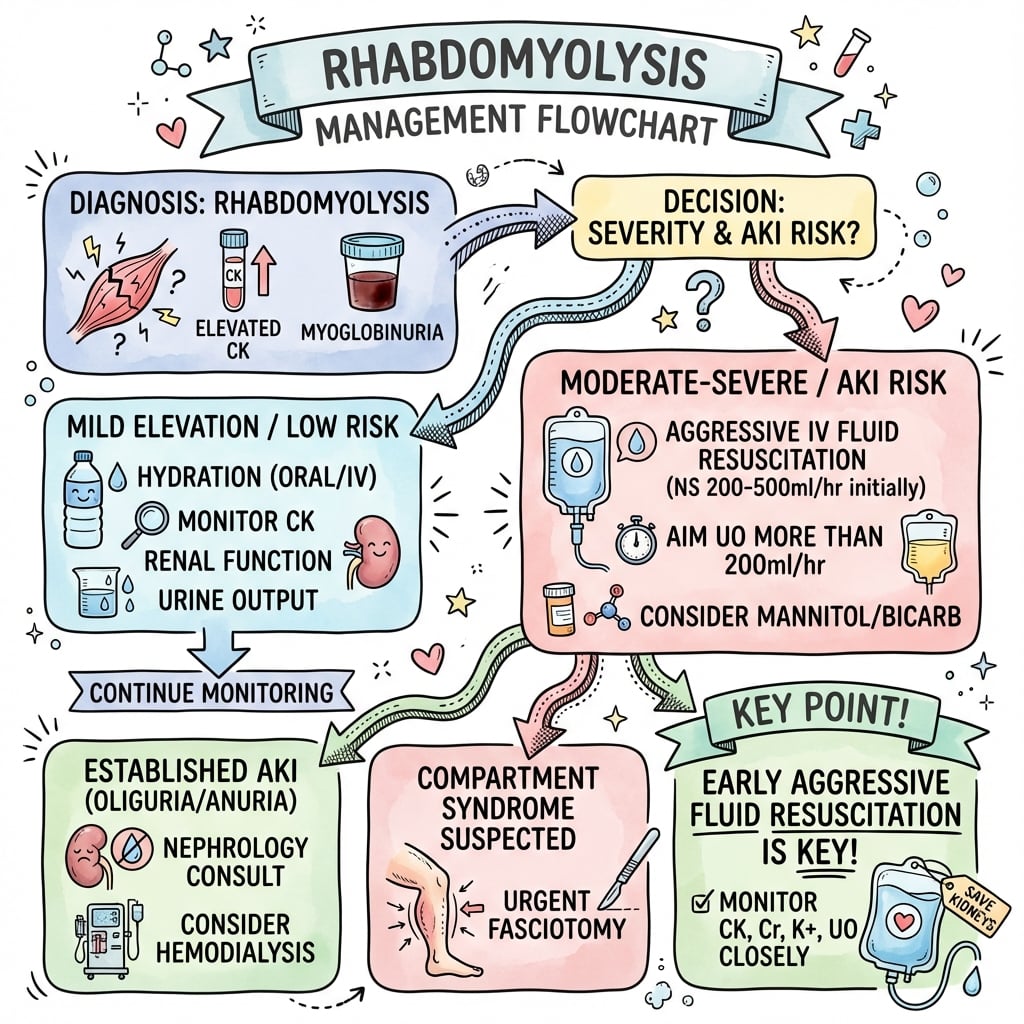
Management Algorithm
CK 5,000-10,000, No Myoglobinuria
- IV fluids: 1-2L over 24 hours
- Monitor CK levels
- Monitor urine output
- Usually resolves without complications
Excellent with simple hydration.
FARMRhabdomyolysis Management
Hook:FARM the patient: Fluids Aggressive, Alkalinize urine, Renal protection, Monitor closely!
BICARBMyoglobinuria Management
Hook:BICARB: Bicarbonate, IV fluids, CK monitoring, Avoid nephrotoxins, Renal function, Bicarbonate target!
Treatment Details
Aggressive IV Fluid Therapy
Maintain urine output over 200ml/hr (or over 1ml/kg/hr)
- Initial: 1-2L bolus if dehydrated
- Maintenance: 4-6L over first 24 hours
- Type: Normal saline or lactated Ringer's
- Monitoring: Hourly urine output, daily weights, clinical assessment
- High urine flow prevents myoglobin precipitation
- Maintains renal perfusion
- Prevents tubular obstruction
Target urine output over 200ml/hr (or over 1ml/kg/hr) is essential to prevent myoglobin tubular obstruction. This requires aggressive IV fluid administration - 4-6L in first 24 hours is common.
Surgical Technique
Note: Rhabdomyolysis itself is a medical condition, not a surgical procedure. However, if compartment syndrome is the underlying cause, fasciotomy is required. This section addresses fasciotomy technique when rhabdomyolysis is present or suspected.
Fasciotomy Technique (When Rhabdomyolysis Present)
- Rhabdomyolysis may already be present if delayed over 6 hours
- Monitor for myoglobinuria post-operatively
- Plan for aggressive fluid resuscitation
- Consider ICU admission if severe
- Standard fasciotomy approach (leg: 2-incision, 4-compartment)
- Release all compartments completely
- Assess muscle viability (pink, contractile = viable; dark, non-contractile = necrotic)
- Debride obviously necrotic muscle
- Leave wounds open
- Immediate aggressive fluid resuscitation
- Monitor for rhabdomyolysis
- Serial CK levels
- Urine output monitoring
Fasciotomy prevents further muscle necrosis and may limit rhabdomyolysis progression.
Complications
- Incidence
- 30-50% if untreated
- Management
- Prevent with aggressive fluids, alkalinization
- Incidence
- Common in severe cases
- Management
- Monitor ECG, treat with calcium/insulin/bicarb
- Incidence
- Common
- Management
- Usually resolves, avoid overcorrection
- Incidence
- During recovery
- Management
- Calcium released from necrotic muscle
- Incidence
- If underlying cause
- Management
- Fasciotomy if present
- Incidence
- Rare
- Management
- Supportive care
- Most serious complication
- Develops in 30-50% if untreated
- Prevention is key: aggressive fluids, urine alkalinization
- Most recover renal function with treatment
- May require temporary dialysis
- Life-threatening complication
- Can cause cardiac arrest
- Requires immediate treatment
- Monitor ECG continuously
Most patients with rhabdomyolysis-induced AKI recover renal function with appropriate treatment. Dialysis is often temporary. Long-term renal impairment is uncommon if treated early and aggressively.
Postoperative Care
After Fasciotomy (if compartment syndrome cause):
Post-Fasciotomy Rhabdomyolysis Management
- Aggressive IV fluids (target 200ml/hr urine output)
- Alkalinize urine (sodium bicarbonate)
- Monitor CK, potassium, creatinine
- ECG monitoring
- Continue aggressive fluids
- Serial CK every 6-12 hours
- Monitor urine output hourly
- Assess for hyperkalemia
- Consider ICU if severe
- CK should peak then decline
- Continue monitoring
- Assess renal function
- Wean fluids as CK normalizes
- CK declining
- Renal function improving
- Can reduce monitoring frequency
- Continue until CK under 5,000
Key Monitoring:
- Urine output (target over 200ml/hr)
- Urine color (should lighten as myoglobinuria resolves)
- CK levels (should decline after 24-48h peak)
- Potassium (hyperkalemia risk)
- Creatinine (AKI progression)
Outcomes and Prognosis
- CK normalization: 3-5 days with treatment
- Renal function: Most recover completely
- Mortality: Low if treated appropriately (under 5%)
- Long-term: Usually no sequelae if treated early
- Early treatment: Better outcomes
- CK level: Higher CK = worse prognosis
- Time to treatment: Delayed treatment = higher AKI risk
- Underlying cause: Treatable causes (compartment syndrome) have better outcomes
Guidelines, Registries & Global Practice
Global epidemiology:
- Crush-related rhabdomyolysis follows mass-casualty events: earthquakes are the archetype (Marmara/Turkey 1999, Wenchuan/China 2008, Haiti 2010, Kahramanmaras/Turkey 2023), where it is a leading cause of preventable post-event death after head injury.
- Non-traumatic causes (statins/drugs, exertion, alcohol, immobility, infection, inherited metabolic myopathies) dominate in routine hospital practice worldwide.
- Acute kidney injury complicates roughly 10-50% of significant rhabdomyolysis depending on cause and resuscitation timing; with early aggressive fluids, AKI and dialysis dependence fall substantially.
Guidelines side by side (where emphasis genuinely differs):
- Emphasis
- Crush-syndrome focus: start IV isotonic saline before/at extrication; large volumes (up to ~6-12 L/day) with close monitoring; alkalinisation optional
- Emphasis
- Recognise and decompress acute compartment syndrome early (delta-P under 30 mmHg); fasciotomy limits ongoing muscle necrosis and myoglobin load
- Emphasis
- General AKI principles: volume optimisation, avoid nephrotoxins, treat hyperkalaemia, RRT for standard indications
- Emphasis
- Exertional rhabdomyolysis: rest, hydration, screen for recurrent/atypical cases and inherited metabolic myopathy before return to sport
High- vs limited-resource practice variation:
- High-resource: point-of-care CK and electrolytes, continuous ECG, ICU-level monitoring, and ready access to haemodialysis/CRRT for refractory AKI.
- Limited-resource / disaster settings: the decisive intervention is early oral and IV fluid; dialysis capacity is the rate-limiting step. International nephrology task forces pre-position dialysis support after earthquakes precisely because crush AKI overwhelms local capacity.
The preventable harms are the same everywhere: missed/late acute compartment syndrome, under-resuscitation, failure to detect rising potassium, and continued nephrotoxin exposure (NSAIDs, contrast). Early fluids and serial CK/potassium/creatinine address all four.
Controversies and Areas of Uncertainty
Much of "classic" rhabdomyolysis management is based on physiology and observational data rather than randomised trials. The exam-relevant debates are:
- Where the evidence sits
- No RCT shows bicarbonate-alkaline diuresis is superior to adequate isotonic saline. Brown et al (J Trauma 2004) found no benefit of bicarbonate/mannitol on renal failure, dialysis or mortality. Most guidelines now favour volume first, bicarbonate optional.
- Where the evidence sits
- Theoretical osmotic diuretic and free-radical scavenger benefit is unproven; risks volume depletion and hyperosmolality. Avoid in oliguria/established AKI. Not supported by trial data.
- Where the evidence sits
- No universal cutoff. Commonly CK over 5x upper limit of normal (~1,000 U/L) defines the syndrome, while AKI risk rises sharply above ~5,000 U/L and especially over 15,000-40,000 U/L. The '10,000' figure is a pragmatic teaching threshold, not an absolute rule.
- Where the evidence sits
- 100-300 ml/hr is variously quoted; ~200-300 ml/hr (or 3 ml/kg/hr) is typical, but the real goal is brisk flow without precipitating fluid overload.
- Where the evidence sits
- RRT is for standard indications (refractory hyperkalaemia, acidosis, fluid overload, uraemia). 'Prophylactic' high-flux dialysis to clear myoglobin is not evidence-based for routine use.
If you remember one thing for the viva: volume, volume, volume. Early, adequate isotonic fluid resuscitation prevents AKI; bicarbonate and mannitol are adjuncts of unproven incremental benefit and must never delay or substitute for fluids.
Exertional Rhabdomyolysis, Recurrent Episodes and Inherited Metabolic Myopathy
The etiology table and Scenario 3 both invoke exertional rhabdomyolysis, and the sports-medicine guidance flags screening for "recurrent/atypical cases and inherited metabolic myopathy before return to sport" - but the underlying disease is never developed. This is a distinct, high-yield strand: most exertional cases are self-limiting, but a minority signal a genetic muscle disorder that changes counselling and return-to-play.
Exertional (exercise-induced) rhabdomyolysis typically follows unaccustomed, intense or eccentric exercise, often compounded by heat, humidity, dehydration, sleep deprivation or stimulant/supplement use. Compared with crush injury, CK elevations are usually lower and acute kidney injury is uncommon (McMahon and colleagues found exercise carried one of the lowest rates of dialysis or death). Sickle cell trait is a recognised risk factor for exertional collapse and rhabdomyolysis under extreme exertion, and drugs (statins, especially with fibrates, and alcohol) lower the threshold.
- Recurrent episodes, or rhabdomyolysis grossly disproportionate to the exertion
- Personal or family history of exercise intolerance, cramps, dark urine or unexplained anaesthetic reactions
- Persistently elevated resting CK that fails to settle toward normal several weeks after the episode (a "hyperCKaemia" that does not resolve)
- Symptoms of exercise intolerance with cramps, and the "second-wind" phenomenon (transient improvement in exercise tolerance after a few minutes of rest, characteristic of McArdle disease)
- McArdle disease (glycogen storage disease type V, myophosphorylase deficiency) - block in glycogenolysis; classic second-wind phenomenon; the ischaemic (or non-ischaemic) forearm exercise test shows a flat lactate with a normal or exaggerated ammonia rise.
- Carnitine palmitoyltransferase II (CPT II) deficiency - a disorder of fatty-acid oxidation and the most common inherited cause of recurrent exertional rhabdomyolysis in adults; episodes are triggered by prolonged exercise, fasting, cold or intercurrent illness rather than short bursts.
- RYR1 variants, which overlap with malignant hyperthermia susceptibility - a reason a personal or family history of an adverse anaesthetic reaction matters.
the acute treatment is identical (fluids, monitor potassium and renal function), but the disposition differs - screen for a metabolic myopathy before clearing an athlete to return, refer for genetic/metabolic work-up when red flags are present, and advise avoidance of malignant-hyperthermia triggers where RYR1 disease is suspected.
A single exertional episode with a rapidly falling CK needs hydration and sensible return-to-training. Recurrence, a family history, a persistently raised resting CK, or rhabdomyolysis out of proportion to the exercise are the triggers to hunt for an inherited metabolic myopathy (McArdle disease, CPT II deficiency, RYR1/malignant-hyperthermia-related disease) rather than simply rehydrating and discharging.
CRUSHRhabdomyolysis Causes
Hook:CRUSH causes: Compartment syndrome, Rhabdomyolysis, Unconscious, Seizures, Heat stroke!
Risk Stratification: The McMahon Score
The "Outcomes and Prognosis" section lists prognostic factors qualitatively ("higher CK = worse prognosis", "delayed treatment = higher AKI risk"). The examinable upgrade is that these have been combined into a validated admission risk score that quantifies who needs intensive monitoring and early nephrology.
The McMahon score was derived and validated in 2,371 patients with CK over 5,000 U/L across two Boston teaching hospitals. It combines eight admission variables: age, female sex, initial creatinine, initial CK, phosphate, calcium, bicarbonate, and the cause of rhabdomyolysis (seizure, syncope, exercise, statin or myositis attracting negative/protective weighting versus other causes). It predicts the composite of renal replacement therapy or in-hospital death - not a treatment threshold - with good discrimination (C-statistic about 0.82-0.83).
- A low score (under 5) carried roughly a 2-3% risk of dialysis or death.
- A high score (over 10) carried roughly a 61% risk - a very different level of care.
- Cause dominates outcome: the highest composite-event rates were after cardiac arrest, sepsis and compartment syndrome, while myositis, exercise and seizures carried the lowest.
This reframes the exam answer: rather than fixating on a single CK cut-off, use the whole admission picture - age, renal function, electrolytes and the cause - to decide who is triaged to ICU, early nephrology and closer surveillance.
A Risk Prediction Score for Kidney Failure or Mortality in Rhabdomyolysis
- Derivation and validation in 2,371 patients with CK over 5,000 U/L (Massachusetts General and Brigham and Women's Hospitals)
- Composite of renal replacement therapy or in-hospital death occurred in 19% overall (8% RRT, 14% died)
- Eight admission variables: age, female sex, initial creatinine, CK, phosphate, calcium, bicarbonate and cause
- Score under 5 gave a 2-3% event rate; score over 10 gave a 61% event rate; C-statistic 0.82-0.83
When asked to risk-stratify, resist quoting a single CK threshold. The McMahon score shows the underlying cause is the strongest driver - cardiac arrest, sepsis and compartment syndrome carry the worst outcomes, while exercise, seizures and myositis carry the best - alongside age, renal function and electrolytes. The score predicts dialysis or death, not a fluid-prescription cut-off.
MCQ Practice Points
Q: What is the diagnostic threshold for rhabdomyolysis? A: CK over 10,000 U/L - this is the widely accepted diagnostic threshold. Levels can exceed 100,000 in severe cases. CK peaks at 24-48 hours after injury, then declines if treated appropriately.
Q: What is the clinical sign of myoglobinuria? A: Dark tea-colored or cola-colored urine - this indicates significant myoglobin release from muscle breakdown. Dipstick will be positive for blood, but microscopy shows no red blood cells (myoglobin, not hemoglobin).
Q: What is the target urine output for rhabdomyolysis management? A: Over 200ml/hr (or over 1ml/kg/hr) - this high urine flow prevents myoglobin precipitation in renal tubules and maintains renal perfusion. Requires aggressive IV fluid administration (4-6L first 24 hours).
Q: Why is urine alkalinization important in rhabdomyolysis? A: Myoglobin precipitates in acidic urine (pH under 5.6), causing tubular obstruction and AKI. Alkaline urine (pH over 6.5) prevents precipitation. Sodium bicarbonate is added to IV fluids to maintain urine pH over 6.5.
Q: Why does rhabdomyolysis cause hyperkalemia? A: Massive muscle necrosis releases intracellular potassium into circulation. This can cause life-threatening hyperkalemia with cardiac arrhythmias. Requires immediate treatment (calcium, insulin/glucose, bicarbonate) and may need dialysis.
Exam Viva Scenarios
Practise clinical reasoning and management decisions out loud
“A 35-year-old man underwent fasciotomy for compartment syndrome 12 hours after injury. Post-operatively, his urine is dark tea-colored, CK is 25,000 U/L, and creatinine is rising. How do you manage this?”
“A 45-year-old construction worker is extracted from a building collapse after 8 hours. He has a crushed leg, is hypotensive, and his urine is dark. CK is 75,000 U/L, potassium is 6.8mmol/L, and ECG shows peaked T-waves. Describe your immediate management.”
“A 28-year-old athlete presents 48 hours after a marathon with severe muscle pain, weakness, and dark urine. CK is 45,000 U/L, creatinine is 250 micromol/L, and urine output is 30ml/hr. How do you manage this?”
Key Facts
- CK over 10,000 U/L is diagnostic threshold
- Dark tea-colored urine = myoglobinuria
- Compartment syndrome is most common orthopaedic cause
- CK peaks at 24-48 hours, then declines
Management (FARM)
- Fluids Aggressive: Target 200ml/hr urine output (4-6L first 24h)
- Alkalinize urine: Sodium bicarbonate to pH over 6.5
- Renal protection: Prevent myoglobin tubular obstruction
- Monitor: CK, potassium, creatinine, ECG
Hyperkalemia Treatment
- IV Calcium (cardioprotective, stabilizes membrane)
- Insulin/Glucose (drives K+ into cells)
- Sodium Bicarbonate (alkalinizes, shifts K+)
- Dialysis if refractory or severe
Complications
- Acute kidney injury: 30-50% if untreated
- Hyperkalemia: Life-threatening, cardiac risk
- Hypocalcemia early, hypercalcemia late
- Most recover with appropriate treatment
Evidence Base
Rhabdomyolysis and Acute Kidney Injury
- Definitive modern review of pathophysiology and management
- Myoglobin causes AKI via tubular cast obstruction, direct heme toxicity and renal vasoconstriction
- Early, aggressive volume expansion is the cornerstone of AKI prevention
- Evidence for routine bicarbonate and mannitol beyond saline is weak
Compartment Monitoring in Tibial Fractures: The Pressure Threshold for Decompression
- Prospective study of 116 tibial diaphyseal fractures with continuous compartment monitoring
- Acute compartment syndrome occurred in 3 patients (2.6%)
- A differential pressure (diastolic minus compartment pressure) under 30 mmHg missed no cases
- Absolute thresholds of 30 or 40 mmHg would have led to many unnecessary fasciotomies
Early Management of Shock and Prophylaxis of Acute Renal Failure in Traumatic Rhabdomyolysis
- Foundational protocol for crush/traumatic rhabdomyolysis
- Early, vigorous volume loading begun before or during extrication prevents oliguric ARF
- Mannitol-alkaline diuresis proposed to reduce cast formation and tubular toxicity
- Hypovolaemia and aciduria are the key drivers of renal injury
Early Fluid Resuscitation in Patients with Rhabdomyolysis
- Early vigorous fluid resuscitation (up to 12 L/day alkaline fluid) can prevent myoglobinuric AKI
- Resuscitation started at the scene mobilises sequestered fluid and corrects hyperkalaemia and acidosis
- In crush syndrome, mortality has fallen from nearly 100% to under 20% over 70 years
- A large positive fluid balance is tolerated in young, monitored patients
Rhabdomyolysis and Myohemoglobinuric Acute Renal Failure
- Classic mechanistic review of myoglobinuric AKI
- Heme proteins precipitate with Tamm-Horsfall protein, favoured by acidic urine
- Iron-catalysed lipid peroxidation drives direct tubular toxicity
- Volume expansion and urinary alkalinisation are rational, mechanism-based interventions
Preventing Renal Failure in Rhabdomyolysis: Do Bicarbonate and Mannitol Make a Difference?
- Review of 2,083 trauma ICU admissions; 85% had abnormal CK
- CK over 5,000 U/L was associated with renal failure (19% vs 8%, p under 0.0001)
- Bicarbonate/mannitol did NOT reduce renal failure, dialysis or mortality
- Authors called for re-evaluation of routine bicarbonate/mannitol use