Inflammatory | Synovitis | Deformity | Multijoint
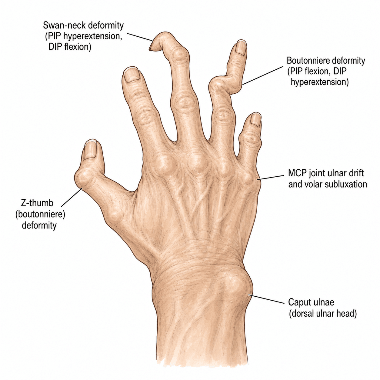
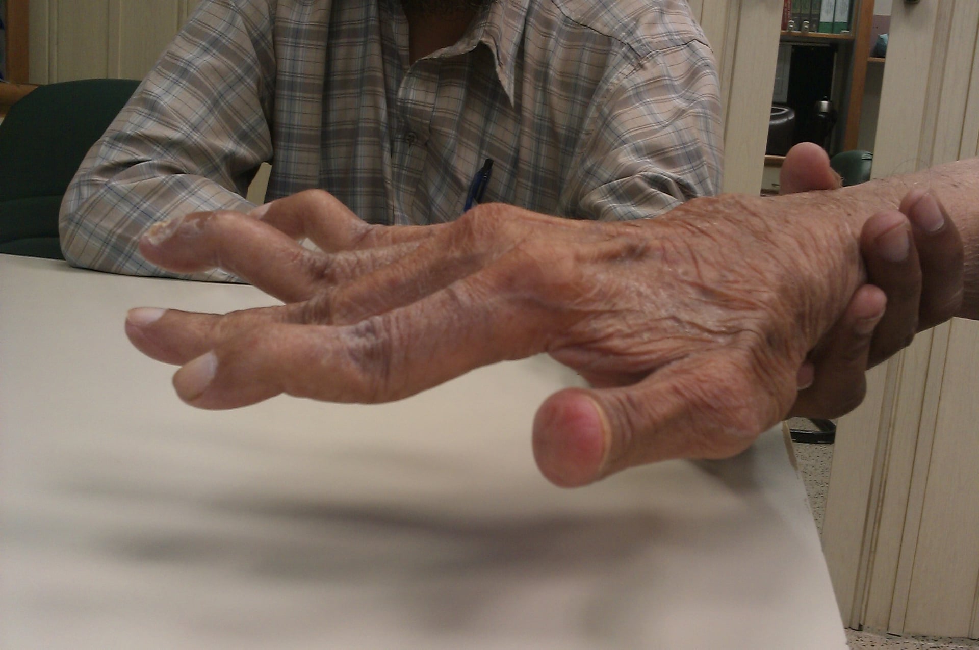
- Ulnar drift from MCP radial collateral destruction + extensor subluxation
- Boutonniere = PIP flexion + DIP extension (central slip rupture)
- Swan-neck = PIP hyperextension + DIP flexion (volar plate laxity)
- Medical optimization before surgery - stop methotrexate 2 weeks pre-op
- Proximal-to-distal surgery - wrist before digits, correct deforming forces first
- “RF positive 70%, anti-CCP more specific (95%) for RA diagnosis
- “Caput ulnae syndrome = dorsal ulnar head prominence from DRUJ destruction
- “Vaughan-Jackson lesion = EDM/EDC rupture from attrition on ulnar head
- “Synovectomy only effective if done under 6 months of synovitis onset
Radial collateral ligament destruction. Extensor tendon subluxates ulnarly. Intrinsics pull fingers into ulnar deviation. Progressive worsening with grip.
Boutonniere vs Swan-neck. Boutonniere = PIP flexion from central slip rupture. Swan-neck = PIP hyperextension from volar plate laxity or FDS rupture.
Vaughan-Jackson lesion. EDM ruptures first, then EDC ring, then EDC middle. Attrition on dorsal ulnar head (caput ulnae). Early Darrach or hemiresection.
Disease control first. Optimize medical management before reconstructive surgery. DMARDs within 2 years critical. Address proximal deformities before distal.
- Stage
- No deformity yet
- Treatment
- Synovectomy + medical optimization
- Key Pearl
- Window of opportunity - DMARDs critical
- Stage
- Passively correctable
- Treatment
- MCP arthroplasty + soft tissue rebalancing
- Key Pearl
- Silicone implants standard, 80% satisfaction
- Stage
- PIP flexion contracture over 40°
- Treatment
- PIP arthrodesis in function
- Key Pearl
- Arthroplasty fails in fixed contracture
- Stage
- EDM/EDC rupture
- Treatment
- Darrach + tendon transfer/graft
- Key Pearl
- Prevent further ruptures with DRUJ excision
BUTTONBoutonniere Deformity Pathomechanics
Hook:BUTTON yourself up wrong - PIP stuck flexed, can't extend like a buttonhole!
Overview and Epidemiology
Rheumatoid arthritis (RA) is a chronic inflammatory autoimmune disease affecting approximately 1% of the population worldwide. The hand is involved in 90% of patients, making it the most common site of RA manifestation. Early diagnosis and treatment with disease-modifying antirheumatic drugs (DMARDs) within the first 2 years significantly impacts disease progression and outcomes.
Hand involvement in RA causes progressive functional disability through synovitis, tendon rupture, and joint destruction. Modern biologic DMARDs have revolutionized medical management, but surgical intervention remains critical for correcting established deformities. The key is medical optimization before surgery and addressing deformities in a proximal-to-distal sequence to correct deforming forces.
- Age: Peak onset 40-60 years
- Gender: 3:1 female predominance
- Genetics: HLA-DR4 association (70%)
- Smoking: Doubles RA risk, worsens severity
- MCP joints: Most commonly affected (95%)
- PIP joints: 80% involvement
- Wrist: 60-70% early in disease
- DIP joints: Rarely affected (under 10%)
- Thumb: Nalebuff deformities in 50%
Pathophysiology
RA begins with immune-mediated synovial inflammation driven by T cells, B cells, and inflammatory cytokines (TNF-alpha, IL-6). Synovial hypertrophy (pannus formation) invades cartilage and bone, causing progressive joint destruction. Unchecked inflammation leads to ligament destruction, tendon attrition, and bone erosion.
Pathophysiologic Sequence in RA Hand
Disease Progression
- Pannus formation: Synovial hypertrophy with inflammatory cells
- Cartilage invasion: Proteases degrade articular cartilage
- Bone erosion: Marginal erosions at joint edges
- Ligament weakening: Collateral and volar plate stretch
- Extensor subluxation: Radial collateral destruction allows ulnar drift
- Central slip attenuation: Boutonniere deformity develops
- Volar plate laxity: Swan-neck deformity from PIP hyperextension
- Tendon rupture: Attrition on roughened bone (Vaughan-Jackson, Mannerfelt)
- Joint destruction: Bone-on-bone arthritis
- Contractures: Soft tissue shortening prevents passive correction
- Ankylosis: Spontaneous fusion in some joints
- Arthritis mutilans: Severe bone resorption (opera-glass hand)
Specific Deformity Mechanisms
- Primary Pathology
- Radial collateral ligament destruction
- Secondary Changes
- Extensor subluxates ulnarly, intrinsics pull fingers ulnar
- Result
- Progressive ulnar deviation, worst with grip
- Primary Pathology
- Central slip rupture/attenuation
- Secondary Changes
- Lateral bands migrate volar to PIP axis, ORL contracts
- Result
- PIP flexion, DIP extension, fixed after 6-12 months
- Primary Pathology
- Volar plate laxity or FDS rupture
- Secondary Changes
- Intrinsic tightness extends PIP, lateral bands tighten
- Result
- PIP hyperextension, DIP flexion, can't hook
- Primary Pathology
- CMC subluxation (Type III) or MCP hyperextension (Type II)
- Secondary Changes
- Compensatory IP flexion for pinch
- Result
- Loss of opposition and pinch strength
SWANSwan-Neck Deformity Causes
Hook:SWAN with elegant neck extended - PIP hyperextends while DIP flexes!
Classification Systems
Nalebuff Classification of RA Thumb Deformities
- Deformity Pattern
- MCP flexion, IP extension
- Primary Pathology
- MCP synovitis → volar plate stretching
- Treatment
- MCP arthroplasty or arthrodesis
- Deformity Pattern
- MCP hyperextension, IP flexion
- Primary Pathology
- MCP ligament laxity → hyperextension
- Treatment
- MCP fusion or volar capsulodesis
- Deformity Pattern
- CMC subluxation, MCP hyperext, IP flex
- Primary Pathology
- CMC synovitis → adduction collapse
- Treatment
- CMC stabilization + MCP fusion
- Deformity Pattern
- CMC arthritis, adduction contracture
- Primary Pathology
- CMC joint destruction
- Treatment
- CMC arthroplasty or arthrodesis
- Deformity Pattern
- Multiple joint involvement
- Primary Pathology
- Progressive pannus at all joints
- Treatment
- Staged reconstruction, proximal to distal
- Deformity Pattern
- Bone resorption, instability
- Primary Pathology
- Severe osteolysis
- Treatment
- Fusion if salvageable, otherwise adaptive
Type I (Boutonniere): MCP is the problem joint (flexed from synovitis), IP compensates with extension. Type II (Swan-neck): MCP ligaments fail allowing hyperextension, IP compensates with flexion for pinch. Type III is swan-neck but driven by CMC subluxation proximally. Knowing the primary pathology determines surgical target.
2010 ACR/EULAR Classification Criteria (Diagnosis)
The 2010 ACR/EULAR criteria replaced the 1987 ACR criteria to enable earlier classification of RA, focusing on features predicting persistent and erosive disease rather than late deformity. A patient with synovitis in at least one joint and no better alternative diagnosis is classified as definite RA with a score of 6 or more out of 10 across four domains.
- Category
- 1 large joint
- Score
- 0
- Category
- 2-10 large joints
- Score
- 1
- Category
- 1-3 small joints
- Score
- 2
- Category
- 4-10 small joints
- Score
- 3
- Category
- Over 10 joints (at least 1 small)
- Score
- 5
- Category
- Negative RF and anti-CCP
- Score
- 0
- Category
- Low-positive RF or anti-CCP
- Score
- 2
- Category
- High-positive RF or anti-CCP
- Score
- 3
- Category
- Normal CRP and ESR
- Score
- 0
- Category
- Abnormal CRP or ESR
- Score
- 1
- Category
- Less than 6 weeks
- Score
- 0
- Category
- 6 weeks or more
- Score
- 1
2010 ACR/EULAR Rheumatoid Arthritis Classification Criteria
- Joint ACR/EULAR initiative replacing the 1987 criteria, which lacked sensitivity in early disease
- Score of 6 or more of 10 across four domains (joints, serology, acute-phase reactants, symptom duration) classifies definite RA
- Requires at least one joint with definite clinical synovitis not better explained by another diagnosis
- Deliberately weights small-joint involvement and high-titre serology to capture patients at risk of persistent, erosive disease early
BMI-CSSNalebuff Thumb Classification
Hook:BMI-CSS classification - B (boutonniere) vs swan-neck types, CMC to catastrophic!
Clinical Presentation
- Morning stiffness: Over 1 hour (vs OA under 30 min)
- Symmetric involvement: Both hands, multiple joints
- Systemic symptoms: Fatigue, fever, weight loss
- Pain pattern: Worse with rest, improves with activity
- Functional loss: Difficulty with grip, pinch, fine motor
- Extra-articular: Nodules, lung involvement, vasculitis
- Look: Swelling (boggy, soft), deformities, nodules, skin thinning
- Feel: Warm, boggy MCP/PIP synovitis, ulnar head prominence
- Move: Reduced ROM, pain at end-range, crepitus
- Function: Pinch/grip strength, ulnar drift with grip
- Tendons: Trigger fingers, palpable ruptures
Key Physical Findings
Caput ulnae syndrome: Dorsal prominence of distal ulna from DRUJ destruction. Ulnar head becomes:
- Prominent dorsally
- Painful with forearm rotation
- Site of extensor tendon attrition (Vaughan-Jackson lesion)
Vaughan-Jackson lesion: Sequential rupture of ulnar-sided extensors:
- EDM (extensor digiti minimi) - first to rupture
- EDC ring finger - second
- EDC middle finger - third
- Pattern: ulnar to radial progression
Mannerfelt lesion: FPL rupture from attrition on volar scaphoid osteophyte. Less common than Vaughan-Jackson but causes complete loss of thumb IP flexion.
- Morning stiffness over 1 hour
- Soft, boggy swelling (synovitis)
- Symmetric involvement
- Spares DIP joints (unlike OA)
- Systemic features
- RF/anti-CCP positive
- Morning stiffness under 30 minutes
- Hard, bony swelling (osteophytes = Heberden's/Bouchard's nodes)
- Asymmetric, wear pattern
- Affects DIP joints (Heberden's nodes)
- No systemic features
Differential Diagnosis of the Inflammatory or Deformed Hand
- Joint Pattern
- Symmetric MCP/PIP/wrist, spares DIP
- Key Discriminators
- Boggy synovitis, ulnar drift, marginal erosions, rheumatoid nodules
- Serology / Investigation
- RF and anti-CCP positive; anti-CCP highly specific
- Joint Pattern
- DIP involvement, asymmetric, ray/dactylitis
- Key Discriminators
- Nail pitting, dactylitis (sausage digit), skin psoriasis, enthesitis
- Serology / Investigation
- RF and anti-CCP usually negative; pencil-in-cup erosions on XR
- Joint Pattern
- DIP and PIP, asymmetric
- Key Discriminators
- Heberden/Bouchard nodes, gull-wing central erosions, bony hard swelling
- Serology / Investigation
- Seronegative; normal or mildly raised CRP
- Joint Pattern
- MCP, reducible deformity
- Key Discriminators
- Deformity passively correctable, non-erosive on XR despite ulnar drift
- Serology / Investigation
- ANA positive, anti-CCP usually negative (distinguishes from RA)
- Joint Pattern
- Asymmetric, mono/oligoarticular
- Key Discriminators
- Tophi, acute attacks, chondrocalcinosis (CPPD)
- Serology / Investigation
- Negatively (urate) or positively (CPPD) birefringent crystals
Anti-CCP is the single most useful serological discriminator: it is approximately 96-98% specific for RA (Schellekens 2000) and is typically negative in psoriatic arthritis, erosive OA and Jaccoud arthropathy of SLE. A correctable (non-erosive) ulnar drift with positive ANA but negative anti-CCP points to lupus rather than RA.
Investigations
Diagnostic Workup
Rheumatoid factor (RF):
- Positive in 70% of RA patients
- Sensitivity 70%, specificity 85%
Anti-CCP antibodies:
- Positive in 70% of RA patients
- More specific (95%) than RF
- Predicts erosive disease
Inflammatory markers:
- ESR, CRP elevated during flares
- Track disease activity
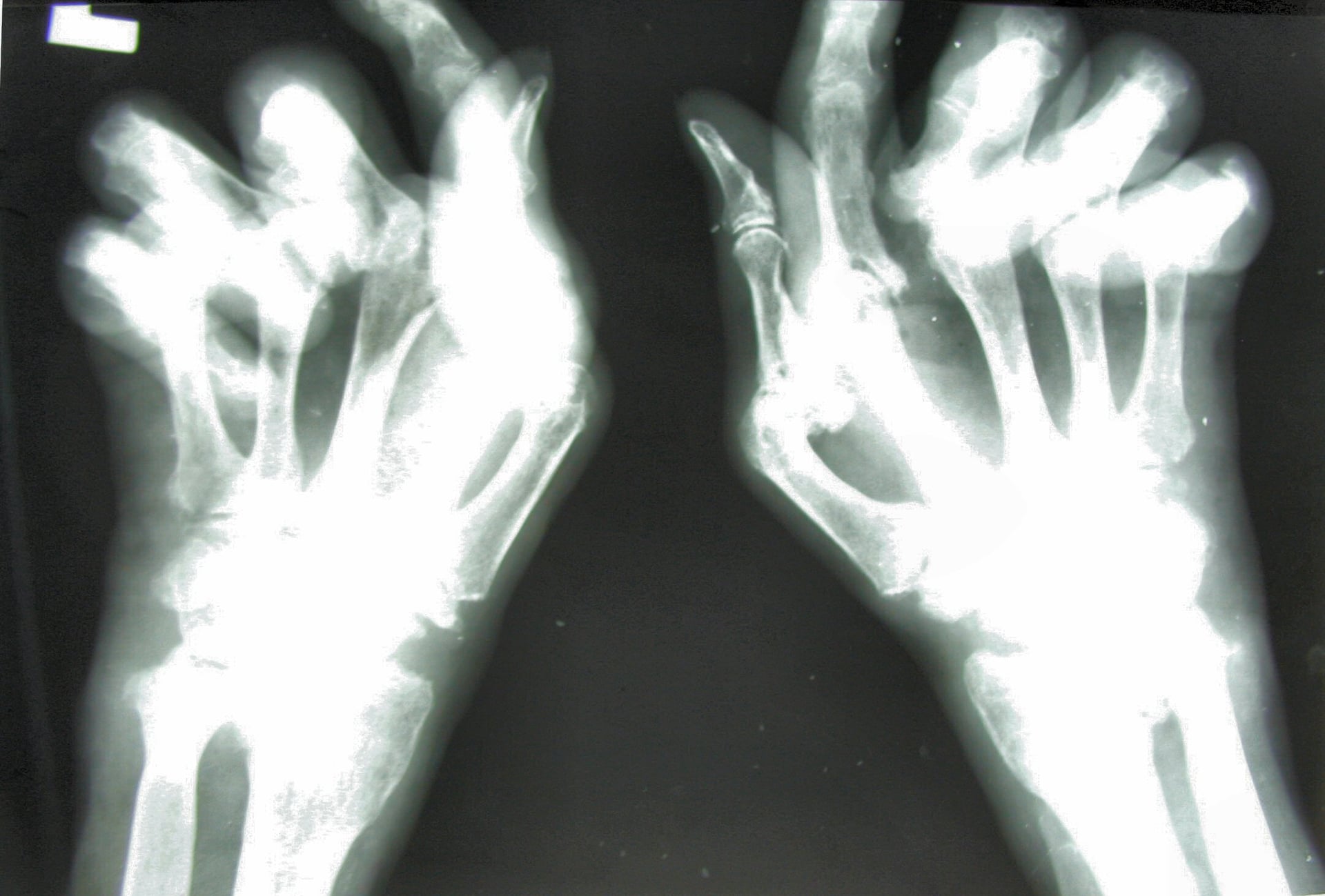
Standard hand series: PA, lateral, oblique views
Early findings:
- Periarticular osteopenia
- Soft tissue swelling
- Joint space narrowing (symmetric)
Late findings:
- Marginal erosions (at joint edges)
- Subluxations and deformities
- Bone resorption (arthritis mutilans)
- Ankylosis
Indications:
- Early disease detection (synovitis before x-ray changes)
- Assess tendon integrity pre-operatively
- Monitor DMARD response
MRI findings:
- Synovial enhancement with contrast
- Bone marrow edema
- Tendon tenosynovitis
- Early erosions
Anti-CCP antibodies are 95% specific for RA compared to RF at 85%. Presence of anti-CCP predicts more aggressive, erosive disease and guides earlier DMARD initiation. Patients with high anti-CCP titers benefit from biologic agents earlier in the disease course.
Management Algorithm
Medical Management (Primary)
RA Medical Management Protocol
Methotrexate:
- Gold standard DMARD
- 10-25mg weekly, oral or subcutaneous
- Add folic acid 5mg daily
- Monitor LFTs, FBC every 3 months
Alternative: Sulfasalazine, hydroxychloroquine
Add biologic DMARD:
- TNF-alpha inhibitors (adalimumab, etanercept)
- IL-6 inhibitors (tocilizumab)
- B-cell depletion (rituximab)
- JAK inhibitors (tofacitinib)
Target: Low disease activity or remission
NSAIDs: Short-term for pain/inflammation
Corticosteroids:
- Prednisolone 5-10mg daily during flares
- Intra-articular injection for single joint synovitis
- Bridge therapy while DMARDs take effect (6-12 weeks)
DMARD initiation within 2 years of symptom onset is critical. Early aggressive treatment prevents erosive disease, preserves joint function, and reduces need for surgery. Delay beyond 2 years results in irreversible joint damage.
Surgical Indications
Surgery indicated when:
- Persistent pain despite optimal medical management
- Functional impairment affecting ADLs or work
- Tendon rupture or impending rupture (tenosynovitis)
- Progressive deformity with joint destruction
- Nerve compression (carpal tunnel syndrome common in RA)
Before ANY operation (including hand surgery) in a rheumatoid patient, the cervical spine is the critical safety consideration that this topic must include - neck manipulation during intubation can cause catastrophic cord injury. RA destabilises the cervical spine in three patterns:
- Atlantoaxial subluxation (AAS) - the commonest: pannus and transverse-ligament incompetence allow anterior C1-on-C2 translation. The anterior atlanto-dental interval (ADI) is abnormal over about 3 mm, but the posterior atlanto-dental interval (PADI) - the actual space available for the cord - is the better predictor of neurological risk, with surgery considered when it falls below about 14 mm.
- Basilar invagination / cranial settling - the most dangerous: vertical migration of the odontoid towards the foramen magnum, risking brainstem compression.
- Subaxial subluxation: multilevel "staircase" instability lower down.
neck pain and occipital headache, and an insidious myelopathy that is easily masked by hand/limb arthritis - actively look for hyperreflexia, gait change, sphincter disturbance and Lhermitte's.
obtain flexion-extension cervical radiographs (and MRI if there is instability or myelopathy) before elective surgery and general anaesthesia; when instability is present use awake fibreoptic intubation with in-line stabilisation and explicitly flag it to the anaesthetist. In any RA patient going to theatre, clear the cervical spine first.
Surgical Technique
MCP Joint Arthroplasty with Soft Tissue Rebalancing
- Implant fracture/failure: 10-20% at 10 years
- Recurrent deformity: 10-15% ulnar drift recurrence
- Infection: 2-3% risk (higher in RA)
- Stiffness: ROM reduced from pre-op in 30%
- Sensory loss: Digital nerve injury rare (under 2%)
- Silicone implants: Swanson or Sutter design, sizes 0-3
- Bone reamers: MCP head and base reamers
- Saw: Sagittal saw for bone resection
- Sutures: 3-0 and 4-0 for soft tissue repair
- K-wires: 0.045" for temporary fixation
Surgical Steps
MCP Arthroplasty Procedure
Dorsal longitudinal incision over MCP joints:
- Can do single incision for index through small (4 joints)
- Or individual incisions per digit
- Elevate skin flaps to expose extensor mechanism
Split extensor hood longitudinally:
- Incise between sagittal band and extensor tendon
- Reflect radial-sided hood ulnarly
- Expose MCP joint capsule
- Excise hypertrophic synovium completely
- Remove pannus from joint and tendon sheath
- Send specimen for culture (rule out infection)
Resect MCP joint:
- Remove metacarpal head with sagittal saw
- Ream intramedullary canal of metacarpal
- Ream proximal phalanx base
- Preserve collateral ligament origins if possible
- Trial silicone implant sizing
- Insert implant into metacarpal canal first
- Reduce MCP joint
- Insert distal stem into phalanx
- Confirm alignment and smooth ROM
Correct ulnar drift:
- Release ulnar sagittal band and intrinsics
- Tighten radial sagittal band (reef)
- Centralize extensor tendon over MCP
- Consider radial collateral ligament reconstruction
Extensor hood repair:
- Close hood with 3-0 braided suture
- Ensure tendon centralizes with finger flexion
- Close skin with 4-0 nylon
- Apply bulky dressing
- Dynamic extension splint with MCP extension, radial deviation
- Outrigger splint worn for 6 weeks
Implant alone will fail without soft tissue rebalancing. The deforming forces (intrinsics pulling ulnar, sagittal band incompetent radially) persist after arthroplasty. Ulnar intrinsic release + radial sagittal band tightening prevents recurrent ulnar drift. 30% of recurrent deformity is from inadequate soft tissue balancing.
The rheumatoid wrist is the proximal driver of the finger deformities and has its own reconstruction ladder beyond the DRUJ - examiners expect both the biomechanics and the surgical options.
radiocarpal synovitis lets the carpus translate ulnarly and the wrist deviate radially; the fingers then compensate by deviating ulnarly (ulnar drift). This is why correcting the radially-deviated wrist is part of correcting MCP ulnar drift - the proximal-to-distal principle. (Volar/ulnar carpal translocation and caput ulnae accompany this collapse.)
- Synovectomy and dorsal tenosynovectomy - early, to relieve synovitis and prevent extensor tendon rupture.
- Partial fusion - radiolunate (Chamay) or radioscapholunate - stabilises the carpus and halts ulnar/volar translocation while preserving some midcarpal motion in the unstable but not-yet-destroyed wrist.
- Total wrist arthrodesis - the durable, reliable, pain-relieving salvage for the destroyed/painful wrist, especially in high-demand patients and those who load the wrist through crutches/wheelchair; it sacrifices motion (acceptable when other joints or the contralateral wrist retain movement).
- Total wrist arthroplasty - preserves motion for the lower-demand patient (valuable in bilateral disease so that not both wrists are fused), but has higher loosening/failure than arthrodesis.
stabilise/correct the wrist and DRUJ before MCP reconstruction. Exam point: the radially-deviated rheumatoid wrist drives finger ulnar drift - fix the wrist first, and run the ladder synovectomy -> radiolunate fusion -> total wrist fusion or arthroplasty.
Complications
- Incidence
- 2-5%
- Risk Factors
- Immunosuppression (MTX, biologics), corticosteroids, diabetes
- Management
- Aggressive antibiotics, consider implant removal if deep
- Incidence
- 10-20% at 10 years
- Risk Factors
- High-demand activities, poor bone quality
- Management
- Observation if painless, revision arthroplasty or fusion if symptomatic
- Incidence
- 10-15%
- Risk Factors
- Inadequate soft tissue rebalancing, poor compliance with splinting
- Management
- Revision soft tissue balancing, intrinsic release
- Incidence
- 20-30%
- Risk Factors
- Prolonged immobilization, poor therapy compliance
- Management
- Hand therapy, dynamic splinting, may need manipulation
- Incidence
- 5-10%
- Risk Factors
- Smoking, corticosteroids, poor bone quality
- Management
- Revision fusion with bone graft if symptomatic
- Incidence
- 3-5%
- Risk Factors
- Unaddressed caput ulnae, continued attrition
- Management
- Tendon transfer or reconstruction, Darrach if not done
RA patients are immunosuppressed from disease and medications (methotrexate, biologics, corticosteroids). Infection risk is 2-5 times higher than non-RA patients. Strategies to reduce infection:
- Stop methotrexate 2 weeks pre-op, restart 2 weeks post-op
- Hold biologics 1 dosing cycle before surgery
- Prophylactic antibiotics (first-generation cephalosporin)
- Meticulous sterile technique
- Monitor closely post-op for signs of infection
Postoperative Care and Rehabilitation
Post-MCP Arthroplasty Rehabilitation
- Splinting: Dynamic extension splint with outrigger
- MCP held in extension and radial deviation
- Passive ROM exercises initiated day 3-5
- Critical: Maintain radial pull to prevent recurrent ulnar drift
- Continue dynamic splint full-time
- Active ROM exercises 5x daily (remove splint)
- Blocking exercises to isolate MCP motion
- Scar massage and edema control
- Wean from splint (nighttime only)
- Progressive grip strengthening
- Functional activities (ADLs)
- Assess for recurrent drift, adjust therapy
- Resume normal activities
- Continue nighttime splint for 3-6 months
- Monitor for implant fracture, recurrent deformity
- Annual follow-up indefinitely
Recurrent ulnar drift occurs in 10-15%, mostly from poor splinting compliance. The dynamic extension splint provides radial-directed force to counteract ulnar pull during healing. Patients must understand that splint wear for 6 weeks full-time, then nighttime for 6 months, is essential to prevent recurrence.
Outcomes and Prognosis
- Pain Relief
- 80-90% significant improvement
- Function
- ROM improved in 60%, reduced in 30%
- Satisfaction
- 80% satisfied
- Revision Rate
- 10-20% at 10 years (implant fracture)
- Pain Relief
- 90% complete relief
- Function
- Stable grip platform, no motion
- Satisfaction
- 85% satisfied
- Revision Rate
- 5-10% (nonunion, malunion)
- Pain Relief
- 70% relief if done early (under 6 months synovitis)
- Function
- Preserves ROM, prevents tendon rupture
- Satisfaction
- 75% satisfied
- Revision Rate
- 30% require repeat synovectomy at 5-10 years
- Pain Relief
- 90% relief of ulnar-sided wrist pain
- Function
- Forearm rotation improves
- Satisfaction
- 80% satisfied
- Revision Rate
- 10% instability/convergence requiring revision
Factors associated with worse surgical outcomes in RA hand:
- Active disease at time of surgery (defer until remission)
- Poor medical compliance (methotrexate, biologics)
- Smoking (impairs wound healing, increases nonunion)
- Advanced age with poor bone quality (implant failure, nonunion)
- Inadequate soft tissue rebalancing (recurrent deformity)
- Poor rehabilitation compliance (stiffness, recurrent drift)
Guidelines, Registries & Global Practice
Global Epidemiology
Rheumatoid arthritis affects approximately 0.5-1% of adults worldwide, with a consistent female predominance of roughly 3:1 and peak onset between 40 and 60 years. Prevalence is highest in some Indigenous North American populations and lowest in parts of sub-Saharan Africa and East Asia, reflecting genetic (HLA-DRB1 shared epitope) and environmental (notably smoking) contributions. The hand is involved in the majority of patients over the disease course, and is the most common site requiring reconstructive surgery. Critically, the incidence of major rheumatoid hand surgery has fallen substantially in the biologic era, mirroring earlier diagnosis (2010 ACR/EULAR criteria) and treat-to-target medical control (TICORA).
Side-by-Side Guideline Guidance
- Scope
- Diagnosis and medical treat-to-target
- Key Position
- Classify and treat early; methotrexate first-line, escalate to biologic/targeted synthetic DMARD if target not met; short-term glucocorticoid bridging only
- Evidence Basis
- RCT and systematic review (Level 1)
- Scope
- Diagnosis, monitoring, referral
- Key Position
- Refer suspected persistent synovitis urgently; treat-to-target with monthly review until target met; offer hand exercise and therapy
- Evidence Basis
- Guideline (Level 1-2 underpinning)
- Scope
- Perioperative DMARD management and hand surgery
- Key Position
- Continue methotrexate through surgery; withhold biologics around surgery timed to dosing cycle; involve hand therapy
- Evidence Basis
- Guideline / consensus
- Scope
- Surgical optimisation
- Key Position
- Methotrexate may be continued perioperatively; biologics held one dosing interval and surgery scheduled at the trough; resume once wound healed and no infection
- Evidence Basis
- Consensus / cohort
Earlier teaching to stop methotrexate before surgery has been superseded: current ACR/EULAR-aligned perioperative guidance supports continuing methotrexate through hand surgery, as it does not increase infection or wound complications and avoids disease flare. Biologics are withheld for one dosing cycle and surgery booked at the trough, then resumed once the wound is healed and infection is excluded. Quote both the traditional and the current position in vivas and state the current evidence-based answer.
Registry and Outcome Evidence
- Falling surgical burden: National cohort and registry data across Scandinavia, the UK and the USA show declining rates of rheumatoid hand and wrist surgery since widespread biologic use
- Implant durability: Long-term series (Goldfarb and Stern) document ~63% silicone MCP implant fracture at 14 years - durability, not early failure, is the key issue
- Treat-to-target benefit: TICORA-type strategies reduce radiographic progression and downstream reconstruction
- DRUJ procedure choice varies: Darrach (older/lower demand) vs Sauve-Kapandji or hemiresection (younger, motion-preserving)
- Implant vs fusion decisions for MCP/PIP differ by surgeon and region
- Access: biologic availability and elective hand-surgery waiting times differ markedly between health systems, shifting the surgical/medical balance
Glucocorticoids in RA - Evidence Informing the 2022 EULAR Update
- Systematic literature review underpinning the 2022 EULAR RA management recommendations
- Confirmed efficacy of short-term glucocorticoid bridging; most patients can stop within 12-24 months
- Dose- and duration-dependent harms confirmed: osteoporotic fracture, serious infection, diabetes and increased mortality
- Supports using the lowest effective steroid dose for the shortest time as a bridge while DMARDs take effect
MCQ Practice Points
Q: What is the primary pathologic mechanism of MCP ulnar drift in rheumatoid arthritis? A: Radial collateral ligament destruction with ulnar subluxation of the extensor tendon. The radial collateral ligament fails from synovitis, allowing the extensor tendon to subluxate ulnarly. Intrinsic muscles then pull the fingers into progressive ulnar deviation, worsened with grip.
Q: What is the typical sequence of extensor tendon rupture in Vaughan-Jackson lesion? A: EDM (extensor digiti minimi) ruptures first, followed by EDC ring, then EDC middle. The sequence progresses from ulnar to radial as tendons attrit on the prominent dorsal ulnar head (caput ulnae). EDM ruptures first because it is most ulnar and has the thinnest cross-section.
Q: What is the primary pathology distinguishing boutonniere from swan-neck deformity? A: Boutonniere: Central slip rupture/attenuation causes PIP flexion and DIP extension. Lateral bands migrate volar to PIP axis. Swan-neck: Volar plate laxity or FDS rupture causes PIP hyperextension and DIP flexion. Intrinsics pull PIP into hyperextension.
Q: What is the specificity of anti-CCP antibodies for rheumatoid arthritis diagnosis? A: 95% specificity for RA (compared to RF at 85%). Anti-CCP antibodies are highly predictive of erosive disease and indicate need for early aggressive DMARD therapy. Combination of RF + anti-CCP positive is 90% predictive of erosive RA.
Q: What is the most important soft tissue procedure to prevent recurrent ulnar drift after MCP arthroplasty? A: Ulnar intrinsic release combined with radial sagittal band tightening. Releasing the ulnar intrinsics removes the ulnar-deviating force, while tightening the radial sagittal band centralizes the extensor tendon over the MCP joint. Implant alone without soft tissue rebalancing has 30% recurrence rate.
Q: In Nalebuff Type III thumb deformity, what is the primary pathology and surgical target? A: CMC joint subluxation is the primary pathology (not MCP). The thumb metacarpal adducts and subluxates at CMC from ligamentous laxity. MCP hyperextension and IP flexion are compensatory to achieve pinch. Surgical treatment must address CMC stabilization first before correcting MCP deformity, otherwise recurrence is inevitable.
Exam Viva Scenarios
Practise clinical reasoning and management decisions out loud
“A 55-year-old woman with 8-year history of seropositive RA presents with progressive ulnar deviation of MCP joints in both hands. She is on methotrexate and adalimumab with good systemic disease control. Her ulnar deviation is approximately 30 degrees and partially correctable passively. What is your assessment and management?”
“A 60-year-old man with longstanding RA presents unable to extend his small and ring fingers at the MCP joint. Examination reveals prominent dorsal ulnar head and loss of EDM and EDC ring finger function. What is your assessment and surgical management?”
“A 50-year-old woman with RA has progressive thumb deformity with CMC joint subluxation, MCP hyperextension to 40 degrees, and compensatory IP flexion. She has weak pinch and difficulty with activities requiring precision grip. Describe your classification and surgical approach.”
Key Pathophysiology
- Synovial inflammation (pannus) invades cartilage and bone causing progressive destruction
- Ulnar drift = radial collateral ligament destruction + ulnar extensor subluxation + intrinsic contracture
- Boutonniere = central slip rupture, PIP flexion, DIP extension (lateral bands volar to PIP axis)
- Swan-neck = volar plate laxity or FDS rupture, PIP hyperextension, DIP flexion
- Vaughan-Jackson = EDM, EDC ring, EDC middle sequential rupture from caput ulnae attrition
Classification
- Nalebuff Thumb Type I = boutonniere (MCP flexion, IP extension) = MCP synovitis
- Type II = swan-neck (MCP hyperext, IP flex) = MCP ligament laxity
- Type III = swan-neck from CMC subluxation (treat CMC first!)
- Boutonniere deformity staging: Stage I (under 30° lag, correctable), Stage II (30-50°), Stage III (over 50°, fixed)
Medical Management
- DMARDs within 2 years critical - window of opportunity to prevent erosions
- Anti-CCP 95% specific for RA, predicts erosive disease
- Methotrexate gold standard, add biologic if inadequate response at 3 months
- Pre-op: stop MTX 2 weeks before, biologics 1 dosing cycle, continue corticosteroids
Surgical Pearls
- Proximal-to-distal surgery sequence - correct wrist before MCP, MCP before PIP
- MCP arthroplasty requires soft tissue rebalancing: ulnar intrinsic release + radial sagittal band reef
- Darrach (distal ulna excision) for caput ulnae prevents further extensor ruptures
- Dynamic extension splint for 6 weeks post-MCP arthroplasty prevents recurrent drift
- Synovectomy only effective if done under 6 months of synovitis onset
Complications
- Infection 2-5% (immunosuppression from RA medications)
- MCP implant fracture 10-20% at 10 years (mostly asymptomatic)
- Recurrent ulnar drift 10-15% (inadequate soft tissue balancing or poor splinting compliance)
- Stiffness 20-30% (ROM may worsen but pain improves)
- Arthrodesis nonunion 5-10% (smoking, corticosteroids, poor bone quality)
Evidence Base and Key Trials
Silicone MCP Arthroplasty vs Medical Treatment (Landmark Prospective Trial)
- Multicentre prospective controlled cohort across 3 referral centres (USA and England), 70 surgical and 93 non-surgical patients with severe ulnar drift or extensor lag
- One-year data analysed for 45 surgical cases and 72 non-surgical controls
- Surgical group: significant improvement in overall Michigan Hand Outcomes Questionnaire (MHQ) score despite worse baseline function; non-surgical group unchanged
- Ulnar deviation and extensor lag improved significantly after silicone MCP arthroplasty (SMPA); non-surgical group did not improve
- Grip strength, pinch strength and Arthritis Impact Measurement Scales showed no significant change in either group
TICORA: Tight Control (Treat-to-Target) in Rheumatoid Arthritis
- Single-blind RCT in 111 patients: intensive monthly tight-control management vs routine outpatient care
- Mean fall in disease activity score (DAS) greater with tight control: -3.5 vs -1.9 (difference 1.6, 95% CI 1.1-2.1, p less than 0.0001)
- Good response achieved in 82% of intensive group vs 44% routine; remission (DAS less than 1.6) in 65% vs 16%
- Tight control substantially reduced radiographic disease progression and improved physical function and quality of life at no additional cost
Anti-CCP Antibodies: Landmark Diagnostic Validation
- Defining study introducing the cyclic citrullinated peptide (anti-CCP) ELISA
- Anti-CCP highly specific for RA: 98% in prevalent sera and 96% in an early-arthritis clinic cohort, with moderate sensitivity (sensitivity 48-68% depending on cohort)
- Anti-CCP significantly more specific than IgM rheumatoid factor (96% vs 91%, p = 0.016)
- Combining anti-CCP with IgM-RF raised positive predictive value to 91%
- Anti-CCP at first visit predicted erosive disease at 2 years (PPV 91%)
Silicone MCP Arthroplasty: 14-Year Durability
- 208 silicone MCP arthroplasties in 52 hands (36 patients), mean 14-year follow-up - single surgeon
- Ulnar drift improved from 26 degrees to under 5 degrees postoperatively but recurred to a mean of 16 degrees
- Extension deficit improved from 57 degrees to 11 degrees but worsened to 23 degrees at final follow-up
- 130 of 208 implants (63%) were fractured and a further 22% deformed; implant fracture correlated with recurrent ulnar drift (p less than 0.001)
- Mean MHQ score 48/100; patients were satisfied with only 38% of hands and only 27% were pain-free at final review
DRUJ Hemiresection-Interposition Arthroplasty: Long-Term Results
- 51 wrists (39 rheumatoid patients) treated with matched hemiresection-interposition arthroplasty and dorsal wrist synovectomy, mean 4.5-year follow-up
- Associated extensor tendon ruptures present in 31% of patients at surgery
- Complete relief of ulnar-sided wrist pain in 84% of wrists, partial relief in 14%
- Supination improved 73 to 81 degrees and pronation 68 to 74 degrees; grip strength improved from 6.1 to 11.5 kgf
The Rheumatoid Wrist: Principles of Surgical Management
- Authoritative JAAOS instructional review of the rheumatoid wrist
- DRUJ is usually affected first; managed with Darrach or Sauve-Kapandji depending on age and demand
- Ruptured extensor/flexor tendons are rarely amenable to direct repair - tendon transfer or grafting is required when the motored joint remains functional
- Partial (radiolunate/radioscapholunate) wrist fusion balances stability with retained midcarpal motion; total wrist fusion gives reliable pain relief at the cost of motion