PI-LL Mismatch | SVA | Pelvic Parameters | Compensation Mechanisms
- PI is fixed - cannot be changed surgically (constant after skeletal maturity)
- PI = PT + SS is the fundamental spinopelvic equation
- LL should match PI within 10 degrees (target: PI - 9 to PI + 9)
- PT increases as compensation for sagittal imbalance (pelvic retroversion)
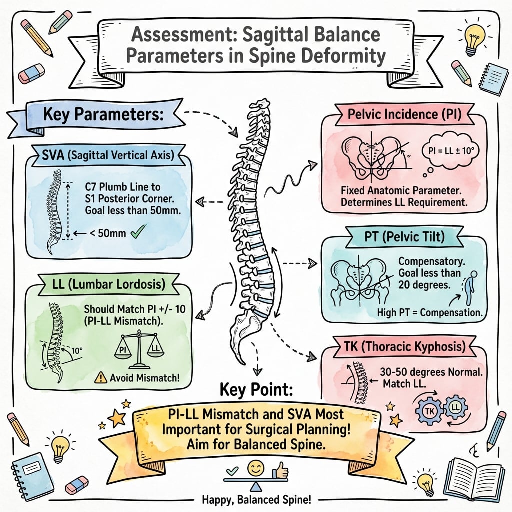
- SVA more than 50mm correlates strongly with pain and disability
- “Know PI-LL mismatch predicts outcomes better than any single parameter
- “Thoracic kyphosis and lumbar lordosis should be balanced (TK ≈ LL - 20)
- “Compensation cascade: thoracic hypokyphosis → pelvic retroversion → hip extension → knee flexion
- “Age-adjusted targets may be appropriate for elderly patients
Pelvic incidence is the master parameter - it is a fixed anatomical constant that determines the lumbar lordosis required for sagittal balance. It cannot be changed surgically. All surgical planning revolves around matching LL to PI.
PI = PT + SS - this equation is always true. As pelvic tilt increases with retroversion (compensation), sacral slope must decrease proportionally. Understanding this relationship is essential for interpreting spinopelvic alignment.
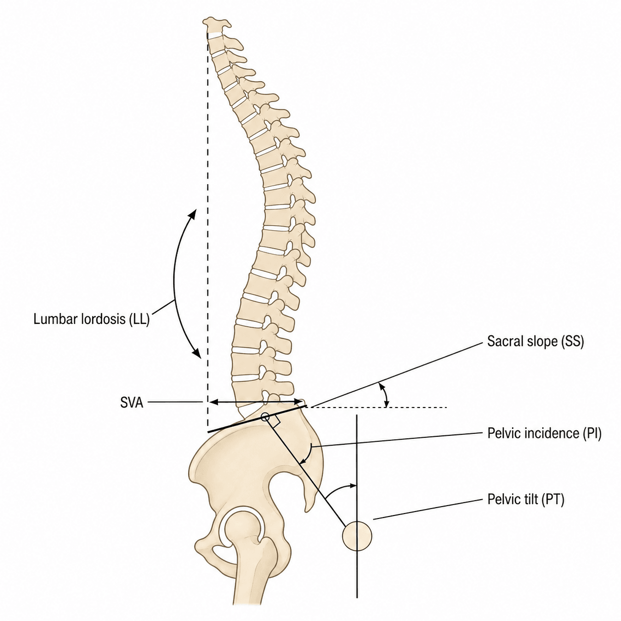
SVA more than 50mm is the critical threshold. This is measured from the C7 plumb line to the posterior-superior corner of S1. Positive SVA (anterior) correlates with disability; negative SVA (posterior) is generally well-tolerated.
PT more than 25° indicates exhausted compensation. When the pelvis has maximally retroverted but SVA remains positive, the patient has decompensated and typically requires surgical correction to restore balance.
- Normal Range
- 40-65°
- Abnormal Threshold
- Fixed - N/A
- Clinical Significance
- Determines required LL
- Normal Range
- Less than 20°
- Abnormal Threshold
- More than 25°
- Clinical Significance
- Compensation indicator
- Normal Range
- 30-50°
- Abnormal Threshold
- Context dependent
- Clinical Significance
- Decreases with retroversion
- Normal Range
- 40-60°
- Abnormal Threshold
- PI-LL more than 10°
- Clinical Significance
- Target: PI ± 9°
- Normal Range
- Less than 50mm
- Abnormal Threshold
- More than 50mm positive
- Clinical Significance
- Disability correlation
- Normal Range
- 20-50° (T4-T12)
- Abnormal Threshold
- Context dependent
- Clinical Significance
- Should balance LL
Overview and Epidemiology
Sagittal balance parameters are radiographic measurements used to assess spinal alignment in the sagittal (lateral) plane. These measurements are fundamental to understanding spinal pathology, planning deformity correction surgery, and predicting clinical outcomes.
Clinical Significance:
Sagittal imbalance is now recognised as the primary driver of disability in adult spinal deformity, surpassing coronal plane deformity in importance (Glassman et al, Spine 2005). Health-related quality of life measures correlate strongly with sagittal parameters, particularly:
- PI-LL mismatch: Strong predictor of disability (Schwab et al, Spine 2012)
- SVA more than 50mm: Strong, near-linear correlation with pain and functional limitation (Glassman et al, Spine 2005)
- PT more than 25°: Indicates progressive pelvic retroversion as compensation (Schwab et al, Spine 2012)
The prevalence of adult spinal deformity rises markedly with age; community studies of older adults report radiographic deformity (including sagittal malalignment) in a substantial proportion of those over 60 years, and demand for corrective surgery is increasing as populations age worldwide. Full epidemiological figures are summarised in the Guidelines, Registries and Global Practice section.
Historical Context:
The importance of sagittal balance was first emphasised by Dubousset, who described the "cone of economy" - the cone of stable standing posture. Pelvic incidence as a fixed morphological parameter was defined by Legaye and Duval-Beaupère, the four normative sagittal morphotypes by Roussouly et al (Spine 2005), and the modern outcome-linked spinopelvic framework by Schwab and Lafage.
Adult spinal deformity management has shifted from a coronal plane focus to a sagittal plane focus. The SRS-Schwab classification emphasizes sagittal modifiers (PI-LL, PT, SVA) because these predict outcomes better than coronal curve magnitude alone.
Pathophysiology and Anatomy
Pelvic Parameters
The pelvis forms the foundation of spinal alignment and transmits forces between the spine and lower extremities. Understanding pelvic morphology is essential for sagittal balance assessment.
- Definition: Angle between the line perpendicular to the sacral endplate at its midpoint and the line connecting this point to the femoral head center
- Characteristic: Fixed anatomical parameter - does not change after skeletal maturity
- Normal range: 40-65 degrees
- Clinical importance: Determines the amount of lumbar lordosis required for sagittal balance
- Definition: Angle between the vertical and the line connecting the midpoint of the sacral endplate to the femoral head center
- Characteristic: Positional parameter - changes with posture
- Normal: Less than 20 degrees
- Pathological: More than 25 degrees indicates compensation
- Definition: Angle between the sacral endplate and the horizontal plane
- Relationship: SS = PI - PT
- Normal range: 30-50 degrees
The Fundamental Equation: PI = PT + SS
This equation always holds true. Since PI is fixed:
- When PT increases (pelvic retroversion), SS must decrease
- When SS increases (anteversion), PT must decrease
- The sum always equals the individual's PI
Spinal Parameters
- Measured from superior endplate of L1 to superior endplate of S1
- Normal range: 40-60 degrees (Cobb method)
- Target: Should match PI within 10 degrees (LL = PI ± 9)
- Distribution: Approximately 2/3 of lordosis in L4-S1 segment
- Measured from T4 (or T5) to T12 superior endplate
- Normal range: 20-50 degrees
- Relationship: TK ≈ LL - 20 (roughly 20 degrees less than LL)
- Distance from C7 plumb line to posterior-superior corner of S1
- Positive: C7 plumb falls anterior to S1 (imbalance)
- Negative: C7 plumb falls posterior to S1
- Normal: Less than 50mm
- Disability threshold: More than 50mm positive
Compensation Mechanisms
When lumbar lordosis is insufficient for a given PI, the body employs a cascade of compensatory mechanisms:
- Mechanism
- Thoracic hypokyphosis
- Effect
- Reduces TK to shift mass posteriorly
- Clinical Observation
- Flat upper back
- Mechanism
- Pelvic retroversion
- Effect
- Increases PT, decreases SS
- Clinical Observation
- Posterior pelvic tilt
- Mechanism
- Hip extension
- Effect
- Extends hip joint
- Clinical Observation
- Standing with hyperextended hips
- Mechanism
- Knee flexion
- Effect
- Flexes knee to shift mass
- Clinical Observation
- Bent-knee gait
- Mechanism
- Decompensation
- Effect
- Exhausted mechanisms
- Clinical Observation
- Forward trunk lean, uses aids
A patient with PT more than 30°, positive SVA despite compensation, and bent-knee gait has exhausted all compensatory mechanisms. This represents surgical-level imbalance that is unlikely to improve with conservative treatment alone.
PI is presented above purely as the master deformity parameter, but a high PI is also a recognised risk factor for developmental L5-S1 spondylolisthesis - a frequently examined link the topic otherwise only mentions in passing. A high PI means a more horizontal sacrum and high sacral slope, which increases the anterior shear force at the lumbosacral junction and predisposes to slip in dysplastic/isthmic spondylolisthesis (the higher the PI, the higher the slip grade tends to be).
This drives the Mac-Thiong / Spinal Deformity Study Group (SDSG) spinopelvic classification of L5-S1 spondylolisthesis, which (for high-grade slips) divides patients by pelvic posture into:
- Balanced pelvis - high SS, low PT (the pelvis is still "anteverted"/balanced); generally better tolerated.
- Retroverted (unbalanced) pelvis - low SS, high PT, the pelvis has retroverted to compensate; a marker that the patient is decompensating and more likely to need realignment/reduction.
- Unbalanced spine - C7 plumb line falls anterior (positive global balance) - the most severe group.
Exam point: a high PI is not just "the number you match LL to" - it also predisposes to spondylolisthesis and worse slip grades, and the SS/PT pattern (balanced vs retroverted pelvis) in the Mac-Thiong/SDSG scheme guides whether a high-grade slip needs reduction.
Classification and Measurement
Radiographic Measurement Protocol
- Full-length standing PA and lateral radiographs
- 36-inch cassette including C2 to femoral heads
- Standardized arm position (hands on clavicles, or fists on shoulders)
- Weight-bearing bilateral stance
- Identify the midpoint of the sacral endplate
- Draw a line perpendicular to the sacral endplate at this point
- Draw a line from this midpoint to the center of the femoral heads
- Measure the angle between these two lines
- Note: PI is measured the same regardless of pelvic position
- Draw a vertical reference line
- Draw a line from the S1 endplate midpoint to femoral head center
- Measure the angle between vertical and this line
- Positive value indicates retroversion (normal position)
- Draw a horizontal reference line
- Draw a line along the sacral endplate
- Measure the angle between horizontal and sacral endplate
- Drop a plumb line from the center of C7 vertebral body
- Measure horizontal distance to posterior-superior corner of S1
- Positive if C7 plumb falls anterior to S1
- Negative if C7 plumb falls posterior to S1
PI measurement is position-independent (can be measured on supine CT), but PT and SS require standing films as they are positional parameters. Always use standing full-length films for complete sagittal assessment.
The parameters above are thoracolumbo-pelvic; a complete sagittal-balance answer also covers the cervical spine, which has its own outcome-linked parameters and mirrors the PI-LL concept at the top of the spine:
- Cervical SVA (cSVA): plumb line from the centre of C2 to the posterosuperior corner of C7; over 40 mm correlates with disability and myelopathy severity (the cervical equivalent of the global SVA threshold).
- T1 slope (T1S): the angle of the T1 superior endplate to horizontal - the cervical analogue of pelvic incidence (it sets the "platform" the cervical spine sits on). A high T1 slope demands more cervical lordosis to keep the head balanced over the trunk, just as a high PI demands more lumbar lordosis.
- T1 slope minus cervical lordosis (T1S-CL): the cervical analogue of PI-LL mismatch; a large mismatch (commonly cited as over about 15-20°) means the neck cannot generate enough lordosis for its T1 slope and predicts cervical malalignment/disability.
- Chin-brow vertical angle (CBVA): already listed - the functional horizontal-gaze parameter, critical in fixed cervicothoracic (e.g. ankylosing spondylitis) deformity.
Cervicothoracic reciprocity: a thoracolumbar deformity that pushes the trunk forward forces a compensatory increase in cervical lordosis and T1 slope to keep horizontal gaze - so correcting a lumbar deformity can unload (or, if overdone, unbalance) the neck.
Exam point: be able to extend "sagittal balance" upward - cSVA over 40 mm, T1 slope as the cervical PI, and T1S-CL as the cervical PI-LL mismatch - and explain horizontal gaze (CBVA) as the functional endpoint.
Clinical Assessment
History
Key Questions:
- Difficulty standing upright or walking distance?
- Need to lean on shopping trolley or walker?
- Back pain location (axial vs. radicular)?
- Can you see the horizon when walking?
- Progressive postural change?
- Prior spinal surgery?
Symptom Patterns:
- Sagittal Implication
- Positive SVA, decompensation
- Sagittal Implication
- Muscle fatigue from compensation
- Sagittal Implication
- Stenosis with imbalance
- Sagittal Implication
- Exhausted compensation
- Sagittal Implication
- Claudication or fatigue
Differential Diagnosis of Sagittal Malalignment
Positive sagittal balance and a forward-stooped posture are signs, not a diagnosis. The key exam skill is distinguishing the underlying cause, because management differs fundamentally.
- Key Distinguishing Feature
- PI-LL mismatch, reducible lordosis on extension
- Flexibility
- Often flexible early
- Typical Management Focus
- Restore LL to match PI
- Key Distinguishing Feature
- Prior lumbar fusion in kyphosis, fixed segment
- Flexibility
- Rigid at fused levels
- Typical Management Focus
- Osteotomy (often PSO)
- Key Distinguishing Feature
- Inflammatory back pain, fused 'bamboo' spine, raised CRP/HLA-B27
- Flexibility
- Rigid (ankylosed)
- Typical Management Focus
- Closing-wedge osteotomy, screen for unstable fracture
- Key Distinguishing Feature
- Disappears when supine, neuromuscular signs
- Flexibility
- Reducible (postural)
- Typical Management Focus
- Treat underlying myopathy/Parkinsonism
- Key Distinguishing Feature
- Forward lean relieves leg symptoms (neurogenic claudication)
- Flexibility
- Voluntary, reducible
- Typical Management Focus
- Decompression; balance often preserved
- Key Distinguishing Feature
- Focal kyphosis, acute pain, marrow oedema on MRI
- Flexibility
- Acute - variable
- Typical Management Focus
- Treat fracture/cause first
- Key Distinguishing Feature
- Pelvic compensation driven by hip, positive Thomas test
- Flexibility
- Hip-dependent
- Typical Management Focus
- Address hip pathology
Physical Examination
- View from side - assess sagittal contour
- Forward trunk lean relative to pelvis
- Hip and knee posture (flexion = compensation)
- Shoulder position relative to hips
- Overall balance and stability
- Plumb line assessment: Drop string from C7, observe position relative to buttock crease
- Finger-floor distance: Assess flexibility
- Wall test: Back against wall, can occiput touch?
- Forward gaze: Can patient look at horizon without neck hyperextension?
- Forward bending: Does spine flex normally?
- Supine over bolster: Assess passive lordosis restoration
- Hip flexion contracture test (Thomas test)
- Knee flexion contracture
- Motor: L2-S1 myotomes
- Sensory: Dermatomal pattern
- Reflexes: Knee and ankle
- Long tract signs if cervical involvement
- Bladder function inquiry
If a patient walks with bent knees, they have exhausted spinal and pelvic compensation and are using knee flexion as a last resort. This indicates severe sagittal imbalance requiring surgical consideration.
Outcome Measures
Standard Assessment Instruments:
- Oswestry Disability Index (ODI)
- Visual Analog Scale (VAS) - back and leg pain
- SF-36 (physical and mental components)
- SRS-22 (Scoliosis Research Society)
- EQ-5D
These correlate with sagittal parameters and guide treatment decisions. Minimum clinically important difference (MCID) for ODI is 12-15 points.
Investigations
Imaging Protocol
Step 1: Full-Length Standing Radiographs (Gold Standard)
- 36-inch (91cm) cassette
- Standing AP and lateral views
- Include C2 to femoral heads
- Standardized arm position
- Bilateral weight-bearing stance
Step 2: Flexibility Assessment
- Supine lateral over bolster (assess lordosis restoration)
- Lateral bending films (coronal flexibility)
- Push-prone films (sagittal flexibility)
Step 3: MRI Whole Spine
- Assess neural compression
- Disc degeneration status
- Spinal cord/cauda equina
- Rule out tumor, infection, other pathology
Step 4: CT (When Indicated)
- Bone quality assessment (Hounsfield units)
- Prior fusion mass evaluation
- Osteotomy planning
- Hardware assessment
Key Radiographic Measurements
- Measurement Method
- S1 endplate perpendicular to femoral head
- Normal Value
- 40-65°
- Surgical Target
- Fixed - measure only
- Measurement Method
- Vertical to S1-femoral head line
- Normal Value
- Less than 20°
- Surgical Target
- Less than 25°
- Measurement Method
- Sacral endplate to horizontal
- Normal Value
- 30-50°
- Surgical Target
- SS = PI - PT
- Measurement Method
- L1 sup to S1 sup endplate (Cobb)
- Normal Value
- 40-60°
- Surgical Target
- PI ± 9°
- Measurement Method
- T4-T12 (or T5-T12)
- Normal Value
- 20-50°
- Surgical Target
- LL - 20° approximately
- Measurement Method
- C7 plumb to S1 posterior corner
- Normal Value
- Less than 50mm
- Surgical Target
- Less than 50mm
- Measurement Method
- PI minus LL
- Normal Value
- Less than 10°
- Surgical Target
- Less than 10°
Bone Density Assessment
- Hip and spine T-scores
- Essential for surgical planning
- Osteoporosis affects fixation strategy
- Hounsfield units from planning CT
- L1 less than 110 HU suggests osteoporosis
- Guides cement augmentation decision
Special Studies
- CT myelogram: If MRI contraindicated
- Flexion-extension radiographs: Assess instability
- Hip-to-ankle films: Limb length, hip OA assessment
- Pulmonary function tests: Severe thoracic deformity
- Cardiac evaluation: For major surgery candidates
Management Algorithm
Non-Operative Treatment
- Mild imbalance with adequate compensation
- Patient preference or surgical contraindication
- High surgical risk with acceptable function
- Asymptomatic or minimally symptomatic
- Core strengthening (abdominals, paraspinals)
- Hip flexor stretching (reduces flexion contracture)
- Hamstring flexibility
- Postural awareness training
- Aerobic conditioning
- Simple analgesics (paracetamol, NSAIDs)
- Neuropathic agents (gabapentin, pregabalin)
- Epidural injections (temporary, diagnostic value)
- Facet injections
- Walking aids (rollator walker with arm rests)
- Bracing (limited role in adults)
- Weight optimization
- Smoking cessation
- Activity modification
- Bone health optimization
Untreated sagittal imbalance with PI-LL mismatch more than 20° tends to progress over time. Curves may progress 1-2 degrees per year on average. The decision for surgery should balance progression risk against operative morbidity.
Complications
Complication Overview
Sagittal balance correction surgery carries significant complication rates. Understanding these risks is essential for patient counseling and surgical planning.
Overall Complication Rates:
- Major complications: 25-50%
- Minor complications: 50-80%
- Neurological: 2-14% (depends on osteotomy type)
- Revision surgery: 15-30% at 5 years
Early Complications
- Incidence
- 2-14%
- Management
- Neuromonitoring, wake-up test, revision
- Incidence
- 5-15%
- Management
- Primary repair, fibrin sealant
- Incidence
- 5-10%
- Management
- Antibiotics, debridement
- Incidence
- 2-5%
- Management
- Prophylaxis, anticoagulation
- Incidence
- 15-30%
- Management
- Multidisciplinary management
- Incidence
- Variable
- Management
- Cell saver, transfusion protocol
Late Complications
- Most common mechanical complication
- Definition: More than 10° kyphosis at UIV
- Risk factors: Age, over-correction, osteoporosis
- May require extension of fusion
- Incidence: 5-20%
- Higher risk at osteotomy site
- May be asymptomatic if fused
- Revision if symptomatic or progressing
- Nonunion at fusion site
- Risk factors: Smoking, diabetes, osteoporosis
- Revision with bone grafting
- Degeneration above/below fusion
- More common with long, rigid constructs
- May require extension
Risk Factor Management
- Impact
- Pseudarthrosis, infection
- Optimization Strategy
- Cessation 6+ weeks before surgery
- Impact
- Hardware failure, PJK
- Optimization Strategy
- Medical treatment, cement augmentation
- Impact
- Infection, nonunion
- Optimization Strategy
- Optimize HbA1c to less than 8%
- Impact
- Wound healing
- Optimization Strategy
- Albumin more than 3.5, pre-habilitation
- Impact
- Multiple complications
- Optimization Strategy
- Weight loss if feasible
Outcomes and Prognosis
Outcome Predictors
- Achievement of PI-LL match (less than 10° mismatch)
- SVA correction to less than 50mm
- PT reduction to less than 25°
- No major complications
- Adequate bone quality
- Under-correction of deformity
- Over-correction (PJK risk in elderly)
- Major complication occurrence
- Revision surgery
- Persistent smoking
- Depression
Expected Results
- SVA correction achieved: 70-85%
- PI-LL correction achieved: 65-80%
- Fusion rate: 85-95%
- Significant pain improvement: 60-75%
- ODI improvement more than MCID: 65-75%
- Patient satisfaction: 70-80%
- Return to desired activities: 50-70%
Long-Term Follow-up
- Key Assessments
- Wound healing, mobilization
- Key Assessments
- Early alignment, function
- Key Assessments
- HRQOL measures, full-length films
- Key Assessments
- Fusion assessment, outcomes
- Key Assessments
- Mechanical complications, ASD
- Key Assessments
- Long-term surveillance
The most consistent predictor of patient satisfaction is achieving appropriate PI-LL alignment (mismatch less than 10°). Under-correction leads to persistent symptoms; over-correction increases PJK risk, especially in elderly patients. Age-adjusted targets may optimize outcomes.
Guidelines, Registries & Global Practice
Sagittal balance is a worldwide concept with a shared evidence base. The parameters, thresholds and surgical targets below are applied across all major boards (FRACS, FRCS (Tr & Orth), EBOT, ABOS, DNB), with only modest regional differences in service organisation.
Global Epidemiology
- Figure
- High prevalence; positive sagittal balance the parameter most linked to disability
- Source
- Glassman et al, Spine 2005 (PMID 16166889)
- Figure
- Positive SVA / PI-LL mismatch, not coronal Cobb angle
- Source
- Schwab et al, Spine 2012 (PMID 22045006)
- Figure
- 6-95% across GAP proportion categories (22-70% in independent cohort)
- Source
- Yilgor 2017 (PMID 28976431); Gupta 2021 (PMID 33857668)
Demand for adult deformity surgery is rising globally as populations age. Across regions the same biomechanical principles apply, because pelvic incidence and the PI-LL relationship are population-independent.
Guideline & Society Guidance, Side by Side
- Position on sagittal alignment
- SRS-Schwab modifiers (PI-LL, PT, SVA) are the standard descriptive and planning framework; aim for grade 0
- Evidence level
- Level III, validated reliability
- Position on sagittal alignment
- Endorses spinopelvic measurement and restoration of PI-LL match and global balance in deformity correction
- Evidence level
- Expert consensus / Level III
- Position on sagittal alignment
- Support individualised, pelvic-incidence-based targets (GAP, Roussouly morphotype) over fixed population means
- Evidence level
- Level III
- Position on sagittal alignment
- No deformity-specific numeric target; recommend specialist multidisciplinary deformity services and shared decision-making
- Evidence level
- Guideline / consensus
- Position on sagittal alignment
- Emphasise restoration of sagittal alignment and patient-reported outcome tracking; no single fixed threshold mandated
- Evidence level
- Consensus / Level III
There is broad international agreement on the targets (PI-LL less than 10°, SVA less than 50mm, PT less than 25°, age-adjusted in the elderly); the main divergence is how strictly fixed thresholds versus individualised proportion-based goals (GAP, Roussouly) are applied.
Registry & Cohort Evidence
There is no dedicated international registry for sagittal alignment, but large multicentre cohorts (International Spine Study Group, European Spine Study Group) underpin the SRS-Schwab and GAP frameworks and the age-adjusted targets (Jalai/Lafage, Spine 2017, PMID 27974739).
Global Practice Variation
- High-resource settings: full-length standing or low-dose biplanar (EOS) imaging, intraoperative neuromonitoring, cell salvage and ICU support are standard for complex correction.
- Limited-resource settings: full-length standing radiographs remain the accessible gold standard; the same PI = PT + SS and PI-LL principles guide planning without specialised equipment.
- Across all settings: pelvic incidence is measured the same way and individualised lordosis targets apply universally.
Referral Principles (Universal)
Patients with suspected sagittal imbalance should be referred to a spinal surgeon with deformity experience. Initial workup should include full-length standing films and patient-reported outcome measures. Complex deformity correction is best performed at centres with neuromonitoring, cell salvage and critical-care support.
MCQ Practice Points
Q: What is the relationship between pelvic incidence, pelvic tilt, and sacral slope?
A: PI = PT + SS - this equation always holds true. Pelvic incidence is a fixed anatomical constant. When pelvic tilt increases (retroversion for compensation), sacral slope must decrease proportionally. This relationship is essential for understanding spinopelvic mechanics.
Q: What is the target lumbar lordosis for sagittal balance?
A: LL = PI ± 9 degrees (or PI-LL mismatch less than 10°). This means lumbar lordosis should approximately equal pelvic incidence. A patient with PI of 55° should have LL between 46-64°. This is the most important correlation with patient outcomes.
Q: What SVA value correlates with disability in sagittal imbalance?
A: SVA more than 50mm (5cm) correlates strongly with pain and disability. The SRS-Schwab classification uses 4cm and 9.5cm as thresholds. Positive SVA means C7 plumb falls anterior to the posterior-superior corner of S1.
Q: What does an elevated pelvic tilt indicate?
A: PT more than 25° indicates pelvic retroversion as compensation for sagittal imbalance. When PT reaches 30-35°, pelvic compensation is typically exhausted. This is a positional parameter that changes with posture, unlike PI which is fixed.
Q: What is the sequence of compensation mechanisms for sagittal imbalance?
A: The compensation cascade is: Thoracic hypokyphosis → Pelvic retroversion → Hip extension → Knee flexion → Decompensation. Pelvic retroversion (increasing PT) is the most powerful mechanism. Bent-knee gait indicates severely exhausted compensation.
At a Glance
Spinopelvic sagittal balance is governed by the fundamental equation PI = PT + SS, where pelvic incidence (PI) is fixed (cannot be surgically altered) and determines the lumbar lordosis required for balance. The target is LL ≈ PI ± 9°; PI-LL mismatch over 10° predicts poor outcomes. Sagittal vertical axis (SVA) over 50mm strongly correlates with pain and disability—measured from the C7 plumb line to posterior-superior S1. When pelvic tilt (PT) exceeds 25°, pelvic compensation is exhausted (maximal retroversion) and surgical correction is typically required. The compensation cascade progresses from thoracic hypokyphosis → pelvic retroversion → hip extension → knee flexion. PT is a positional parameter that increases with compensation as sacral slope correspondingly decreases.
PI = PT + SSPI = PT + SS - The Spinopelvic Equation
Hook:PI never changes - when PT goes up (retroversion), SS must go down to maintain PI = PT + SS
PI-LLPI-LL MATCH - Target Alignment
Hook:LL = PI ± 9 degrees - the 'golden formula' for sagittal balance
TPHKDCOMPENSATION CASCADE
Hook:Thoracic-Pelvic-Hip-Knee-Decompensation: the body's orderly attempt to maintain balance
Clinical Decision Scenarios
Practise clinical reasoning and management decisions out loud
“A 58-year-old woman presents with low back pain and difficulty standing upright. Full-length standing radiographs show: PI = 55°, PT = 32°, SS = 23°, LL = 25°, TK = 45°, SVA = +85mm. She reports increasing difficulty walking more than one block.”
“You are teaching a registrar about sagittal balance. They ask why some patients with loss of lumbar lordosis can stand upright while others cannot.”
“A 62-year-old man has iatrogenic flatback syndrome after L3-S1 posterior fusion 8 years ago. Current measurements: PI = 60°, PT = 28°, LL = 15°, SVA = +95mm. He cannot walk more than 50 meters without resting.”
“A medical student asks you to explain why pelvic incidence determines the required lumbar lordosis. They want to understand the biomechanical basis for the PI-LL relationship.”
Key Equations
- PI = PT + SS (fundamental spinopelvic equation)
- LL = PI ± 9° (target lumbar lordosis)
- PI-LL less than 10° (target mismatch)
- TK ≈ LL - 20° (thoracolumbar relationship)
Normal Values
- PI: 40-65° (fixed anatomical parameter)
- PT: less than 20° (less than 25° acceptable)
- SS: 30-50° (decreases with retroversion)
- LL: 40-60° (match to PI)
- SVA: less than 50mm (positive = anterior)
Compensation Cascade
- 1. Thoracic hypokyphosis (reduce TK)
- 2. Pelvic retroversion (PT increases, SS decreases)
- 3. Hip extension (hyperextend hips)
- 4. Knee flexion (bent-knee gait)
- 5. Decompensation (positive SVA, needs aids)
SRS-Schwab Modifiers
- PI-LL: 0 (less than 10°), + (10-20°), ++ (more than 20°)
- PT: 0 (less than 20°), + (20-30°), ++ (more than 30°)
- SVA: 0 (less than 4cm), + (4-9.5cm), ++ (more than 9.5cm)
- ++ in any modifier = severe disability
Exam Triggers
- Cannot stand upright = positive SVA
- High PT (more than 25°) = exhausted compensation
- Bent-knee gait = severe decompensation
- Prior fusion + flatback = consider PSO
- PI-LL mismatch = key outcome predictor
Evidence and Guidelines
Positive Sagittal Balance and Health Status (Landmark)
- Multicentre study of 752 adult deformity patients; positive sagittal balance was the radiographic parameter most strongly correlated with adverse health status
- All health-status measures (SRS, SF-12, ODI) worsened in a linear fashion as C7 plumb line deviation increased
- Even mildly positive sagittal balance was detrimental; symptoms increased with progressive imbalance
- Lumbar (regional) kyphosis was poorly tolerated, whereas upper-thoracic kyphosis was better tolerated
SRS-Schwab Classification Validation (Landmark)
- Revised the prior Schwab classification to incorporate pelvic parameters; modifier cut-offs were derived from HRQOL analysis of a multicentre adult deformity database
- Excellent inter-rater reliability: Fleiss kappa 0.97-0.98 for PT and 0.96 for SVA, and 0.75-0.86 for PI-LL
- Intra-rater kappa averaged 0.88 (PI-LL), 0.97 (PT) and 0.97 (SVA)
- Three sagittal modifiers (PI-LL, PT, SVA) correlate with disability and define deformity severity
GAP Score for Individualised Targets (Landmark)
- Developed and validated a pelvic-incidence-based proportional score from 222 patients fused over 4 or more levels
- Area under the curve for predicting mechanical complications was 0.92 in the validation cohort
- Mechanical complication rate was 6% in a proportioned spinopelvic state versus 47% (moderately) and 95% (severely) disproportioned
- Components: relative pelvic version, relative lumbar lordosis, lordosis distribution index, relative spinopelvic alignment, and an age factor
Roussouly Classification of Normal Sagittal Alignment (Landmark)
- Prospective radiographic study of 160 asymptomatic volunteers in standardised standing posture
- Defined four sagittal morphotypes of the lumbar spine and pelvis based on sacral slope
- Demonstrated reciprocal relationships between sacral slope, pelvic incidence and the shape of the lumbar lordosis
- Provides the normative basis for matching restored lordosis to an individual's pelvic morphology
Age-Adjusted Alignment Goals (Lower-Limb Compensation)
- Full-body analysis of 778 adult deformity patients across age cohorts (under 40, 40-65, 65 and over)
- Ideal PT, PI-LL, SVA and T1 pelvic angle targets increase with age; SVA and TPA offsets decreased significantly with age
- Greater deviation from age-adjusted ideals recruited progressively more lower-limb compensation (knee flexion correlated across all ages)
- Older patients tolerated larger SVA and PT, supporting age-specific rather than fixed targets
Independent Validation of the GAP Score
- Independent cohort of 322 patients fused 7 or more levels to the pelvis, mean follow-up 69.7 months
- Mechanical complication rates were 21.8% (proportioned), 55.1% (moderately) and 70.4% (severely disproportioned)
- Discrimination improved with longer follow-up (AUC rose from 0.68 at 2 years toward 0.91 at 12 years)
- Disproportioned states carried 2.5-3.2 fold relative risk of mechanical complication versus proportioned