Genetic Disorders of Increased Bone Density
- The SCLEROSING BONE DYSPLASIAS are a group of GENETIC disorders of increased bone density that, following the Vanhoenacker target-site approach, are classified into three groups: dysplasias of ENDOCHONDRAL bone formation (osteopetrosis, pycnodysostosis, enostosis, osteopoikilosis, osteopathia striata), dysplasias of INTRAMEMBRANOUS bone formation (Camurati-Engelmann progressive diaphyseal dysplasia; Van Buchem disease and sclerosteosis), and MIXED sclerosing dysplasias (melorheostosis and overlap syndromes); the radiologist (and orthopaedic surgeon) plays a pivotal role in diagnosis from the pattern and distribution.
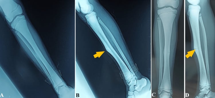
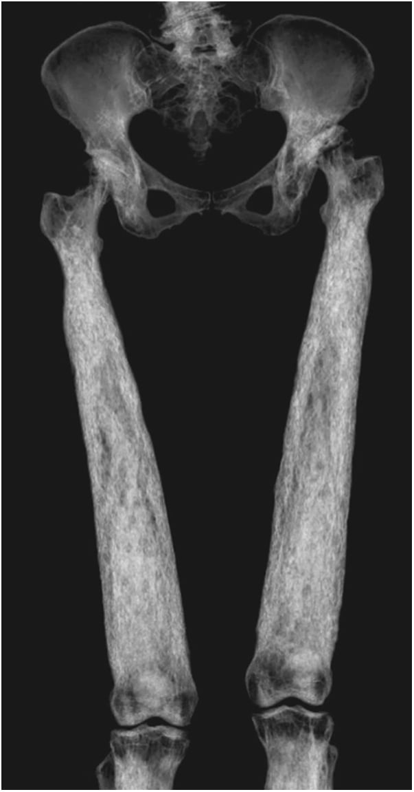
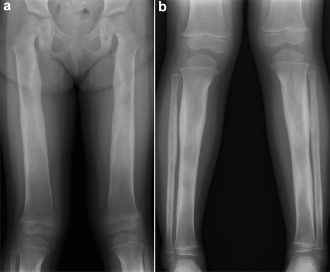
- OSTEOPETROSIS ('marble bone disease', Albers-Schonberg) is the prototype: defective OSTEOCLAST resorption produces dense, sclerotic bone with a 'BONE-IN-BONE' appearance and Erlenmeyer-flask metaphyses; the bone is paradoxically BRITTLE (transverse 'chalk-stick' fractures), and the severe (infantile/autosomal-recessive) form causes MARROW FAILURE (anaemia, pancytopenia, hepatosplenomegaly), CRANIAL-NERVE compression and osteomyelitis (classically of the mandible) - the adult/dominant form is milder.
- PYCNODYSOSTOSIS (cathepsin-K deficiency - the 'Toulouse-Lautrec' disease) combines dense, brittle bone with SHORT STATURE, ACRO-OSTEOLYSIS of the distal phalanges, an open anterior fontanelle/wormian bones, hypoplastic mandible (obtuse mandibular angle) and a tendency to fracture - distinguishing it from osteopetrosis by the short stature and acro-osteolysis.
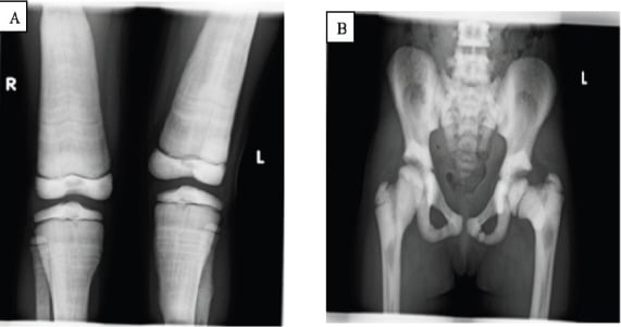
- Several dysplasias are HARMLESS INCIDENTAL findings that must NOT be mistaken for sinister disease: OSTEOPOIKILOSIS (multiple small round periarticular densities - can mimic osteoblastic metastases), OSTEOPATHIA STRIATA (Voorhoeve - linear metaphyseal striations), and the ENOSTOSIS (solitary 'bone island'); recognising their characteristic, symmetric, asymptomatic patterns avoids unnecessary biopsy or treatment.
- The INTRAMEMBRANOUS group features bone OVERGROWTH: SCLEROSTEOSIS and VAN BUCHEM disease result from loss of SCLEROSTIN (SOST) signalling, causing progressive thickening of the skull and jaw with raised intracranial pressure and CRANIAL-NERVE palsies (facial palsy, deafness, optic compression) - and have driven the development of anti-sclerostin (romosozumab) osteoporosis therapy; CAMURATI-ENGELMANN causes painful, symmetric DIAPHYSEAL cortical thickening of the long bones; MELORHEOSTOSIS causes asymmetric 'flowing candle-wax' cortical hyperostosis with pain and contracture.
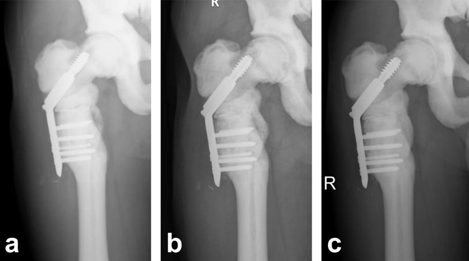
- ORTHOPAEDIC MANAGEMENT is largely supportive and complication-directed: fractures are common and FIXATION IS DIFFICULT in dense, brittle bone - plan carefully, sharp tools, pre-drill. Be precise about the evidence: NO COHORT STUDY of fracture fixation in osteopetrosis exists, so no nonunion or complication rate can be quoted. Case reports show union IS achievable (one author found union ability similar to normal) but slow - 12 months in a reported subtrochanteric fracture - and the documented hazards are largely TECHNICAL: broken drills and screws, inability to find the medullary canal, very difficult hardware removal, refracture, and intraoperative fracture during arthroplasty.
- HAEMATOPOIETIC STEM-CELL TRANSPLANT is the only cure for malignant infantile osteopetrosis, supplying donor-derived osteoclasts: 2-year overall survival 84.2% (disease-free 73.7%) in one prospective series of 19, and 100% overall survival in a 31-patient reduced-intensity series - though with a median follow-up of only 363 days. Morbidity is real: acute GvHD 38.7%, hypercalcaemia 25.8%, viral reactivation in almost 80%.
- TRANSPLANT EARLY, BECAUSE IT DOES NOT REVERSE ESTABLISHED DAMAGE. Of 19 transplanted children, visual acuity improved in only 2 and hearing partially in only 4 - cranial-nerve injury from compression is largely irreversible. The urgency is about preserving sight and hearing, not just correcting marrow failure. Mixed chimerism (45-70% donor cells) was still sufficient for disease-free status.
- CRANIAL-NERVE compression (sclerosteosis/osteopetrosis) may need decompression; and the benign incidental dysplasias need only recognition and reassurance.
- “Classify by target site (Vanhoenacker): endochondral (osteopetrosis, pycnodysostosis, osteopoikilosis, osteopathia striata, enostosis), intramembranous (Camurati-Engelmann, Van Buchem, sclerosteosis), mixed (melorheostosis).
- “Osteopetrosis = osteoclast failure → dense BRITTLE bone (chalk-stick #), 'bone-in-bone', Erlenmeyer flask, marrow failure (severe form), mandibular osteomyelitis. Pycnodysostosis = cathepsin-K, short stature + acro-osteolysis (Toulouse-Lautrec).
- “Don't mistake benign osteopoikilosis/osteopathia striata/bone island for metastases. Sclerosteosis/Van Buchem (SOST) = overgrowth + cranial-nerve palsies. Fixation in dense bone is HARD (predrill, sharp tools, broken-screw kit) - but no cohort exists, so quote no nonunion rate.
- “HSCT is the only cure for infantile osteopetrosis: 2-yr overall survival 84.2%, GvHD 38.7%, hypercalcaemia 25.8%. Transplant EARLY - sight/hearing improved in only 2 and 4 of 19, so established cranial-nerve damage does not reverse.
Osteopetrosis/pycnodysostosis: dense but brittle - transverse 'chalk-stick' fractures; fixation is difficult (drilling, hardware failure, nonunion).
Osteopoikilosis / osteopathia striata / bone island are benign incidental - do not mistake for osteoblastic metastases or biopsy them.
Sclerosteosis / Van Buchem (overgrowth) and severe osteopetrosis cause cranial-nerve palsies and raised ICP - may need decompression.
Classification (by Target Site)
The sclerosing bone dysplasias are classified by the bone-formation pathway affected: dysplasias of endochondral bone (osteopetrosis, pycnodysostosis, enostosis, osteopoikilosis, osteopathia striata), of intramembranous bone (Camurati-Engelmann; Van Buchem and sclerosteosis), and mixed (melorheostosis and overlap syndromes). The diagnosis is made largely from the radiographic pattern and distribution, refined by clinical features and inheritance.
- Defect / feature
- Osteoclast failure; 'bone-in-bone', Erlenmeyer flask
- Clinical clue
- Dense brittle bone, marrow failure (severe), mandibular osteomyelitis
- Defect / feature
- Cathepsin-K deficiency
- Clinical clue
- Short stature, acro-osteolysis, open fontanelle (Toulouse-Lautrec)
- Defect / feature
- Multiple small round periarticular densities
- Clinical clue
- Benign/incidental - mimics osteoblastic metastases
- Defect / feature
- Linear metaphyseal striations
- Clinical clue
- Benign/incidental
- Defect / feature
- Progressive diaphyseal cortical thickening
- Clinical clue
- Painful long bones, waddling gait
- Defect / feature
- Loss of sclerostin (SOST)
- Clinical clue
- Bone overgrowth, raised ICP, cranial-nerve palsies
- Defect / feature
- 'Flowing candle-wax' cortical hyperostosis
- Clinical clue
- Asymmetric, painful, contracture/limb deformity
Management
- Benign incidental dysplasias (osteopoikilosis, osteopathia striata, enostosis): recognise and reassure - no biopsy/treatment; do not mistake for metastases.
- Fractures in dense, brittle bone: difficult fixation - heat/difficulty drilling, hardware failure, high nonunion/infection risk; plan carefully, use sharp tools and pre-drilling, and anticipate problems.
- Severe osteopetrosis: haematopoietic stem-cell transplant is the only cure - 2-year survival 84.2% in one prospective series, with acute GvHD in 38.7% and hypercalcaemia in 25.8% in another. Transplant EARLY: it does not reverse established visual or hearing loss. Manage anaemia, infection (mandibular osteomyelitis), and fractures.
- Sclerosteosis / Van Buchem / severe osteopetrosis: decompress symptomatic cranial-nerve compression / raised intracranial pressure as needed.
- Symptomatic relief for painful dysplasias (Camurati-Engelmann, melorheostosis); manage deformity/contracture.
The sclerosing bone dysplasias pose two opposite traps. First, several are entirely benign incidental findings - osteopoikilosis, osteopathia striata and the solitary bone island (enostosis) - whose symmetric, asymptomatic, characteristic patterns must be recognised so they are not mistaken for osteoblastic metastases and subjected to needless biopsy or treatment. Second, the clinically significant disorders (osteopetrosis, pycnodysostosis) make the bone dense yet brittle, so it fractures transversely with low energy and is then very hard to fix: drilling generates heat and is difficult, implants fail, and nonunion and infection (including the classic mandibular osteomyelitis of osteopetrosis) are common. Operate on these bones with careful planning, sharp instruments and pre-drilling, and keep the systemic dimensions in mind - marrow failure in severe osteopetrosis (which may need stem-cell transplant) and cranial-nerve compression in osteopetrosis and the sclerostin disorders.
Stem-Cell Transplant: The Numbers, and Why Timing Decides the Outcome
Haematopoietic stem-cell transplantation is the only curative treatment for malignant infantile osteopetrosis - it supplies donor-derived osteoclasts. Two modern series give the figures the recommendation needs.
- Survival is now good. A prospective series of 19 children (median age 17 months) using busulfan-based myeloablative conditioning reported 2-year overall survival 84.2% and disease-free survival 73.7%; the three deaths were from infection, graft-versus-host disease and disease progression. A later series of 31 children using reduced-intensity conditioning (fludarabine, treosulfan, thiotepa and antithymocyte globulin) reported 100% overall survival - but with a median follow-up of only 363 days (range 74-1891), so that figure describes early survival, not cure, and should not be quoted as a long-term result.
- The morbidity is substantial. In that 31-patient series: acute graft-versus-host disease in 38.7%, hypercalcaemia in 25.8%, and viral reactivation in almost 80%, with two cases of Epstein-Barr-virus-driven post-transplant lymphoproliferative disease. All were successfully treated, but this is not a low-risk intervention.
- THE KEY POINT FOR THE EXAM: transplant does not reverse established damage. In the 19-patient series, visual acuity improved in only 2 patients and conductive hearing loss partially reversed in only 4. Cranial-nerve damage from compression is largely irreversible, which is precisely why both groups conclude that transplantation should be performed as early as possible after diagnosis, before severe sequelae develop. The urgency is about preserving sight and hearing, not only about marrow failure.
- Partial engraftment may be enough. Three children with mixed chimerism (45%, 45% and 70% donor cells) remained disease-free, suggesting full donor chimerism is not required to resolve the disease.
The Gene and Mechanism Map
- Endochondral group. Osteopetrosis - the osteoclast cannot acidify the resorption lacuna: TCIRG1 (the proton-pump a3 subunit; commonest severe autosomal-recessive form), CLCN7 (the chloride channel; AR-intermediate and the AD Albers-Schonberg type), and CAII (carbonic anhydrase II; the syndrome of osteopetrosis + renal tubular acidosis + cerebral calcification). Pycnodysostosis - CTSK (cathepsin K, the protease that degrades collagen after demineralisation). Osteopoikilosis (and Buschke-Ollendorff) - LEMD3. Osteopathia striata - WTX/AMER1 (X-linked, with cranial sclerosis).
- Intramembranous group. Camurati-Engelmann - an activating TGFB1 mutation (enhanced TGF-beta signalling). Sclerosteosis / Van Buchem - loss of SOST/sclerostin (a Wnt antagonist), so unopposed Wnt drives overgrowth.
- Mixed. Melorheostosis - LEMD3 or somatic MAP2K1 mutations.
- The unifying axis. Density comes either from failed resorption (osteopetrosis, pycnodysostosis) or enhanced formation (the sclerostin/Wnt disorders) - which is why anti-sclerostin (romosozumab) was developed from the Van Buchem/sclerosteosis biology.
Q: Map the sclerosing dysplasias to their genes.
A: Osteopetrosis = the osteoclast-acidification genes TCIRG1 (proton pump), CLCN7 (chloride channel; AD Albers-Schonberg), CAII (+ renal tubular acidosis + cerebral calcification). Pycnodysostosis = CTSK (cathepsin K). Osteopoikilosis/Buschke-Ollendorff = LEMD3. Osteopathia striata = WTX/AMER1. Camurati-Engelmann = TGFB1. Sclerosteosis/Van Buchem = SOST/sclerostin (Wnt antagonist). Melorheostosis = LEMD3 / MAP2K1. Theme: failed resorption (osteopetrosis/pycnodysostosis) vs enhanced formation (sclerostin/Wnt).
The Radiographic Sign Map
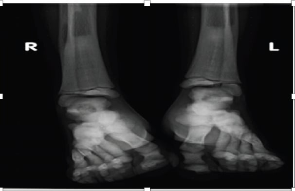
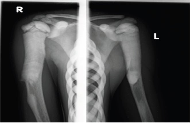
- Osteopetrosis: bone-in-bone (endobone), Erlenmeyer-flask metaphyses, and the 'rugger-jersey' / sandwich vertebra (dense superior and inferior end-plate bands) - with transverse 'chalk-stick' fractures.
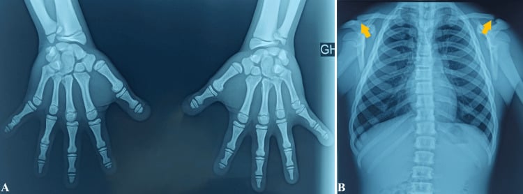
- Pycnodysostosis: acro-osteolysis of the distal phalanges, wormian bones / open fontanelle, and an obtuse (increased) mandibular angle.
- Osteopoikilosis: multiple small round periarticular densities (symmetric); osteopathia striata: linear metaphyseal striations.
- Camurati-Engelmann: symmetric diaphyseal cortical thickening (sparing the epiphyses); melorheostosis: 'flowing candle-wax' hyperostosis along one side of a bone (monomelic/asymmetric).
Q: Match the radiographic sign to the sclerosing dysplasia.
A: Bone-in-bone + Erlenmeyer flask + rugger-jersey/sandwich vertebra + chalk-stick fracture = osteopetrosis. Acro-osteolysis + wormian bones + obtuse mandibular angle = pycnodysostosis. Multiple small round periarticular densities = osteopoikilosis. Linear metaphyseal striations = osteopathia striata. Symmetric diaphyseal cortical thickening = Camurati-Engelmann. Flowing candle-wax cortical hyperostosis (monomelic) = melorheostosis.

Mnemonics & Memory Aids
DENSE
Hook:DENSE: Dysplasias by target site, Endochondral group, Nerve compression (SOST), Strong-looking but brittle, Easy to over-call benign lesions.
Clinical Decision Scenarios
Practise clinical reasoning and management decisions out loud
“A radiograph shows diffusely dense bones with a 'bone-in-bone' appearance and a transverse femoral fracture. What is the likely diagnosis and what are the management challenges?”
Classification (target site)
- Endochondral: osteopetrosis, pycnodysostosis, enostosis, osteopoikilosis, osteopathia striata
- Intramembranous: Camurati-Engelmann; Van Buchem; sclerosteosis
- Mixed: melorheostosis ('flowing candle wax') and overlap syndromes
The serious ones
- Osteopetrosis: dense brittle bone, bone-in-bone, marrow failure (severe), mandibular osteomyelitis
- Pycnodysostosis: cathepsin-K, short stature, acro-osteolysis (Toulouse-Lautrec)
- Sclerosteosis/Van Buchem: SOST loss, overgrowth, cranial-nerve palsies/raised ICP
The benign ones (don't over-call)
- Osteopoikilosis: small round periarticular densities (mimics osteoblastic metastases)
- Osteopathia striata (Voorhoeve): linear metaphyseal striations
- Enostosis (bone island): solitary dense focus
Management
- Recognise/reassure benign lesions; symptomatic care for painful dysplasias
- Fractures: difficult fixation (predrill, sharp tools, broken-screw kit) - NO cohort exists, so quote no rate
- Union IS achievable but slow (12 months in a reported case); hazards are mainly technical
- Severe osteopetrosis: HSCT is the only cure - 2-yr survival 84.2%; GvHD 38.7%, hypercalcaemia 25.8%
- Transplant EARLY: sight and hearing loss are largely irreversible (improved in only 2 and 4 of 19)
- Decompress symptomatic cranial-nerve compression
Fixing Fractures in Dense Bone: What the Evidence Actually Says
Be honest about the evidence base here, because it is weaker than the confident tone of most teaching. No cohort study of fracture fixation in osteopetrosis exists. The literature is case reports and small reviews, and it is subject to obvious publication bias toward successes. That has two consequences for a viva answer.
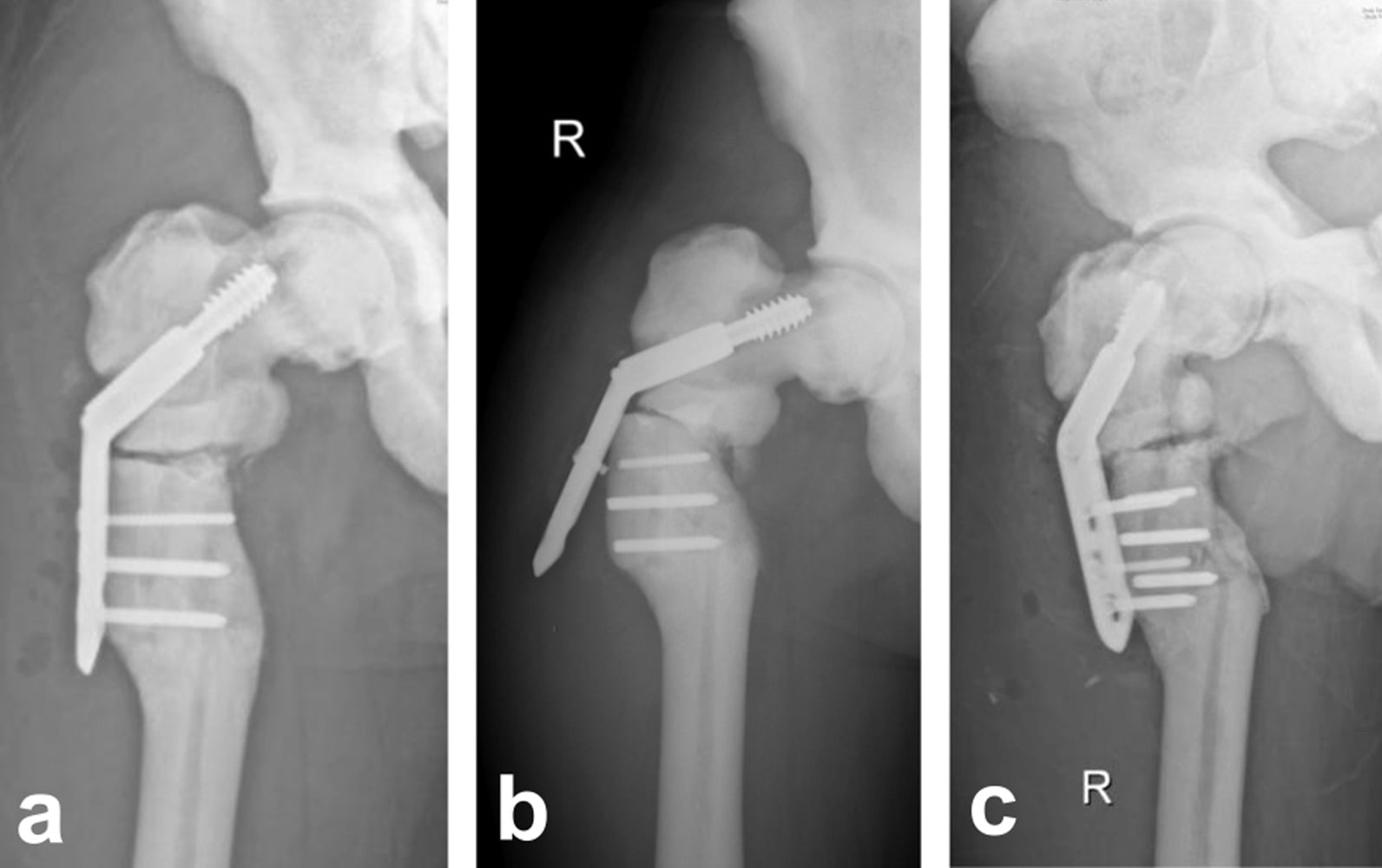
- Union is achievable, and "nonunion is inevitable" overstates it. Reported cases of open reduction and internal fixation have united, one author concluding that treated patients showed union ability similar to normal. But union can be slow - 12 months in one reported subtrochanteric fracture - so judge fixation stability against a longer timeline than usual and do not mistake slow union for failure.
- The documented hazards are TECHNICAL rather than biological. Reported difficulties include drill-bit breakage and heat generation, screws breaking and being left in the canal, difficulty identifying the medullary canal at all, very difficult hardware removal, and intraoperative fracture during arthroplasty (an acetabular fracture in one conversion hip replacement). Refracture and displacement after fixation are recurring themes.
- Failed fixation compounds itself. One reported patient had sustained four previous femoral fractures inadequately treated before definitive surgery, and conversion total hip arthroplasty after failed fixation was described as substantially more complex and unpredictable. The first operation is the one that matters most.
- Practical implications. Plan preoperatively with CT, have multiple sharp drill bits and broken-screw removal equipment available, pre-drill and irrigate to limit thermal necrosis, accept longer operating times, and anticipate that intramedullary options may be impossible if no canal can be found.
If asked for the nonunion or complication rate of fracture fixation in osteopetrosis, the correct answer is that no series large enough to generate one has been published - the evidence is case reports. Quoting a specific percentage would be inventing it. What you can say is which complications are repeatedly described, that union is achievable but slow, and that the difficulties are predominantly technical and therefore mitigated by planning.
Evidence & Key Studies
Sclerosing bone dysplasias: genetic and radioclinical features
- A NARRATIVE REVIEW, and now 26 years old - it is the source of the classification framework, not of any outcome data. The genetics it describes have been substantially extended since publication.
- The sclerosing bone dysplasias are classified by a target-site approach into three groups: dysplasias of endochondral bone formation (osteopetrosis, pycnodysostosis, enostosis, osteopoikilosis, osteopathia striata), dysplasias of intramembranous bone formation (Camurati-Engelmann and variants; Van Buchem disease and variants), and mixed sclerosing dysplasias (melorheostosis and overlap syndromes).
- Within each group, further differentiation is made by distinctive clinical findings and the mode of inheritance.
- Despite advances in basic genetics, the radiologist plays a pivotal role in diagnosing this relatively poorly understood group of disorders from the radiographic pattern and distribution.
Non-total-body-irradiation myeloablative conditioning with intravenous busulfan and cyclophosphamide in HSCT for malignant infantile osteopetrosis
- Prospective series of 19 children (median age 17 months) transplanted 2008-2013: 14 from HLA-matched related donors, 2 haploidentical related, 2 partially matched cord blood, 1 matched unrelated.
- TWO-YEAR OVERALL SURVIVAL 84.2% and disease-free survival 73.7%. All but one patient achieved primary engraftment; two had secondary graft failure. The three deaths were from infection, graft-versus-host disease and disease progression.
- CRITICAL FOR TIMING: established sensory damage largely did not reverse - visual acuity improved in only 2 patients and conductive hearing loss partially reversed in only 4, out of 19. This is the argument for transplanting early, before cranial-nerve compression has done its damage.
- Three patients with MIXED chimerism (45%, 45% and 70% donor cells) remained disease-free, suggesting complete donor chimerism is not required to resolve the disease.
- Limitations: 19 patients at a single centre with a predominance of matched related donors, which flatters outcomes relative to settings where such donors are unavailable; two-year survival is not lifelong cure.
Successful haematopoietic stem-cell transplantation for osteopetrosis using reduced-intensity conditioning
- Retrospective review of 31 children with infantile malignant osteopetrosis transplanted 2012-2017 with fludarabine, treosulfan, thiotepa and antithymocyte globulin; 26 from 10/10 matched donors.
- OVERALL SURVIVAL 100% - but read the follow-up before quoting it: median 363 days, range 74-1891. This is excellent early survival, not evidence of long-term cure.
- Morbidity was substantial: acute graft-versus-host disease in 12 (38.7%), hypercalcaemia in 8 (25.8%), and viral reactivation in almost 80%, including two cases of Epstein-Barr-virus-driven post-transplant lymphoproliferative disease. No veno-occlusive disease occurred. All complications were successfully treated.
- The authors conclude transplantation should be performed as early as possible after diagnosis, before severe disease sequelae develop.
- Limitations: retrospective, single centre, no comparison group against myeloablative conditioning, and short follow-up for a condition whose complications accrue over decades.
Fracture fixation in osteopetrosis - the published experience
- THERE IS NO COHORT STUDY. The entire evidence base for fracture fixation in osteopetrosis consists of case reports and small literature reviews, so no nonunion rate, complication rate or implant-survival figure can legitimately be quoted.
- Union is achievable: reported open reduction and internal fixation cases united, with one author concluding that union ability was similar to that of normal bone - though union took 12 months in a reported subtrochanteric fracture, so slow union should not be mistaken for failure.
- Repeatedly described technical hazards: difficulty and heat generation when drilling, broken drill bits and screws (sometimes retained in the canal), difficulty identifying the medullary canal, very difficult hardware removal, refracture and displacement after fixation, and intraoperative acetabular fracture during conversion hip arthroplasty.
- Conversion total hip arthroplasty after failed internal fixation was described as markedly more complex and unpredictable - so the quality of the index procedure matters disproportionately.
- Limitations: publication bias toward successful cases is severe in this literature; failures are systematically under-reported, so the apparent reassurance about union should be treated cautiously.
The target-site classification and its constituent entities come from the Vanhoenacker review (DOI) - a narrative review from 2000. The transplant survival figures (2-year overall survival 84.2%, disease-free 73.7%), the limited reversal of visual and hearing loss and the mixed chimerism observation come from Behfar (DOI), 19 children. The 100% overall survival at a median 363 days, the acute GvHD rate of 38.7%, hypercalcaemia 25.8% and the near-80% viral reactivation come from Shadur (DOI), 31 children. The fracture-fixation observations come from case reports and small reviews, principally Ding (DOI), Huang (DOI) and Poursalehian (DOI). The gene assignments, the radiographic signs, the mandibular osteomyelitis association and the benign nature of osteopoikilosis, osteopathia striata and the bone island are standard, well-established teaching.
What does not exist: there is no cohort study, and therefore no nonunion rate, complication rate or implant-survival figure, for fracture fixation in osteopetrosis - any percentage quoted for this would be fabricated. There is no randomised comparison of conditioning regimens in osteopetrosis, no agreed age threshold for transplantation, no validated screening interval for cranial-nerve compression, and no disease-specific fracture classification. Both transplant series are single-centre with short-to-medium follow-up, and the fixation literature is dominated by publication bias toward successful cases.