Lateral Tibial Plateau Avulsion | Anterolateral Ligament | Pathognomonic for ACL Tear
- Pathognomonic for ACL tear - Segond fracture = small avulsion lateral tibial plateau = anterolateral ligament avulsion
- 75-100% association with ACL injury - if you see Segond fracture, look for ACL tear on MRI
- Anterolateral ligament (ALL) avulses from lateral tibial plateau - this is the Segond fracture
- High-grade pivot shift - Segond fracture associated with more severe rotational instability
- Treatment: Focus on underlying ACL injury - Segond fracture itself rarely needs fixation
- “Segond fracture = pathognomonic for ACL tear - small avulsion lateral tibial plateau
- “Anterolateral ligament (ALL) avulses from lateral tibial plateau - this creates the Segond fracture
- “75-100% of Segond fractures have associated ACL tears - always order MRI
- “Reverse Segond fracture (medial) = MCL avulsion = associated with PCL injury
Segond fracture = pathognomonic for ACL tear. Small avulsion lateral tibial plateau represents anterolateral ligament (ALL) avulsion. 75-100% of Segond fractures have associated ACL tears. Always order MRI if Segond fracture seen on X-ray.
ALL avulses from lateral tibial plateau - this creates the Segond fracture. The ALL is a secondary stabilizer to internal rotation. Its avulsion indicates high-grade rotational instability (pivot shift).
Segond fracture associated with high-grade pivot shift - more severe rotational instability than isolated ACL tears. This may influence decision for lateral extra-articular tenodesis (LET) in ACL reconstruction.
Treat underlying ACL injury - Segond fracture itself rarely needs fixation. The avulsed fragment is small and non-articular. Focus on ACL reconstruction. Consider LET for high-grade pivot shift.
- Location
- Lateral tibial plateau
- Associated Injury
- ACL tear (75-100%)
- Treatment
- ACL reconstruction
- Location
- Medial tibial plateau
- Associated Injury
- PCL tear (MCL avulsion)
- Treatment
- PCL reconstruction
- Location
- Lateral tibial plateau
- Associated Injury
- No ACL tear (less than 5%)
- Treatment
- Conservative if stable
- Location
- Lateral tibial plateau
- Associated Injury
- ACL tear + high-grade pivot shift
- Treatment
- ACL reconstruction + LET consideration
SEGONDSegond Fracture Features
Hook:SEGOND: Small avulsion, Examine ACL (75-100%), Grade pivot shift high, Order MRI, Non-articular fragment, Don't fix - treat ACL!
ALLAnterolateral Ligament
Hook:ALL: Anterolateral Ligament avulses from Lateral tibia, creating the Segond fracture!
ACL MENAssociated Injuries
Hook:ACL MEN: ACL tear (most common), Cartilage bruise, Lateral meniscus tear, MCL injury, Edema pattern, Non-articular fragment!
Overview and Epidemiology
Segond fractures are small avulsion fractures of the lateral tibial plateau, representing avulsion of the anterolateral ligament (ALL). They are pathognomonic for ACL tears, with 75-100% association. The fracture itself is small and non-articular, but its presence indicates significant rotational instability.
Mechanism of Injury
Classic mechanism: Same as ACL injury
- Non-contact: Cutting/pivoting on planted foot with knee near extension, valgus collapse
- Contact: Direct blow causing valgus/hyperextension
- Internal rotation: Combined with valgus and extension
- Peak force: Near full extension with valgus and internal rotation
The anterolateral ligament (ALL) is a secondary stabilizer to internal rotation. When the ACL tears, the ALL experiences excessive force and avulses from the lateral tibial plateau, creating the Segond fracture.
Segond fracture = pathognomonic for ACL tear. Small avulsion lateral tibial plateau represents anterolateral ligament (ALL) avulsion. 75-100% of Segond fractures have associated ACL tears. If you see Segond fracture on X-ray, always order MRI to assess ACL.
Epidemiology
- Incidence: Seen on injury radiographs in roughly 7-9% of ACL tears; up to about 15% when healed lesions are included (Slagstad et al, AJSM 2020)
- Age: Peak 20-30 years (athletic population); skiing mechanisms over-represented
- Gender: Male predominance (approximately 2:1)
- Laterality: Usually unilateral
- Associated injuries: ACL tear (the lesion is highly specific for ACL injury), lateral meniscal tear (around 30-40%), MCL injury (around 20-30%)
The phrase "pathognomonic" is traditional teaching: a Segond fracture is highly specific for ACL injury and should be treated as an ACL tear until proven otherwise, but rare isolated cases without ACL rupture do exist.
Anatomy and Pathophysiology
Anterolateral Ligament (ALL) Anatomy
The anterolateral ligament (ALL) is a distinct ligament structure:
- Origin: Lateral femoral epicondyle (proximal and posterior to LCL origin)
- Insertion: Lateral tibial plateau (anterolateral aspect, 5mm below joint line)
- Course: Oblique, anterior to LCL
- Function: Secondary stabilizer to internal rotation
- Relationship: Works with ACL to resist rotational instability
Discovery: The ALL was formally described in 2013, though Segond described the fracture in 1879. The ligament was previously thought to be part of the iliotibial band.
Pathophysiology
ACL injury mechanism:
- Valgus + internal rotation + extension
- ACL tears first (primary restraint)
- ALL experiences excessive force
- ALL avulses from lateral tibial plateau
- Creates Segond fracture
Why ALL avulses:
- ALL is weaker than ACL
- Excessive internal rotation force
- Bone (tibial plateau) is stronger than ligament-bone interface
- Avulsion occurs at insertion site
High-grade pivot shift:
- Segond fracture indicates more severe rotational instability
- ALL avulsion = loss of secondary rotational restraint
- Results in higher-grade pivot shift than isolated ACL tears
Reverse Segond fracture (medial tibial plateau avulsion) = MCL avulsion = associated with PCL injury, not ACL. This is the opposite pattern - medial avulsion indicates PCL tear with MCL involvement.
Classification Systems
Location-Based Classification
Classic Segond (lateral):
- Lateral tibial plateau avulsion
- Anterolateral ligament (ALL) avulsion
- Associated with ACL tear (75-100%)
- Most common type
Reverse Segond (medial):
- Medial tibial plateau avulsion
- MCL avulsion
- Associated with PCL injury
- Less common (5-10% of Segond fractures)
Location determines which ligament is involved and which cruciate ligament injury to expect.
Clinical Assessment
History
Mechanism: Same as ACL injury
- Cutting/pivoting on planted foot
- Knee near extension with valgus collapse
- "Pop" or "snap" sensation
- Immediate pain and swelling
- Inability to continue activity
Symptoms:
- Immediate pain and swelling
- Knee "giving way" (instability)
- Inability to bear weight
- Locking (if meniscal injury)
Physical Examination
Inspection:
- Knee effusion (hemarthrosis)
- Antalgic gait
- Knee held in slight flexion
Palpation:
- Tenderness over lateral tibial plateau (Segond fracture site)
- Joint line tenderness (if meniscal injury)
- MCL tenderness (if MCL injury)
Range of Motion:
- Limited flexion (pain, effusion)
- Limited extension (pain, effusion)
Ligament Testing:
- Lachman test: Positive (anterior translation)
- Anterior drawer: Positive (anterior translation)
- Pivot shift: Positive (rotational instability) - high-grade with Segond fracture
- Valgus stress: May be positive (if MCL injured)
Pivot shift test is high-grade with Segond fracture - the ALL avulsion indicates more severe rotational instability. This may influence decision for lateral extra-articular tenodesis (LET) in ACL reconstruction.
Associated Injuries
- ACL tear: 75-100% (most common)
- Lateral meniscal tear: 30-40% (posterolateral)
- MCL injury: 20-30%
- Bone bruises: Posterolateral tibial plateau, lateral femoral condyle (kissing contusion)
- Cartilage injury: Lateral femoral condyle (30-40%)
Grading the Pivot Shift: What 'High-Grade' Means
This topic repeatedly makes treatment decisions on the basis of a high-grade pivot shift, yet the pivot shift is a graded — not a binary — sign, and the topic never defines the grades its own classification tab relies on. Because a Segond fracture is a bony flag for anterolateral rotatory instability, pinning down the grade is what converts that radiographic clue into an operative decision (whether to add a lateral procedure).
The pivot shift reproduces the anterior subluxation-then-reduction of the lateral tibial plateau as the knee moves from extension into flexion — the iliotibial band, converting from an extensor to a flexor at roughly 20-30 degrees, snaps the subluxated plateau back. The IKDC grading is:
- Grade 0: no pivot (equal to the normal contralateral knee)
- Grade I (glide): a smooth, subtle reduction — low-grade
- Grade II (clunk): a distinct jerk or clunk as the plateau reduces — high-grade
- Grade III (gross): a gross clunk with momentary impingement or transient locking of the plateau — high-grade
"High-grade" therefore means grade II (clunk) or grade III (gross). This is not academic. The STABILITY randomised trial used a grade 2 pivot shift or greater as one of its enrolment criteria for adding lateral extra-articular tenodesis, and this is precisely the population a Segond fracture tends to identify. Because the anterolateral ligament and the wider anterolateral complex are secondary restraints to internal tibial rotation, their bony avulsion (the Segond fragment) correlates with a larger, higher-grade pivot shift.
A practical caveat for the viva: guarding and pain make the pivot shift unreliable in the acute, awake knee, so a low-grade or absent pivot shift on the day of injury does not exclude high-grade instability. The examination under anaesthesia at the time of surgery is the definitive grading, and the Segond fragment on the plain film is your early warning of what you will find.
Grade the pivot shift by IKDC: a glide (I) is low-grade; a clunk (II) or a gross clunk with transient locking (III) is high-grade. STABILITY enrolled knees with a grade 2 pivot shift or greater for lateral augmentation — the same high-risk phenotype a Segond fracture flags. Acute guarding masks the true grade, so weight the radiographic Segond sign and confirm the grade on examination under anaesthesia.
Investigations
Standard X-ray Protocol
Views: AP and lateral knee.
Key findings:
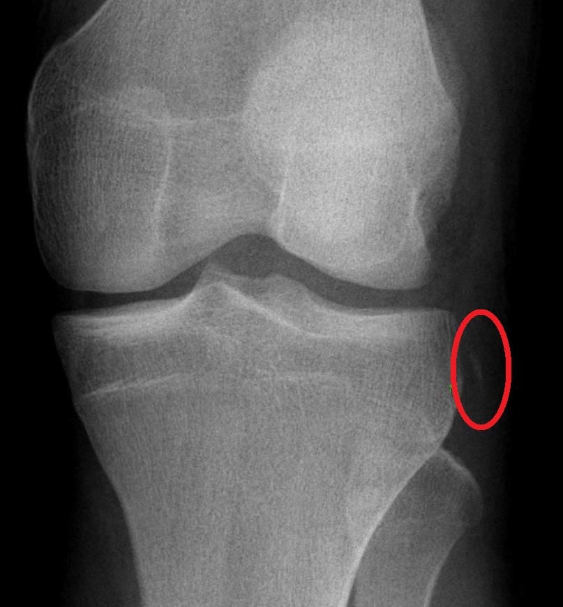
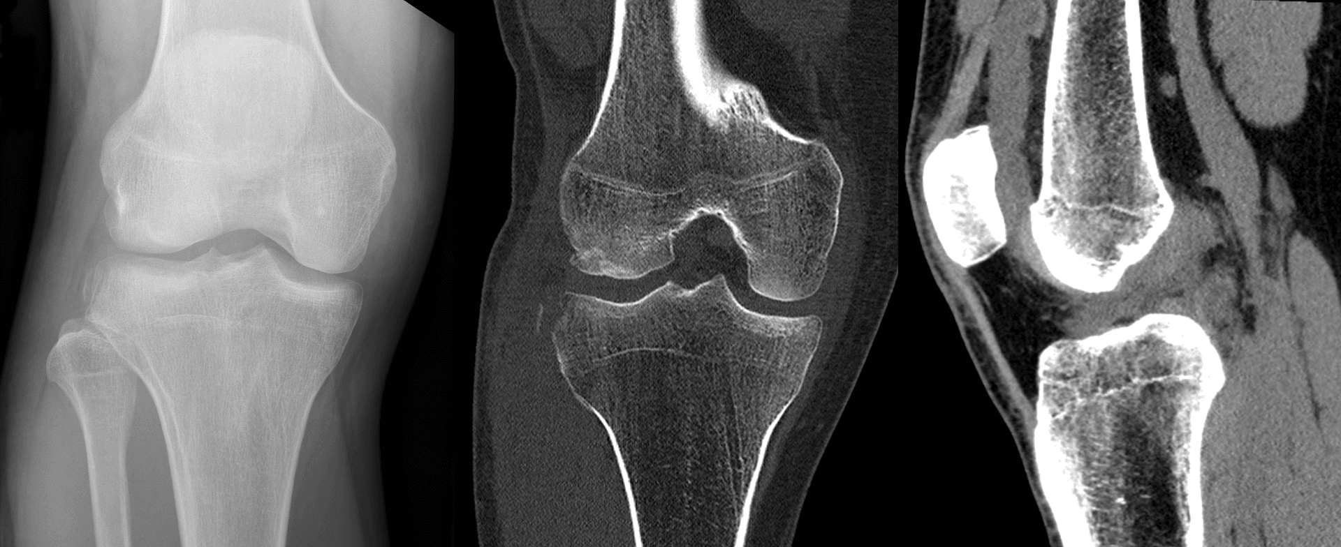
- Segond fracture: Small avulsion lateral tibial plateau (anterolateral aspect)
- Fragment size: Usually 5-10mm, non-articular
- Location: Lateral tibial plateau, 5mm below joint line
- Associated findings: Deep lateral notch sign (greater than 1.5mm), tibial spine fracture (pediatric)
Lateral view: May show fragment, but AP view is diagnostic.
If Segond fracture seen: Always order MRI to assess ACL and associated injuries.
The Deep Lateral Femoral Notch Sign: A Companion Bony Marker of ACL Rupture
The Investigations section lists the deep lateral femoral notch sign as an associated finding, but it deserves development because it is the other bony surrogate of ACL rupture you can read off the very same plain radiograph as a Segond fragment — and, unlike the Segond fleck, it sits on the femur, not the tibia.
The lateral femoral condyle normally carries a shallow groove — the condylopatellar sulcus (sulcus terminalis) — separating its patellar and tibiofemoral articular surfaces, seen end-on as a small notch on a true lateral radiograph. During the pivot-shift subluxation event of an ACL rupture, the lateral femoral condyle impacts against the posterolateral tibial plateau; the resulting osteochondral impaction deepens this sulcus. A sulcus depth greater than 1.5 mm (some authors use a 2 mm threshold) is abnormal and is an indirect sign of a torn ACL, described in the radiology literature as the "deep lateral femoral notch sign."
Why it matters alongside the Segond fragment:
- Two bony clues, one mechanism. Both the Segond avulsion and a deep notch are skeletal footprints of the same rotatory subluxation event; finding either on plain films should trigger dedicated ACL assessment on MRI.
- The notch localises the bone bruise. The impaction that deepens the notch is the femoral half of the classic "kissing contusion" bone-bruise pattern (lateral femoral condyle plus posterolateral tibial plateau) seen on MRI, and it is a clue to associated lateral meniscal and chondral injury.
- Look on the lateral, not just the AP. The Segond fleck is an AP-view finding, whereas the deep notch is best appreciated on the true lateral view — so a complete plain-film read for suspected ACL injury interrogates both projections.
The notch sign is specific but insensitive: its absence does not exclude an ACL tear. Its presence, like a Segond fracture, is a high-value plain-radiograph pointer that saves you from missing the cruciate injury while cross-sectional imaging is awaited.
A Segond fracture (tibial, AP view) and a deep lateral femoral notch (femoral, lateral view — sulcus deeper than 1.5 mm) are the two bony surrogates of ACL rupture on plain radiographs. Both arise from the pivot-shift impaction of the lateral compartment, and either one mandates MRI. The deepened notch marks the femoral pole of the lateral-condyle / posterolateral-tibia "kissing" bone bruise.
Differential Diagnosis of a Proximal Tibial Avulsion
A small fleck of bone around the proximal tibia is the radiographic crux. Each fleck points to a different ligament and a different associated injury, so localising it precisely is the exam-winning step.
- Fragment location
- Anterolateral tibial rim, just below joint line
- Structure avulsed
- Anterolateral ligament / capsule
- Key association
- ACL tear + anterolateral rotatory instability
- Discriminator
- Lateral fleck + positive Lachman/pivot shift
- Fragment location
- Medial tibial rim
- Structure avulsed
- Deep MCL tibial attachment
- Key association
- PCL / posteromedial corner injury
- Discriminator
- Medial fleck + posterior sag
- Fragment location
- Fibular styloid / head
- Structure avulsed
- Arcuate complex (PLC)
- Key association
- Posterolateral corner, common peroneal nerve
- Discriminator
- Fibular fleck + varus/dial test positive
- Fragment location
- Intercondylar eminence (central)
- Structure avulsed
- ACL tibial insertion (bony)
- Key association
- ACL functional disruption (paediatric/adolescent)
- Discriminator
- Central fragment, often a child after a fall from bike
- Fragment location
- Anterolateral tibia at Gerdy tubercle
- Structure avulsed
- Iliotibial band insertion
- Key association
- Direct ITB traction; not pathognomonic for ACL
- Discriminator
- More anterior/lateral, at the ITB footprint
- Fragment location
- Lateral plateau, articular
- Structure avulsed
- Bony - articular surface
- Key association
- Valgus/axial load; bumper fracture
- Discriminator
- Larger, intra-articular, joint depression on CT
Management Algorithm
Management Pathway
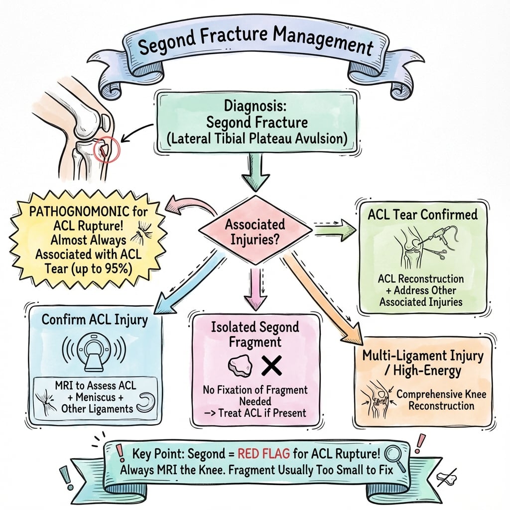
Segond Fracture Management
Recognize Segond fracture on X-ray. Order MRI to assess ACL and associated injuries. Classify as with ACL tear (75-100%) or isolated (less than 5%).
Most common (75-100%). Treat underlying ACL injury with ACL reconstruction. Consider lateral extra-articular tenodesis (LET) for high-grade pivot shift. Segond fracture itself rarely needs fixation.
Less than 5% of cases. If stable (no ACL tear, no instability), treat conservatively with brace and rehabilitation. Monitor for instability.
Address associated injuries: meniscal repair if indicated, MCL treatment if needed, cartilage management.
Surgical Technique
Standard ACL Reconstruction
Primary treatment for Segond fracture with ACL tear:
Graft selection:
- BTB autograft: Gold standard, bone-to-bone healing
- Hamstring autograft: Good outcomes, less morbidity
- Allograft: Older patients, revision cases
Tunnel placement:
- Standard anatomic ACL reconstruction
- Anteromedial portal technique
- Avoid Segond fracture site (lateral tibial plateau)
Considerations:
- High-grade pivot shift: Consider lateral extra-articular tenodesis (LET)
- Associated injuries: Address meniscal tears, MCL if needed
- Timing: Standard (4-12 weeks post-injury)
ACL reconstruction is the primary treatment - Segond fracture does not change technique.
Complications
- Incidence
- 4-11% at 2y (STABILITY)
- Risk Factors
- High-grade pivot shift, young age, return to pivoting sport
- Prevention/Management
- Consider LET in young high-risk patients (rupture 11% to 4% with LET)
- Incidence
- 10-15%
- Risk Factors
- Inadequate ACL reconstruction, no LET
- Prevention/Management
- Anatomic ACL reconstruction, consider LET
- Incidence
- Less than 5%
- Risk Factors
- Large fragment, displacement
- Prevention/Management
- Fixation if large fragment (rare)
- Incidence
- 10-20%
- Risk Factors
- Untreated meniscal tear
- Prevention/Management
- Repair meniscal tears at time of ACL reconstruction
- Incidence
- 5-10%
- Risk Factors
- Early surgery, delayed ROM
- Prevention/Management
- Wait for ROM recovery, early ROM postoperatively
ACL Graft Failure
5-10% incidence:
- Cause: High-grade pivot shift, young age, inadequate reconstruction
- Prevention: Consider LET for high-grade pivot shift, anatomic ACL reconstruction
- Management: Revision ACL reconstruction with LET
Residual Pivot Shift
10-15% incidence:
- Cause: Inadequate ACL reconstruction, no LET for high-grade pivot shift
- Prevention: Anatomic ACL reconstruction, consider LET for high-grade cases
- Management: Revision ACL reconstruction with LET if symptomatic
Segond Fracture Nonunion
Less than 5% incidence:
- Cause: Large fragment, displacement, inadequate fixation
- Prevention: Fixation if large fragment (rare)
- Management: Fixation if symptomatic (rare)
Postoperative Care
Immediate Postoperative
- Immobilization: Hinged knee brace locked in extension (2-4 weeks)
- Weight bearing: Non-weight bearing initially (2-3 weeks)
- ROM: Begin passive ROM at 2-4 weeks (unlock brace)
- PT: Quadriceps sets, straight leg raises (immediate)
Rehabilitation Protocol
Weeks 0-2:
- Brace locked in extension
- Non-weight bearing
- Quadriceps sets, straight leg raises
- Ice and elevation
Weeks 2-4:
- Unlock brace for passive ROM (0-90 degrees)
- Progressive weight bearing (partial to full)
- Stationary bike (when ROM allows)
- Continue quadriceps strengthening
Weeks 4-6:
- Full ROM
- Full weight bearing
- Progressive strengthening
- Balance and proprioception
Weeks 6-12:
- Sport-specific training
- Return to sport (when strength and ROM normal)
- Continue PT for 3-6 months
Return to Sport
Criteria:
- Full ROM (equal to contralateral)
- Quadriceps strength greater than 90% of contralateral
- No instability (negative Lachman, pivot shift)
- Functional testing passed (hop test, agility)
Timeline: Usually 6-9 months postoperatively.
Outcomes and Prognosis
Overall Outcomes
ACL reconstruction outcomes:
- Success rate: 85-90% (same as standard ACL reconstruction)
- Functional outcomes: 80-85% return to pre-injury level
- Complications: 10-15% (graft failure, residual pivot shift)
Segond fracture outcomes:
- Healing: Usually heals without fixation (fragment is small)
- Nonunion: Less than 5% (rare, usually asymptomatic)
- Fixation: Rarely needed
Functional Outcomes
Return to sport:
- Timeline: 6-9 months postoperatively
- Rate: 80-85% return to pre-injury level
- Factors: Age, sport level, rehabilitation compliance, LET use
Functional testing:
- Quadriceps strength: 90%+ of contralateral
- No instability (negative Lachman, pivot shift)
- Full ROM
Long-Term Prognosis
Graft rupture risk (STABILITY trial, young high-risk patients, 2 years):
- Isolated hamstring ACL reconstruction: 11%
- ACL reconstruction + LET: 4% (number needed to treat 14.3)
- Composite clinical failure (rupture or residual rotatory laxity): 40% vs 25% favouring LET
Residual rotatory laxity:
- Persistent anterolateral rotatory laxity correlates with poorer outcomes and graft failure; adding LET reduces the composite failure that includes residual rotatory laxity.
- A Segond fracture itself does not independently raise revision risk (Slagstad et al, AJSM 2020) - it flags the high-risk phenotype rather than causing failure.
Factors Affecting Outcomes
Positive factors:
- Anatomic ACL reconstruction
- LET for high-grade pivot shift
- Complete rehabilitation
- Early ROM (2-4 weeks)
Negative factors:
- High-grade pivot shift without LET
- Inadequate ACL reconstruction
- Incomplete rehabilitation
- Early return to sport
Prevention and Return to Sport
Prevention
Primary prevention:
- Proper landing technique (knee flexion, not hyperextension)
- Strength training (quadriceps, hamstrings)
- Balance and proprioception training
- Sport-specific conditioning
Secondary prevention (after injury):
- Complete rehabilitation before return to sport
- Bracing (controversial - may not prevent reinjury)
- Continued strength and conditioning
Return to Sport Criteria
Clinical:
- Full ROM (equal to contralateral)
- Quadriceps strength greater than 90% of contralateral
- No effusion
- No instability (negative Lachman, pivot shift)
Functional:
- Single-leg hop test (greater than 90% of contralateral)
- Agility testing passed
- Sport-specific drills completed
Timeline: Usually 6-9 months postoperatively, depending on sport and level.
Guidelines, Registries & Global Practice
Global Epidemiology
- Segond fracture is seen on injury radiographs in roughly 7-9% of ACL tears; including healed lesions the figure rises to about 15% (Slagstad et al, AJSM 2020).
- Demographics mirror ACL injury: young, athletic, pivoting/cutting sports and skiing; the Segond subgroup is associated with downhill skiing mechanisms.
- The lesion is a radiographic surrogate, not a separate disease - so there is no dedicated guideline for "Segond fracture"; guidance is embedded within ACL and anterolateral-complex recommendations.
Side-by-Side Guidance (ACL / Anterolateral Complex)
- Position on the Segond / anterolateral lesion
- Segond fracture supports a clinical diagnosis of ACL tear; MRI confirms ligamentous injury
- Position on lateral augmentation (LET/ALL)
- Acknowledges anterolateral procedures as an adjunct; reserved for selected high-risk knees
- Position on the Segond / anterolateral lesion
- Treat the ACL injury, not the small avulsion; image to define associated injury
- Position on lateral augmentation (LET/ALL)
- Lateral augmentation considered in revision and high-grade rotatory laxity
- Position on the Segond / anterolateral lesion
- Anterolateral injury (incl. Segond) marks rotatory instability
- Position on lateral augmentation (LET/ALL)
- Recommends lateral augmentation for young, high-risk, pivoting, revision, high-grade pivot shift
- Position on the Segond / anterolateral lesion
- Fragment is extra-articular and rarely needs fixation
- Position on lateral augmentation (LET/ALL)
- Focus on cruciate and associated structures
Registry & Trial Evidence
- High-quality randomised data (STABILITY, Getgood et al 2020) and the SANTI group series (Sonnery-Cottet) underpin lateral augmentation in young high-risk patients; this is the population a Segond fracture identifies.
- ACL-reconstruction registries (e.g. the Scandinavian Knee Ligament registries - Norwegian, Swedish, Danish - and the UK NLR) track graft survival and revision but do not separately code the Segond fragment; revision risk is driven by age, graft choice, and rotatory laxity rather than the avulsion itself.
High- vs Limited-Resource Practice
- Well-resourced settings: routine MRI, arthroscopic anatomic ACL reconstruction, selective LET/ALL reconstruction for high-risk knees.
- Limited-resource settings: the plain radiograph alone is a powerful clue - a Segond sign should trigger referral and ACL-focused management even where MRI is delayed or unavailable; structured rehabilitation remains the default first step and may be definitive in low-demand patients.
Segond fractures are a common viva topic. Know that Segond fracture = pathognomonic for ACL tear (75-100% association), represents anterolateral ligament (ALL) avulsion, indicates high-grade pivot shift, and treatment focuses on ACL reconstruction (not fixing the Segond fracture). Be prepared to discuss LET for high-grade pivot shift and the STABILITY trial.
Controversies and Areas of Uncertainty
The Segond fragment has been attributed to the anterolateral ligament, the mid-third lateral capsular ligament, the iliotibial band's capsulo-osseous layer, and the anterior arm of the short head of biceps. The ALL re-description (Claes 2013) is the dominant modern view, but the precise soft-tissue origin of the fleck remains debated.
"Pathognomonic" is the classic teaching, but pooled radiographic series show the lesion in only roughly 7-9% of ACL tears, and rare isolated Segond fractures without ACL rupture exist. The defensible statement is that a Segond fracture is highly specific for, and strongly suggestive of, ACL injury - not that it is universal.
Most surgeons leave it: it is small, extra-articular, and only about a third heal radiographically yet revision risk is not increased (Slagstad 2020). Whether fixing or reconstructing the anterolateral structure adds value beyond standard ACL reconstruction is unsettled.
STABILITY supports adding a lateral procedure in young, high-risk patients, but the optimal technique (modified Lemaire LET vs anatomic ALL reconstruction), graft, and exact indication thresholds are still being defined, as is whether routine LET over-constrains the lateral compartment long term.
MCQ Practice Points
Q: What does a Segond fracture indicate? A: ACL tear (pathognomonic) - Segond fracture = small avulsion lateral tibial plateau = anterolateral ligament avulsion. 75-100% of Segond fractures have associated ACL tears.
Q: What ligament avulses to create a Segond fracture? A: Anterolateral ligament (ALL) - The ALL avulses from the lateral tibial plateau, creating the Segond fracture. The ALL is a secondary stabilizer to internal rotation.
Q: What percentage of Segond fractures have associated ACL tears? A: 75-100% - Segond fracture is pathognomonic for ACL tear. Always order MRI if Segond fracture seen on X-ray to assess ACL integrity.
Q: What does Segond fracture indicate about pivot shift severity? A: High-grade pivot shift - Segond fracture indicates more severe rotational instability than isolated ACL tears. This may influence decision for lateral extra-articular tenodesis (LET).
Q: What is the primary treatment for Segond fracture? A: ACL reconstruction - Segond fracture itself rarely needs fixation (fragment is small and non-articular). Focus on treating the underlying ACL injury. Consider LET for high-grade pivot shift.
Q: What does a reverse Segond fracture (medial tibial plateau avulsion) indicate? A: PCL injury - Reverse Segond fracture = MCL avulsion = associated with PCL injury, not ACL. This is the opposite pattern.
Exam Viva Scenarios
Practise clinical reasoning and management decisions out loud
“A 25-year-old athlete presents to ED after a non-contact knee injury during a soccer game. He felt a 'pop' and cannot continue playing. X-ray shows a small avulsion fracture of the lateral tibial plateau. He has a swollen, painful knee and cannot bear weight.”
“A 22-year-old elite athlete presents 6 weeks after ACL injury with Segond fracture. He has persistent instability and cannot return to sport. Examination shows positive Lachman and high-grade pivot shift. MRI confirms ACL tear and shows the Segond fracture (anterolateral ligament avulsion).”
“A 30-year-old is brought in after a high-energy road traffic accident with a hyperextension knee injury. Radiograph shows a small avulsion fragment off the MEDIAL tibial rim, not the lateral side. The knee is grossly swollen with a positive posterior sag sign.”
Key Anatomy
- Anterolateral ligament (ALL): Secondary stabilizer to internal rotation
- ALL origin: Lateral femoral epicondyle
- ALL insertion: Lateral tibial plateau (anterolateral, 5mm below joint line)
- ALL avulsion from tibia = Segond fracture
Classification
- Classic Segond: Lateral tibial plateau avulsion = ACL tear (75-100%)
- Reverse Segond: Medial tibial plateau avulsion = PCL injury (MCL avulsion)
- With ACL tear: Most common (75-100%)
- Isolated: Rare (less than 5%)
Treatment Algorithm
- Segond fracture seen on X-ray: Always order MRI
- With ACL tear (75-100%): ACL reconstruction
- High-grade pivot shift: Consider LET (STABILITY trial)
- Segond fracture itself: Rarely needs fixation (small, non-articular)
Surgical Pearls
- Focus on ACL reconstruction - Segond fracture rarely needs fixation
- High-grade pivot shift: Consider LET (reduces failure rate)
- LET technique: ITB strip, attach to lateral femur, tension in 30° flexion
- Standard ACL reconstruction timing: 4-12 weeks post-injury
Complications
- ACL graft rupture: 11% isolated vs 4% with LET at 2y (STABILITY, young high-risk)
- Residual rotatory laxity reduced by LET (composite failure 40% to 25%)
- Segond fracture nonunion: Less than 5% (rare, usually asymptomatic)
- Meniscal tear progression: 10-20% (repair at time of ACL reconstruction)
Evidence Base
Anatomy of the Anterolateral Ligament (Modern Re-description)
- ALL identified as a discrete ligament in 40 of 41 cadaveric knees (97%)
- Origin at the lateral femoral epicondyle (slightly anterior to LCL); tibial insertion midway between Gerdy's tubercle and the fibular head, separate from the iliotibial band
- Explicitly linked to Segond's 1879 'pearly band' and the eponymous Segond fracture; hypothesised to control internal tibial rotation (pivot shift)
Incidence and Prognostic Significance of Segond Fractures in ACL Reconstruction
- In 1364 ACL-reconstruction patients, acute Segond fracture incidence was 7.4%; including healed fractures, 15.2% showed the lesion
- Spontaneous radiographic healing rate from injury to surgery was only 35.6% (so most are NOT fixed yet do not need fixation)
- Presence of a Segond fracture did NOT increase the risk of revision ACL surgery; associated with downhill skiing and shorter injury-to-surgery interval
Anterolateral Ligament: Diagnosis, Indications, Technique, Outcomes
- Segond fracture on radiograph is listed as a clinical marker of combined ALL injury alongside a positive pivot shift
- ALL controls internal tibial rotation, predominantly at knee flexion over 35 degrees
- Established indications for lateral augmentation: ACL revision, high-grade pivot shift, chronic rupture, young patients, pivoting sports, concomitant medial meniscus repair
STABILITY Trial - LET in Hamstring ACL Reconstruction
- 618 high-risk patients aged 25 or under randomised to hamstring ACLR with or without LET (iliotibial band strip)
- Composite clinical failure 40% (ACLR) vs 25% (ACLR+LET); graft rupture 11% vs 4% at 2 years
- Number needed to treat with LET to prevent one graft rupture was 14.3 over 2 years
Reverse Segond Fracture - Medial Avulsion Pattern
- Reverse Segond fracture is an avulsion of the tibial attachment of the deep medial collateral ligament (medial tibial rim)
- Classically described in association with PCL and posteromedial corner injury, though combinations vary
- Recognising the medial-sided pattern directs MRI assessment toward PCL and medial structures