Progressive Degeneration | Posterior Wear | Cuff Status Critical
- Posterior glenoid wear (retroversion) is the hallmark deformity
- Axillary lateral X-ray is mandatory for diagnosis
- Cuff status determines surgical option (Anatomic vs Reverse)
- Non-operative management is first line but has limits
- Inflammatory arthritis erodes centrally (medialization)
- “External Rotation block = Pathognomonic for OA
- “Functional preserved longer than Hip/Knee OA
- “'Goat's Beard' osteophyte endangers axillary nerve
- “Young patients have high failure rates with arthroplasty
Overview and Epidemiology
Definition Glenohumeral osteoarthritis (GHOA) is a chronic, degenerative condition characterized by the progressive loss of articular cartilage, subchondral sclerosis, cyst formation, and osteophyte development. It leads to significant pain, stiffness, and functional limitation. While often considered a disease of "wear and tear", the pathophysiology involves a complex interplay of mechanical, biochemical, and genetic factors. It is the end-stage of joint failure.
Epidemiology
- Prevalence: Symptomatic glenohumeral OA affects roughly 3-5% of people over 60 years; radiographic changes are far more common (up to ~30% of the elderly).
- Sex: Primary GHOA affects men and women in broadly similar numbers (approximately 1:1), in contrast to the female predominance of hand and knee OA.
- Bilateralism: A substantial minority develop clinically significant bilateral disease over time.
- Trend: Shoulder arthroplasty is among the fastest-growing joint replacements globally, driven largely by the expansion of reverse TSA indications.
- Age Distribution: An increasing younger cohort (50-60 years) presents with end-stage disease ("the young OA patient"), posing a biological and mechanical dilemma for reconstruction longevity.
Pathophysiology of Progression The disease follows a predictable pattern of degeneration, often described in four stages:
- Cartilage Fibrillation: Initial softening and focal loss of cartilage, usually starting on the glenoid center. This asymptomatic phase can last years.
- Posterior Erosion: As the anterior capsule tightens and the posterior capsule stretches, the humeral head subluxates posteriorly. This eccentric loading causes preferential wear of the posterior glenoid rim. This creates the classic Walch B2 (Biconcave) deformity, where the humeral head carves a second articular surface ("Neoglenoid") while the anterior native surface ("Paleoglenoid") remains relatively preserved.
- Osteophyte Formation: Large osteophytes form in the inferior recess of the joint. The inferior humeral osteophyte, known as the "Goat's Beard", effectively increases the articular surface area to distribute load but mechanically blocks adduction and rotation. These osteophytes can become massive, obliterating the axillary pouch and wrapping around the surgical neck.
- Contracture and Stiffness: The subscapularis muscle and anterior capsule become fibrotic and contracted. This leads to the characteristic fixed internal rotation contracture (loss of External Rotation). The patient loses the ability to "cock the arm" for throwing or reach behind the head. The posterior capsule becomes attenuated and lax.
Etiology Details
- Primary: Idiopathic osteoarthritis is the most common form. There is a strong genetic component.
- Secondary Causes:
- Post-traumatic: Intra-articular fractures (Head split, impression fractures) or previous dislocation damage. Malunion of tuberosities can also alter mechanics leading to eccentric wear.
- Capsulorraphy Arthropathy: A specific iatrogenic form of OA caused by historical instability surgeries (Putti-Platt, Magnuson-Stack) where the anterior structures were over-tightened, forcing the head posteriorly and grinding out the joint (like a mortar and pestle).
- Metabolic: Hemochromatosis ("Iron Fist, Iron Shoulder"), Gout, CPPD (Chondrocalcinosis).
- Osteonecrosis: Collapse of the humeral head leading to secondary glenoid wear. Common after prolonged steroid use or alcohol abuse.
- Post-infectious: Cartilage destruction from previous septic arthritis (Chondrolysis). Prior history of staph aureus infection is a red flag.
- Inflammatory: Rheumatoid Arthritis (RA), Psoriatic Arthritis, Ankylosing Spondylitis.
- RA Specifics: RA causes concentric central erosion ("Acetabularization") due to pannus, rather than posterior wear. The bone is osteopenic. The cuff is often thin or torn.
- Neuropathic: Charcot arthropathy (Syringomyelia, Diabetes). Characterized by massive destruction and debris.
Molecular Pathogenesis At a cellular level, OA is an active metabolic disorder, not just passive wear.
- Cytokines: IL-1 and TNF-alpha drive catabolic enzymes (MMPs) which degrade the collagen type II matrix.
- Chondrocytes: Undergo senescence and apoptosis.
- Subchondral Bone: Increases in stiffness (sclerosis), transferring more load to the cartilage, accelerating wear (a vicious cycle).
- Osteophytes: Driven by TGF-beta and BMPs in an attempt to stabilize the joint surface area.
Anatomy and Biomechanics
Normal Anatomy
- Version: The glenoid is naturally retroverted ~2-8 degrees relative to the scapular body, but the scapula is anteverted on the chest wall.
- Mismatch: The radius of curvature of the glenoid is larger than the humeral head (Mismatch ratio), allowing translation. In OA, this mismatch is lost as the joint becomes congruent and stiff.
Pathoanatomy of OA
- Glenoid: Retroversion is the hallmark. The posterior lip wears down, creating a slope that pushes the head out the back.
- Humerus: Flattens and enlarges (hypertrophic OA).
- Soft Tissues:
- Subscapularis: Shortened/Contracted.
- Posterior Capsule: Stretched/Attenuated.
- Biceps: Often frayed or subluxed/dislocated medially.
Classification Systems
Walch Classification
Used to classify glenoid morphology on CT. Helps planning for Anatomic TSA.
- Description
- Centered Head
- Features
- A1: Minor central erosion A2: Major central erosion (Protrusio)
- Description
- Posterior Subluxation
- Features
- B1: Posterior narrowing/sclerosis B2: Biconcave (Paleo/Neo glenoid) - The 'Bad' one
- Description
- Dysplastic
- Features
- Native retroversion over 25 degrees (Hypoplasia of neck)
Clinical Assessment
- Pain: Deep, posterior shoulder pain. "Toothache". Worse at night.
- Stiffness: Difficulty reaching back pocket (IR) or washing hair (ER/Abd).
- Timeline: Chronic, progressive course over years.
- Function: Often surprisingly well preserved until late stages compared to hip/knee.
- Inspection: Muscle wasting (supra/infra) due to disuse. Anterior prominence (head subluxation).
- Palpation: Posterior joint line tenderness.
- ROM: Loss of External Rotation is the most sensitive sign.
- Crepitus: Coarse grinding throughout range.
Always consider infection in patients with rapid progression of symptoms, rest pain out of proportion, or systemic symptoms. Gout and CPPD can also present acutely ("Pseudogout"). Aspiration is required if effusion + warmth are present.
Investigations
Imaging Protocol and Interpretation
Diagnostic Workup
Plain radiographs are the cornerstone of diagnosis.
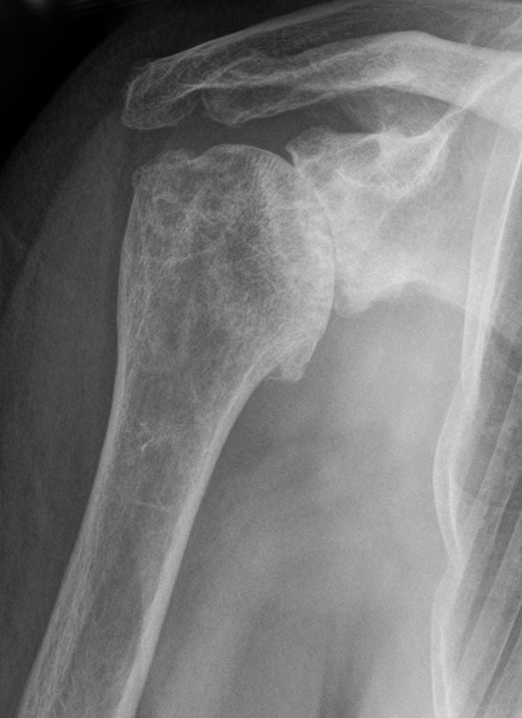
- AP (Grashey - True AP): Shows the true joint space. In OA, this space is obliterated ("Bone on Bone"). Look for subchondral cysts and sclerosis.
- Axillary Lateral: The most important view.
- Posterior Subluxation: The head sits behind the glenoid center line.
- Version: Estimating retroversion.
- Biconcavity: The tell-tale sign of B2 glenoids.
- Outlet: Assess the acromial shape (Type 1-3) and acromiohumeral distance (under 7mm suggests cuff tear).
- Indication: Mandatory for all arthroplasty planning in modern practice.
- Glenoid Version: Measured at the mid-glenoid level (Friedman method).
- Glenoid Inclination: Assessing superior/inferior tilt.
- Bone Stock: Assessing the depth of the glenoid vault to ensure it can support peg fixation (needs over 25mm usually).
- Classification: Defines the Walch type (A, B, or C).
- 3D Reconstructions: Used to generate Patient Specific Instrumentation (PSI) guides.
- Indication: If cuff strength is equivocal, or there is history of tear, or high-riding head on X-ray.
- Finding: Rotator cuff integrity is the binary switch for surgical decision making.
- Fatty Infiltration: Goutallier Grade 3 or 4 (fat greater than muscle) indicates an irreparable cuff. In this scenario, an Anatomic TSA is contraindicated because the "concavity compression" mechanism is lost.
Measuring Glenoid Version: Friedman vs Corrected Methods
The Investigations section states that version is "measured at the mid-glenoid level (Friedman method)," but how version is measured — and why the number can mislead — is a favourite examiner probe in B2 planning.
Friedman (2D axial) method. On an axial CT slice at the mid-glenoid, a reference line is drawn from the tip of the medial scapular border (the vault apex) to the centre of the glenoid face. Version is the angle between the glenoid articular line and the line perpendicular to that scapular axis; posterior tilt is reported as retroversion. It is simple and reproducible on a single slice.
Why it can mislead in arthritic glenoids:
- 2D versus 3D. A single axial slice is only truly accurate if the scan plane matches the scapular plane. Patient positioning tilts the scapula in the gantry, so raw 2D axial measurements tend to overestimate retroversion relative to scapular-plane-corrected 3D measurements. Correcting to the true scapular plane (3D reconstruction or reformatting) gives smaller, more reliable version values and underpins patient-specific planning.
- Paleoglenoid versus neoglenoid. In a biconcave B2 glenoid the surface is split into the native anterior paleoglenoid and the worn posterior neoglenoid. Measuring to the paleoglenoid rim (as the classic Friedman line tends to) captures the premorbid version, whereas measuring to the intermediate/neoglenoid surface captures the functional version the humeral head actually articulates against. The "corrected" or intermediate-glenoid technique references this functional surface, which is what determines how much retroversion must be neutralised.
This is why modern practice combines the modified Walch type with 3D, scapular-plane-corrected version and inclination rather than a single 2D number — it changes whether eccentric reaming alone will suffice or whether an augment or reverse TSA is needed.
Q: Why can a 2D axial (Friedman) retroversion measurement mislead in a B2 glenoid? A: It overestimates retroversion versus scapular-plane-corrected 3D measurement, and referencing the native anterior paleoglenoid reports premorbid rather than the functional version at the worn posterior neoglenoid. Plan from 3D-corrected version to decide eccentric reaming vs augment vs reverse TSA.
Management Algorithm

Conservative Management
First line for all patients, particularly those with mild symptoms or significant comorbidities.
- Education/Modification: Activity modification (avoiding heavy overhead lifting, push-ups).
- Physiotherapy:
- Range of Motion: Gentle stretching (pulleys, stick exercises) to prevent capsular contracture.
- Strengthening: Focus on periscapular stabilizers (Trapezius, Rhomboids, Serratus) and Deltoid.
- Avoid: Aggressive internal rotation stretching if painful.
- Analgesia:
- Oral: Paracetamol (Osteo), NSAIDs (Naproxen/Celecoxib) during flares.
- Topical: Diclofenac gel.
- Injections:
- Corticosteroid: Potent anti-inflammatory. Provides 3-6 months relief. Caution: Multiple injections can degrade soft tissue/bone quality. Do not inject within 3 months before elective arthroplasty — meta-analysis shows injection within 3 months raises periprosthetic joint infection risk, with no excess risk beyond 3 months (Akhtar et al, Cureus 2024).
- Hyaluronic Acid (Viscosupplementation): Lubricant effect. Evidence is mixed (AAOS Guidelines: Inconclusive). expensive.
- PRP (Platelet Rich Plasma): Growth factors. Current Level 1 evidence is weak/conflicting and it is not considered standard of care.
Pearl: Decision making regarding injections should be shared with the patient, balancing short term relief against long term risks.
Joint-Preserving Surgery: Comprehensive Arthroscopic Management (CAM)
The young, high-demand patient with end-stage glenohumeral OA is repeatedly flagged in this topic as the hardest reconstructive problem, and Comprehensive Arthroscopic Management (the CAM procedure) is the joint-preserving option named in that setting — yet its individual steps are what examiners probe. CAM is a single-sitting arthroscopic bundle that addresses every pain generator in the arthritic shoulder without burning arthroplasty bridges.
The components (all in one setting):
- Chondroplasty and debridement of unstable cartilage flaps, with removal of loose bodies.
- Synovectomy of inflamed synovium.
- Humeral osteoplasty — excision of the inferior humeral osteophyte (the "goat's beard") to restore clearance and relieve the mechanical adduction block.
- Capsular release (especially anteroinferior) to regain the external rotation lost to the fixed internal-rotation contracture.
- Axillary nerve neurolysis / decompression — the nerve lies immediately deep to the inferior osteophyte and capsule, so it is identified and protected during the osteoplasty and inferior release.
- Biceps tenotomy or tenodesis when the long head is a frayed pain source.
- Subacromial decompression / bursectomy where there is concomitant impingement, plus selective microfracture of small contained focal defects.
Selection matters more than technique. CAM relieves pain and delays arthroplasty best when there is preserved joint space and a concentric joint; a severely narrowed "bone-on-bone" glenohumeral space (in the region of 2 mm or less), large bipolar Walch-B posterior wear, or a grossly incongruent joint predicts early failure and progression to arthroplasty. Because it removes no bone stock that a later replacement needs, CAM is best framed to the patient as "buying time," not a cure.
Q: Name the key steps of Comprehensive Arthroscopic Management for young glenohumeral OA. A: Debridement/chondroplasty, loose-body removal, synovectomy, inferior humeral osteophyte excision (goat's beard) with axillary nerve neurolysis, capsular release for external rotation, biceps management, and selective subacromial decompression. A preserved joint space predicts success; a space of 2 mm or less predicts early failure.
Surgical Management
Anatomic Total Shoulder Arthroplasty
Gold Standard for primary OA with an intact rotator cuff.
Components:
- Humeral Head: Polished cobalt-chrome (or ceramic) head. Stemmed (standard), Short-stem, or Stemless (metaphyseal fixation).
- Glenoid: Ultra-high molecular weight polyethylene (UHMWPE). Usually cemented (pegged or keeled). Metal-backed glenoids have higher failure rates in aTSA.
Mechanism: Replicates anatomy. Relies on "Concavity Compression" by the intact rotator cuff to center the ball in the socket.
Survivorship: Excellent (90-95% at 10 years).
Patients under 50 years old are the most challenging. Arthroplasty has high failure rates (Poly wear, Loosening). Exhaust all non-operative measures. Consider Hemi or Arthroscopic Debridement (CAM).
Surgical Technique
Deltopectoral Approach
Standard approach for shoulder arthroplasty.
- Interval: Between Deltoid (Axillary N) and Pectoralis Major (Lat/Med Pectoral N).
- Vein: Cephalic vein retracted laterally with deltoid. Can be ligated if needed but best preserved.
- Landmarks: Coracoid (medial), Deltoid insertion (lateral), Bicipital groove (guides subscapularis).
- Incision Length: 12-15cm for anatomic TSA, can be extended for complex cases.
Pearl: The cephalic vein is the lighthouse of the interval. Always preserve it to minimize edema.
Complications
- Risk
- under 1%
- Management
- Washout vs Revision
- Pearl
- C. acnes is #1 cause
- Risk
- 1-2% per year
- Management
- Revision to Reverse
- Pearl
- Radiolucent lines common
- Risk
- 1-3%
- Management
- Repair or Reverse
- Pearl
- Avoid early active IR
- Risk
- Rare
- Management
- Observation
- Pearl
- Axillary N. most at risk
Deep Infection (PJI)
- Incidence: Under 1% for primary cases, higher for revision.
- Organism: Cutibacterium acnes (formerly Propionibacterium) is the dominant pathogen. It is a slow-growing anaerobe that colonizes the hair follicles (dermoglandular unit).
- Diagnosis: Difficult. ESR/CRP often normal. X-rays may show nonspecific loosening. Definitive diagnosis requires tissue culture held for 14 days.
- Prevention: Benzoyl peroxide prep pre-op, minimizing traffic, laminar flow, vancomycin powder (debated).
Glenoid Loosening
- Incidence: The most common mode of long-term failure in aTSA.
- Mechanism: "Rocking horse" phenomenon. If the head is not centered (due to cuff imbalance or uncorrected retroversion), eccentric loading occurs at the glenoid edge, toggling the component until the cement bond fails.
- Radiology: Radiolucent lines over 2mm around the pegs/keel.
Subscapularis Failure
- Mechanism: Rupture of the repair or failure to heal.
- Consequence: Anterior instability (dislocation), loss of active internal rotation, pain.
- Risk Factors: Aggressive early rehab, poor tissue quality, over-tensioning.
- Pectoralis Major Transfer: Salvage option for irreparable subscapularis.
Postoperative Care
Rehab Phases
- Sling: Worn day and night. Removed for hygiene and exercises.
- Restrictions:
- No active Internal Rotation (Protect Subscap repair).
- No lifting over 1kg ("Cup of tea").
- External Rotation limited to neutral or 30 degrees (depending on intra-op tension).
- Exercises:
- Pendulums.
- Passive Elevation (pulley/supine) to tolerance.
- Elbow/Wrist/Hand ROM.
- Sling: Wean off.
- Goals: Restore functional range.
- Exercises:
- Active Assist → Active Elevation.
- Hydrotherapy.
- Isometrics for cuff.
- Scapular control.
- Goals: Restore power and endurance.
- Exercises:
- Theraband resistance (IR/ER/Abd).
- Late return to gym (Chest press, Row etc).
- Return to Sport: Golf (Chip/Putt at 3-4m, Drive at 6m). Swimming. Tennis (Doubles preferred over Singles). Avoid heavy contact sports.
Outcomes and Prognosis
- Survival: ~90% at 10 years, 80% at 15 years.
- Function: aTSA provides better range of motion (internal/external rotation) compared to Reverse TSA.
- Constraint: aTSA allows the patient to do more "normal" activities but heavy loading is discouraged to protect the glenoid.
Guidelines, Registries & Global Practice
Global Epidemiology
- Symptomatic GHOA affects ~3-5% of adults over 60; radiographic disease is far more prevalent. Demand for shoulder arthroplasty is rising faster than for hip or knee, driven chiefly by expanding reverse TSA indications across high-income health systems.
- Younger, higher-demand patients (under 60) form a growing share of arthroplasty referrals worldwide and consistently show higher revision rates.
Society Guidance, Side by Side
- Position on Shoulder OA Management
- Evidence-based glenohumeral OA work supports NSAIDs and physiotherapy first; intra-articular corticosteroid offers short-term relief; viscosupplementation and PRP have inconclusive/limited evidence. TSA recommended over hemiarthroplasty when glenoid bone stock allows.
- Position on Shoulder OA Management
- Stepwise care: analgesia, activity modification and physiotherapy before surgery. Anatomic TSA for intact-cuff OA; reverse TSA for cuff-deficient or complex glenoid deformity. Emphasis on shared decision-making and realistic expectations.
- Position on Shoulder OA Management
- Supports 3D CT planning and the modified Walch classification for glenoid morphology; growing endorsement of reverse TSA for B2/B3/C deformity where anatomic glenoid fixation is at risk.
- Position on Shoulder OA Management
- Restore version and centre the head; correct B2 retroversion with eccentric reaming (limited) or augments; protect the axillary nerve during inferior release; robust subscapularis repair after anatomic TSA.
Registry Evidence (Joint Picture)
- Revision rate: National joint registries (AOANJRR, NJR UK, the Nordic registries) report cumulative revision of primary shoulder arthroplasty for OA in the region of ~5-10% at 10 years.
- Age effect: Younger patients (under 55) have roughly double the revision risk of patients over 75 — consistent across registries.
- Prosthesis choice: Total shoulder arthroplasty has lower revision rates than hemiarthroplasty for OA. Reverse TSA revision rates are broadly comparable to anatomic TSA in the short-to-medium term, but very long-term (over 15 years) data remain richest for anatomic TSA.
- Fixation: Cemented all-polyethylene glenoids show superior survivorship to uncemented metal-backed glenoids in anatomic TSA (metal-backed designs failed through polyethylene dissociation and construct over-stiffening).
High- vs Limited-Resource Practice Variation
- Well-resourced settings: Routine preoperative 3D CT planning, patient-specific instrumentation or navigation, and an increasing default to reverse TSA for elderly and deformed glenoids.
- Resource-limited settings: CT and modern implants may be scarce; hemiarthroplasty or non-operative management retains a larger role, and stemmed cemented implants are often preferred for availability and cost.
Exam-Ready Practice Points
- Quote a revision figure in the "~5-10% at 10 years" range and note the strong age effect.
- Understand why metal-backed glenoids fell out of favour (polyethylene dissociation, construct over-stiffening).
- Reverse TSA usage is expanding rapidly, but anatomic TSA remains the functional benchmark for the intact-cuff patient with adequate glenoid bone stock.
- Higher-volume surgeons and centres have lower complication rates (volume-outcome relationship).
- Indications for hemiarthroplasty are now narrow (very young patients for bone preservation, insufficient glenoid bone stock, or AVN with a spared glenoid).
Mnemonics
BADWalch B Glenoid
Hook:Walch B is BAD: Biconcave, Augment needed, Difficult case.
SIXAxillary Nerve Risk
Hook:Remember SIX: Subscap, Inferior capsule, X-ray.
RIPContraindications to aTSA
Hook:RIP: Don't do an anatomic TSA if the shoulder mechanisms are dead (Cuff/Deltoid).
MCQ Practice Points
Q: What is the primary deformity of the glenoid in Osteoarthritis? A: Retroversion (Posterior wear). This leads to posterior subluxation of the humeral head.
Q: Which X-ray view is essential for assessing glenoid version? A: Axillary Lateral. An AP view often underestimates posterior wear.
Q: What clinical sign suggests a rotator cuff tear in the setting of OA? A: Weakness/Lag or superior migration (high riding head) on X-ray. Stiffness is typical of OA; Weakness (Pseudoparalysis) suggests Cuff Tear Arthropathy.
Q: What is the most common organism in shoulder PJI? A: Cutibacterium acnes. It requires extended culture incubation (14 days).
Q: Why is active infection a contraindication for TSA? A: High recurrence. Infection must be cleared (antibiotics/debridement) before implantation.
At a Glance
- X-ray Features
- Osteophytes, Sclerosis, Posterior Wear
- Cuff Status
- Intact
- Key Pearl
- Stiff, grinding, posterior pain
- X-ray Features
- Femoralization of acromion, High riding head
- Cuff Status
- Torn (Massive)
- Key Pearl
- Weakness, Pseudoparalysis
- X-ray Features
- Central erosion (Medialization), Osteopenia
- Cuff Status
- Variable (often thinning)
- Key Pearl
- Bilateral, systemic symptoms
- X-ray Features
- Crescent sign, Collapse, Glenoid spared
- Cuff Status
- Intact
- Key Pearl
- Risk factors: Steroids, Alcohol
Clinical Decision Scenarios
Practise clinical reasoning and management decisions out loud
“A 60M presents with global loss of ROM. X-rays show mild OA. Discuss your differential.”
“You are planning a TSA for a B2 Glenoid. How do you manage the retroversion?”
“Counsel a patient on the specific risks of Anatomic TSA.”
“A 45M heavy labourer presents with severe primary OA. Intact cuff. He cannot work due to pain. Discuss your management strategy.”
Diagnosis
- Night Pain
- Loss of ER
- Crepitus
- Axillary X-ray (Posterior wear)
Classification
- Walch A (Concentric)
- Walch B (Posterior/Biconcave)
- Walch C (Dysplastic/Retroverted)
- Samilson-Prieto (Osteophytes)
Treatment
- Non-op first
- Intact Cuff → Anatomic TSA
- Cuff Tear → Reverse TSA
- Young → Hemi/Preservation
Complications
- Subscap Failure
- Glenoid Loosening
- Infection (C. acnes)
- Periprosthetic Fracture
Pearls
- B2 Glenoid needs correction
- Axillary nerve at risk inferiorly
- Protect subscap post-op
- Stiffness = OA; Weakness = Cuff
Evidence
- Total beats Hemi
- Pegged beats Keeled
- Osteotomy best for subscap
- Augments for B2
Evidence Base
Walch Glenoid Classification (Original Description)
- Type A (centered head, symmetric wear) in 59 percent, Type B (posterior subluxation, asymmetric posterior wear) in 32 percent, Type C (dysplastic retroversion over 25 degrees) in 9 percent.
- Humeral head position relative to the glenoid predicted the pattern of glenoid erosion.
- Posterior subluxation drives the exaggerated posterior wear of Type B glenoids.
Modified Walch Classification (3D CT)
- Added the B3 (monoconcave, retroversion at least 15 degrees or subluxation 70 percent) and D (anteversion) types and refined A2.
- Interobserver reliability improved from kappa 0.39 (original) to 0.70 with 3D CT and the modified system.
- Intraobserver reliability improved from 0.61 to 0.88.
Augmented Glenoid for B2/B3 Bone Loss
- Stepped augmented components restored anatomy in B2 glenoids with central peg osteolysis (10 percent) equivalent to non-augmented A1 glenoids (5 percent).
- B3 glenoids had significantly higher central peg osteolysis (29 percent) and more component medialization.
- Version and inclination were corrected without significant difference between groups.
Anatomic vs Reverse TSA for OA with Intact Cuff
- At a mean of 41 months, aTSA and rTSA had similar outcome scores, motion and satisfaction.
- aTSA achieved greater external rotation (exceeding the MCID).
- Complications were significantly higher with aTSA (4.9 vs 2.2 percent); revision rates were similar.
Subscapularis Management: LTO vs Peel
- Lesser tuberosity osteotomy (95 percent) and subscapularis peel (100 percent) had statistically equivalent CT healing rates.
- No significant difference in subscapularis fatty infiltration, strength or clinical scores at 1 year.
- Both techniques showed a small postoperative increase in fatty infiltration.
Shoulder Arthroplasty in Patients 50 Years or Younger
- Estimated survival was 84 percent (TSA) and 75 percent (hemi) at 20 years.
- Glenoid arthrosis after hemiarthroplasty and motion-limiting soft-tissue problems drove unsatisfactory outcomes.
- Pain relief and motion improved reliably, but many results were rated unsatisfactory long term.
Hemiarthroplasty vs Total Shoulder Arthroplasty (RCT)
- TSA provided significantly greater pain relief (p=0.002) and internal rotation (p=0.003) than hemiarthroplasty.
- 3 of 25 hemiarthroplasty patients required revision to resurface the glenoid.
- TSA added cost, operative time and blood loss but no TSA had been revised at follow-up.
Preoperative Corticosteroid Injection and PJI Risk
- Injection within 3 months of TSA raised periprosthetic joint infection risk (RR 1.12, 95% CI 1.04-1.20).
- No increased risk when the injection was given more than 3 months before surgery (RR 1.02).
- Three months is the safest interval between injection and arthroplasty.