Skeletal Fluorosis, Hypervitaminosis A/D, Aluminium Bone Disease
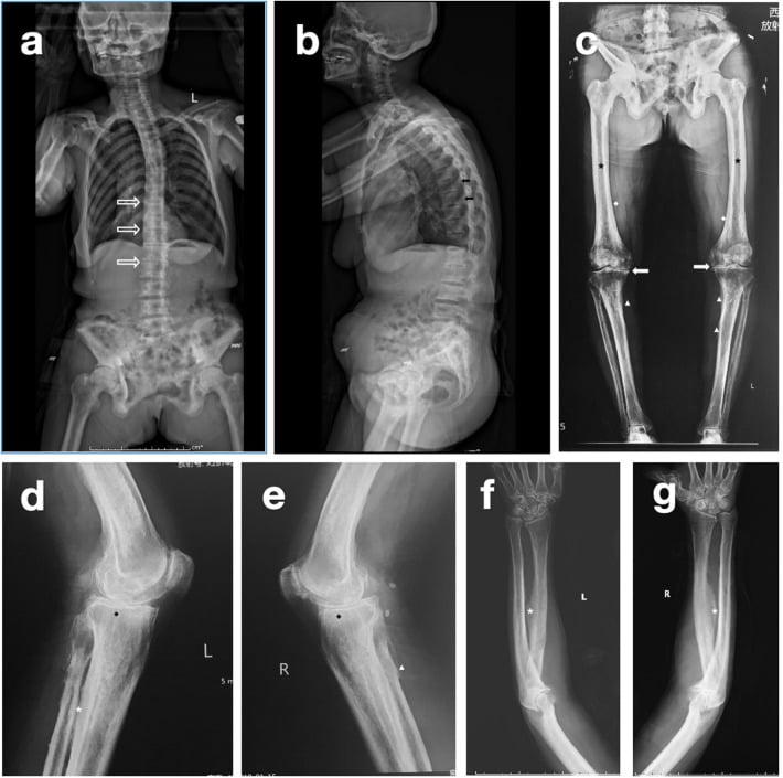
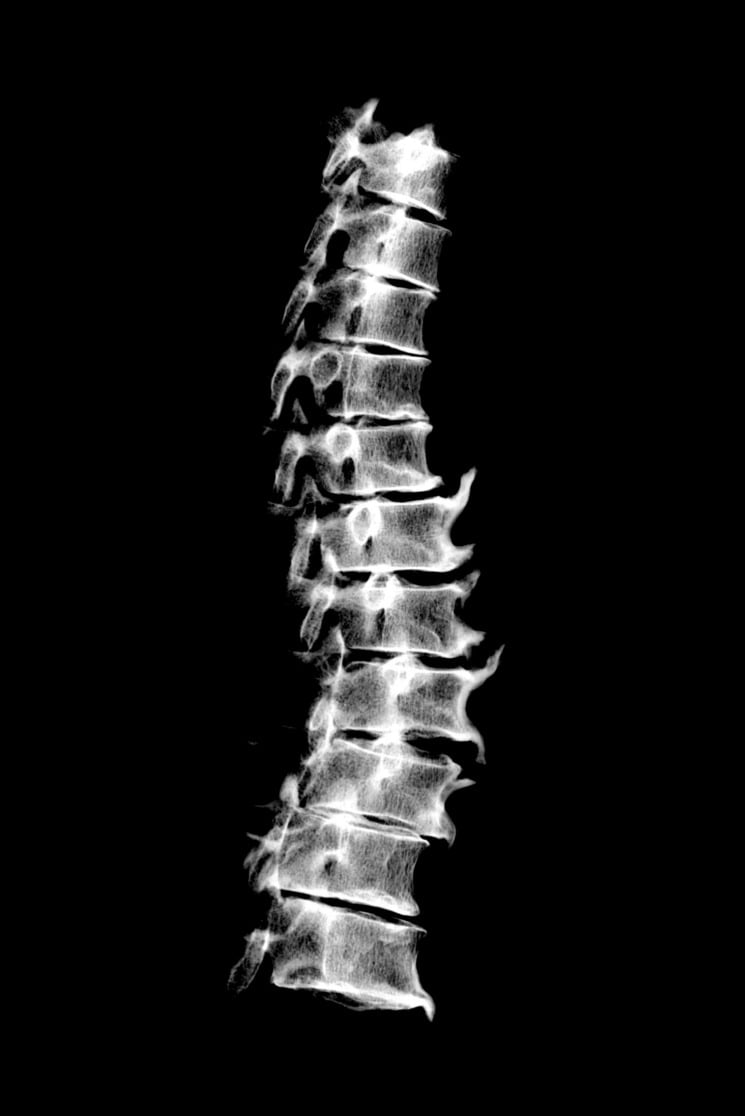
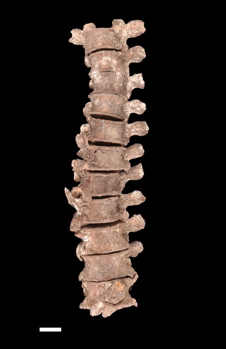
- SKELETAL FLUOROSIS is a toxic osteopathy from chronic EXCESS FLUORIDE - most often ENDEMIC (high-fluoride drinking water or coal-smoke exposure in certain regions), occasionally industrial or from surreptitious ingestion - characterised by dense OSTEOSCLEROSIS, OSSIFICATION of ligaments, tendons and INTEROSSEOUS MEMBRANES, periosteal new bone and osteophytosis, which can cause spinal canal/neural-foraminal stenosis and stiffness.
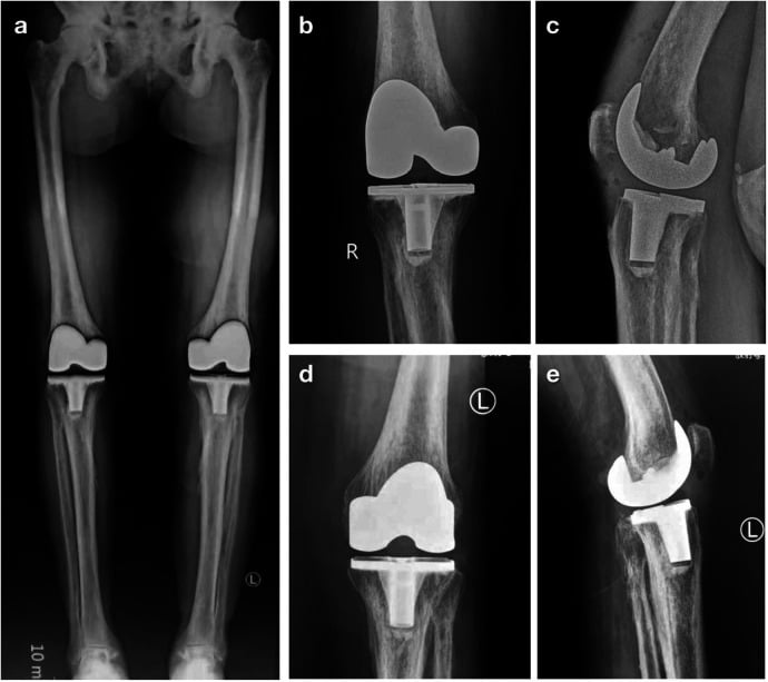
- The crucial PITFALL is that fluorotic bone, although radiographically DENSE (sclerotic), is STRUCTURALLY FRAGILE and of poor quality - there is a coexisting OSTEOMALACIC component - so 'dense' does not mean 'strong': Patients can show radiographic sclerosis yet have intra-operatively osteoporotic, fragile bone with calcified capsule/ligaments, increasing exposure difficulty and the risk of periprosthetic fracture during arthroplasty.
- HYPERVITAMINOSIS A causes bone pain with CORTICAL HYPEROSTOSIS and PERIOSTOSIS (particularly in children, with painful periosteal new bone), and can cause premature physeal closure and hypercalcaemia; HYPERVITAMINOSIS D causes HYPERCALCAEMIA and hyperphosphataemia with METASTATIC CALCIFICATION of soft tissues, vessels and kidneys (nephrocalcinosis) - the two vitamin excesses are distinguished by hyperostosis (A) versus hypercalcaemia/metastatic calcification (D).
- ALUMINIUM-related BONE DISEASE is a HEAVY-METAL osteopathy seen historically in renal-failure patients exposed to aluminium-containing phosphate binders or dialysate: aluminium deposits at the mineralisation front producing a LOW-TURNOVER (adynamic/osteomalacic) bone disease with bone pain, fractures and resistance to vitamin D - it is now rare with modern, aluminium-free dialysis but remains a classic teaching point in renal osteodystrophy.
- DIAGNOSIS rests on the EXPOSURE history plus the imaging pattern (axial osteosclerosis and ligament/interosseous ossification in fluorosis; hyperostosis in vitamin-A excess; hypercalcaemia/metastatic calcification in vitamin-D excess; osteomalacia in aluminium disease), supported by biochemistry (elevated serum/urine FLUORIDE in fluorosis; vitamin/calcium levels; and bone biopsy with aluminium staining where needed) - the differential of diffuse osteosclerosis also includes the sclerosing bone dysplasias, renal osteodystrophy, myelofibrosis and metastases.
- MANAGEMENT is to IDENTIFY and REMOVE the offending TOXIN - the definitive treatment - together with supportive/symptomatic care, correction of any coexisting osteomalacia, and surgical caution: because fluorotic 'dense' bone is fragile and the soft tissues ossified, arthroplasty and other surgery require careful planning, special exposure/release techniques and awareness of fracture risk.
- BE PRECISE ABOUT REVERSIBILITY - it is DECADES, not years. In the one case followed for a decade, urine fluoride fell within 3 months, serum fluoride normalised by 8 months and turnover markers by 14 months, and arthralgias resolved within 2 years - but at 9 years the lumbar Z-score had only fallen from +14.3 to +9.3, and bone fluoride at 8.5 years was still about TEN TIMES normal. The mineralisation defect (the osteomalacic component) does normalise; the sclerosis largely does not.
- WATCH THE KIDNEY DURING RECOVERY: new-onset NEPHROLITHIASIS developed within 9 months of removing the fluoride source and became chronic, as the mobilised fluoride load is excreted. Monitor for stones and renal function.
- KNOW THE DXA TRAP: in that patient Z-scores were +14.3 lumbar spine and +6.6 femoral neck but −0.6 at the DISTAL ONE-THIRD RADIUS. The sclerosis is AXIAL, so a peripheral cortical measurement can read as normal and falsely reassure.
- “Skeletal fluorosis = chronic fluoride excess → osteosclerosis + ligament/tendon/interosseous-membrane ossification + spinal stenosis. PITFALL: bone is DENSE but FRAGILE (osteomalacic component) - 'dense ≠ strong'.
- “Hypervitaminosis A = cortical hyperostosis/periostosis + bone pain (premature physeal closure in kids); hypervitaminosis D = hypercalcaemia + metastatic calcification. Aluminium = low-turnover osteomalacia in renal failure (now rare).
- “Diagnose by exposure history + imaging pattern + biochemistry (serum/urine fluoride). Treatment = REMOVE THE TOXIN; operate with caution on dense-but-fragile, ossified tissues.
- “Reversibility is measured in DECADES: serum fluoride normal by 8 months and markers by 14, but lumbar Z-score only fell +14.3 to +9.3 by 9 years and bone fluoride was still ~10x normal at 8.5 years. NEPHROLITHIASIS appeared within 9 months of removing the source.
- “DXA trap: sclerosis is AXIAL - Z-scores +14.3 spine and +6.6 femoral neck but −0.6 at the distal one-third radius. Endemic in 25+ countries (China, India worst); genetic background modifies susceptibility at equal exposure.
Fluorotic bone looks dense/sclerotic on radiographs but is structurally fragile (osteomalacic component), with ossified ligaments/capsule. At surgery: difficult exposure, fragile bone, high periprosthetic-fracture risk.
Fluoride = osteosclerosis + ligament/interosseous ossification. Vitamin A = hyperostosis. Vitamin D = hypercalcaemia + metastatic calcification. Aluminium = low-turnover osteomalacia (renal failure).
Skeletal Fluorosis
Skeletal fluorosis results from chronic excess fluoride (endemic high-fluoride water or coal smoke; occasionally industrial or surreptitious ingestion). It produces dense osteosclerosis, ossification of ligaments, tendons and interosseous membranes, periosteal new bone and osteophytosis, which can cause spinal stenosis and stiffness. The key pitfall: the dense bone is structurally fragile (a coexisting osteomalacic, low-quality component), so it can be radiographically sclerotic yet intra-operatively osteoporotic and fragile, with calcified capsule/ligaments - increasing exposure difficulty and periprosthetic-fracture risk during arthroplasty. Elevated serum/urine fluoride confirms exposure. The disease is reversible but over decades once the source is removed - biochemistry corrects within months, but bone fluoride remained about ten times normal at 8.5 years in the one case followed that long, and nephrolithiasis can appear during the recovery phase.
Management
- Identify and remove the offending toxin - the definitive treatment (stop the fluoride source/vitamin supplement; switch to aluminium-free binders/dialysate). Skeletal fluorosis is reversible over decades, not years: biochemistry corrects in months, symptoms within about 2 years, but bone fluoride was still roughly ten times normal at 8.5 years in the one case followed that long.
- Monitor for NEPHROLITHIASIS during recovery - new stones appeared within 9 months of removing the source in that case and became a chronic problem; check renal function too.
- Correct coexisting osteomalacia and treat symptoms (analgesia; physiotherapy for stiffness).
- Surgical caution: dense-but-fragile fluorotic bone and ossified soft tissues make exposure difficult and fractures more likely - plan carefully, use special exposure/release techniques, and anticipate periprosthetic fracture risk in arthroplasty.
- Address complications: decompress symptomatic spinal stenosis; manage hypercalcaemia (vitamin-D excess).
The orthopaedic trap in skeletal fluorosis is to equate radiographic density with mechanical strength. Fluorotic bone is sclerotic on imaging but has a coexisting osteomalacic component that leaves it structurally fragile, and the surrounding capsule, ligaments and tendons are often calcified or ossified. As reported, a patient with preoperative radiographic sclerosis can prove to have osteoporotic, fragile bone at operation, with stiff, calcified soft tissues that make exposure difficult and raise the risk of periprosthetic fracture during, for example, total knee arthroplasty. The practical lessons are to evaluate bone quality and soft-tissue flexibility preoperatively in patients from endemic areas, to anticipate difficult exposure and use appropriate release techniques, and to handle the bone gently - treating it as fragile despite its dense appearance.
Why Fluoride Makes Bone Dense Yet Weak
- Fluoride stimulates osteoblasts. Fluoride is a direct osteoblast mitogen, driving bone formation and the increased density - which is why it was once trialled for osteoporosis; but the bone laid down is abnormal in quality (fragile), so density rises without a matching gain in strength.
- Fluorapatite and the mineralisation defect. Fluoride substitutes into hydroxyapatite to form fluorapatite - larger, less soluble, more acid-resistant crystals (the basis of caries prevention) - but the rapid, abnormal mineralisation leaves a defect with excess osteoid (the osteomalacic component), giving the 'dense but weak' paradox.
- Secondary hyperparathyroidism and the dose spectrum. Fluoride also binds calcium, tending to secondary hyperparathyroidism. Lower chronic exposure during tooth development causes dental fluorosis (mottled enamel) in children; higher cumulative exposure over years causes skeletal fluorosis in adults.
Q: Why does fluoride make bone dense yet weak?
A: Fluoride is an osteoblast mitogen → more bone formation/density (once trialled for osteoporosis), but the bone is abnormal-quality/fragile. Fluoride substitutes into hydroxyapatite → fluorapatite (larger, less soluble, acid-resistant - caries prevention), but the abnormal rapid mineralisation leaves a defect with excess osteoid (the osteomalacic component) = 'dense but weak'. Fluoride also binds calcium → secondary hyperparathyroidism. Dose: dental fluorosis (children, tooth development) vs skeletal fluorosis (adults, cumulative).
Lead Lines and the Heavy-Metal Osteopathies
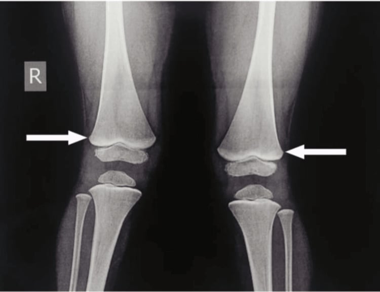
- Lead lines. In chronically lead-exposed growing children, dense transverse metaphyseal bands appear at the fastest-growing metaphyses (distal femur, proximal tibia, distal radius). The density is not deposited lead but disordered metaphyseal remodelling - lead inhibits osteoclastic resorption of the calcified cartilage, leaving a band of retained mineral; adults with mature skeletons do not form them. They are a radiographic marker of chronic exposure (plumbism).
- Plumbism. Lead lines accompany the systemic features of lead poisoning: abdominal colic, encephalopathy (children), motor neuropathy (wrist/foot drop in adults), microcytic anaemia with basophilic stippling, a gum (Burton) line and nephropathy.
- Other heavy metals. Historic examples include phosphorus ('phossy jaw' - osteonecrosis of the mandible in match workers) and bismuth (metaphyseal bands like lead lines).
Q: What are lead lines, and how do they differ from fluorosis?
A: Lead lines = dense transverse metaphyseal bands in growing children (fast-growing metaphyses - distal femur/proximal tibia/distal radius); the density is not deposited lead but disordered metaphyseal remodelling (lead inhibits osteoclastic resorption → retained mineral); adults do not form them - a marker of chronic plumbism (colic, encephalopathy, motor neuropathy, basophilic-stippling anaemia, Burton gum line). Contrast fluorosis = diffuse axial osteosclerosis + ligament/interosseous ossification in adults. (Historic: phosphorus 'phossy jaw', bismuth lines.)
How Reversible Is It Really? The Decade of Follow-Up
"Partially reversible over years" is the usual teaching, and the one case with a decade of documented follow-up shows it is true but far slower and less complete than that phrase suggests. A 52-year-old man with axial osteosclerosis and neck immobility, whose fluoride source proved to be surreptitious toothpaste ingestion, was monitored for ten years after the source was removed.
- Biochemistry corrects in months. Urine fluoride fell from 26 mg/L to 16 within 3 months and 3.9 at 14 months; serum fluoride normalised within 8 months; bone turnover markers corrected by 14 months. So the laboratory picture improves quickly and can mislead you into thinking the skeleton has recovered.
- The skeleton takes decades. At 9 years, lumbar spine bone density had fallen 23.6% and femoral neck 15.1% - yet the Z-scores were still +9.3 and +4.8, having started at +14.3 and +6.6. Bone fluoride at 8.5 years was still 1.15% against a reference of less than 0.1% - a 36% reduction, but still roughly ten times normal. Radiographs showed decreased trabecular sclerosis and some reduction in sacrospinous ligament ossification, not resolution.
- The osteomalacia is quantifiable and it does recover. The initial biopsy showed cancellous bone volume 4.5 times, cortical width 3.2 times, and osteoid thickness 25 times the reference mean, with wide diffuse tetracycline uptake confirming osteomalacia. By 8.5 years osteoid surface and thickness had normalised with distinct double labels - so the mineralisation defect, the part that makes the bone fragile, is the component that actually reverses.
- Symptoms resolved and he never fractured. All arthralgias resolved within 2 years.
- But watch the kidney. New-onset nephrolithiasis appeared within 9 months of stopping the fluoride and became a chronic problem, with creatinine drifting from 1.0 to 1.3 mg/dL over the decade. Patients should be monitored for stones during the recovery phase - the mobilised fluoride load has to be excreted.
Everything in this section comes from a single case report, which the authors published precisely because no data on reversibility existed. It establishes the sequence and the order of magnitude - biochemistry in months, skeleton over decades, stones as a recovery-phase hazard - but it cannot tell you what proportion of patients recover, how completely, or how often nephrolithiasis follows. No cohort of treated skeletal fluorosis with serial imaging has been published.
Why Two People Drinking the Same Water Differ
Exposure is necessary but not sufficient, and this is worth knowing because it explains the clinical observation that individuals in the same village with the same water supply are affected to very different degrees.
- The scale of the problem. Fluorosis is endemic in at least 25 countries, with China and India the worst affected, and dental, skeletal and non-skeletal forms affect millions of people. Skeletal fluorosis is rare in the United States and western Europe - which is why it is easily missed there, and why the exposure history matters more than the radiograph.
- Genetic susceptibility modifies risk. Genetic-epidemiological studies have associated polymorphisms in candidate genes - COL1A2, the calcitonin receptor, the oestrogen receptor, COMT, GSTP1, MMP-2, prolactin, the vitamin D receptor and myeloperoxidase - with differing susceptibility among individuals in the same community under the same environmental exposure.
- How firm is this? These are association studies in candidate genes, a design notoriously prone to false-positive findings and poor replication. Treat the principle - that host genetic background modifies susceptibility at equal exposure - as reasonable, and treat any individual gene on that list as unconfirmed. No genetic test is used clinically, and susceptibility genotype does not change management.
The Other Toxic Osteopathies
- Bone/imaging pattern
- Osteosclerosis + ligament/interosseous ossification; dense-but-fragile
- Biochemistry / key point
- Elevated serum/urine fluoride; endemic exposure; partially reversible
- Bone/imaging pattern
- Cortical hyperostosis/periostosis; premature physeal closure (children)
- Biochemistry / key point
- Bone pain; +/- hypercalcaemia; stop the supplement
- Bone/imaging pattern
- Metastatic (soft-tissue/vascular/renal) calcification
- Biochemistry / key point
- Hypercalcaemia + hyperphosphataemia; nephrocalcinosis/stones
- Bone/imaging pattern
- Low-turnover (adynamic/osteomalacic) bone; fractures
- Biochemistry / key point
- Aluminium at mineralisation front; now rare; bone biopsy/aluminium stain
Diffuse increased bone density is not only toxic: also consider the sclerosing bone dysplasias (osteopetrosis, pycnodysostosis, melorheostosis), renal osteodystrophy ('rugger-jersey' spine), myelofibrosis, and osteoblastic metastases/lymphoma. The exposure history, ligament/interosseous ossification and biochemistry point to fluorosis.
Mnemonics & Memory Aids
FLUORIDE
Hook:FLUORIDE: Fluoride excess, Ligament ossification, Unstrong bone, Osteosclerosis, Remove the source, Intra-op caution, Distinguish vit A/D/aluminium, Elevated fluoride.
Clinical Decision Scenarios
Practise clinical reasoning and management decisions out loud
“A patient from a region with high-fluoride water has dense, sclerotic bones and ossified forearm interosseous membranes. What is the diagnosis, and what does it mean for surgery?”
Skeletal fluorosis
- Chronic fluoride excess (endemic water/coal smoke)
- Osteosclerosis + ligament/tendon/interosseous-membrane ossification + spinal stenosis
- Trap: dense but fragile (osteomalacic); confirm with serum/urine fluoride
- Sclerosis is AXIAL - a distal-radius DXA can read normal (Z −0.6) while the spine is +14.3
- Endemic in at least 25 countries (China and India worst affected); genetic background modifies susceptibility
Vitamin excesses
- Hypervitaminosis A: cortical hyperostosis/periostosis, bone pain, premature physeal closure
- Hypervitaminosis D: hypercalcaemia + metastatic calcification (nephrocalcinosis/stones)
- Distinguish: hyperostosis (A) vs hypercalcaemia/metastatic calcification (D)
Aluminium bone disease
- Renal failure + aluminium binders/dialysate (now rare)
- Low-turnover (adynamic/osteomalacic) bone, fractures, vitamin-D resistance
- Bone biopsy with aluminium stain
Management
- Identify and REMOVE the toxin (definitive treatment)
- Fluorosis reverses over DECADES: serum F normal by 8 months, markers by 14, symptoms by 2 years
- But lumbar Z-score only +14.3 to +9.3 by 9 years; bone fluoride still ~10x normal at 8.5 years
- MONITOR FOR NEPHROLITHIASIS during recovery (appeared within 9 months) and check renal function
- Correct coexisting osteomalacia; treat symptoms/complications
- Operate with caution on dense-but-fragile, ossified tissues (fracture risk)
Evidence & Key Studies
Radiographic sclerosis with intra-operative fragile bone in skeletal fluorosis
- Skeletal fluorosis is a rare toxic osteopathy from prolonged ingestion/inhalation of large amounts of fluoride; radiographs show increased bone density, thickened/fused trabeculae, thickened cortex and ossification of many ligaments and interosseous membranes.
- Despite radiographic sclerosis, severe osteoporosis with calcification of the capsule, ligaments and tendons was found intra-operatively, increasing exposure difficulty and periprosthetic-fracture risk during total knee arthroplasty.
- It is easily misdiagnosed; bone quality and soft-tissue flexibility should be evaluated preoperatively in patients from endemic areas, with special exposure and release techniques.
Recovery from skeletal fluorosis after removal of the fluoride source
- A SINGLE CASE REPORT with a decade of follow-up - published because, as the authors state, there were NO data on the reversibility of skeletal fluorosis. It gives a timeline and orders of magnitude, not rates.
- A 52-year-old American man with 7 years of neck immobility and joint pain had axial osteosclerosis. Fluoride was elevated in serum (0.34 and 0.29 mg/L, reference less than 0.20), urine (26 mg/L, reference 0.2-1.1) and iliac crest bone (1.8%, reference less than 0.1%). Tap and bottled water were negative - surreptitious TOOTHPASTE ingestion was the most plausible source.
- STRIKING SITE DISSOCIATION ON DXA: Z-scores were +14.3 at the lumbar spine and +6.6 at the femoral neck but −0.6 at the distal one-third radius - the sclerosis is axial, and a peripheral cortical site can read as entirely normal.
- Biopsy quantified the paradox: cancellous volume 4.5 times, cortical width 3.2 times, and OSTEOID THICKNESS 25 TIMES the reference mean, with wide diffuse tetracycline uptake documenting osteomalacia - dense bone with a gross mineralisation defect.
- RECOVERY RAN AT TWO SPEEDS. Urine fluoride fell 26 to 16 mg/L within 3 months and to 3.9 at 14 months, normalising (1.2) only at 9 years; serum fluoride normalised in 8 months; markers corrected by 14 months; arthralgias resolved within 2 years and he never fractured. But at 9 years lumbar and femoral BMD had fallen only 23.6% and 15.1%, with Z-scores still +9.3 and +4.8, and bone fluoride at 8.5 years was still 1.15% - a 36% fall but STILL ABOUT TEN TIMES the reference. Osteoid surface and thickness did normalise, with double labels.
- HARM DURING RECOVERY: new-onset nephrolithiasis appeared within 9 months of stopping fluoride and became a chronic problem; creatinine drifted from 1.0 to 1.3 mg/dL over the decade. The authors conclude fluorosis is reversible but likely impacts for DECADES, and that patients should be monitored for stones.
The genetic influence in fluorosis
- A NARRATIVE REVIEW of genetic association studies - it aggregates candidate-gene findings and performs no primary analysis.
- Fluorosis is endemic in at least 25 countries, with China and India the worst affected; dental, skeletal and non-skeletal forms affect millions of people.
- Genetic association studies report that polymorphisms in candidate genes - COL1A2, calcitonin receptor, oestrogen receptor, COMT, GSTP1, MMP-2, prolactin, vitamin D receptor and myeloperoxidase - may increase or decrease fluorosis risk among individuals with the SAME environmental exposure in the same community.
- Limitations that matter: candidate-gene association studies are prone to false-positive results and frequently fail replication, and no effect sizes, populations or replication status are given in the abstract. The general principle that host genetic background modifies susceptibility is reasonable; individual gene claims should be treated as unconfirmed. No genetic test is used clinically and genotype does not alter management.
The radiographic pattern and the 'dense but fragile' intra-operative reality with its arthroplasty implications come from Hou (DOI). The reversibility timeline, the DXA Z-scores (+14.3 lumbar, +6.6 femoral neck, −0.6 distal radius), the biopsy figures including osteoid thickness 25 times reference, the bone-fluoride persistence at 8.5 years and the new-onset nephrolithiasis come from Kurland (DOI). The endemicity in at least 25 countries and the candidate-gene susceptibility associations come from Pramanik (DOI). The features of hypervitaminosis A and D, aluminium-related low-turnover renal bone disease, the lead lines and heavy-metal osteopathies, the fluorapatite mechanism, and the differential of diffuse osteosclerosis are standard, well-established teaching.
What does not exist: both fluorosis papers cited here are single case reports, and no cohort of skeletal fluorosis with serial imaging or treated follow-up has been published - so there is no published reversal rate, no proportion who recover, no incidence of nephrolithiasis during recovery, and no validated staging system. There is no fracture-risk figure and no evidence base for the timing or technique of arthroplasty in fluorotic bone; the surgical advice is extrapolated from a single operated patient plus general principles. The genetic associations are candidate-gene studies that have not been consistently replicated and are not used clinically.