Component-Based Z-Deformity
- Triad: Forefoot Adduction, Midfoot Abduction, Hindfoot Valgus
- Differentiation: From Clubfoot (Hindfoot Varus) and MA (Hindfoot Neutral)
- Radiographic Signs: Increased Talocalcaneal Angle + Adducted Metatarsals
- Surgical principle: correct only the components that remain symptomatic, rigid and structurally abnormal after full assessment
- Talonavicular relationship: quantify midfoot translation and uncoverage rather than assuming one universal subluxation pattern
- “Beware the 'Metatarsus Adductus' that doesn't get better with casting
- “Check the hindfoot! If it's valgus, it's Skewfoot
- “Avoid simple metatarsal osteotomies alone - must address hindfoot
- “Look for the 'Z' shape on weight-bearing X-ray
Overview/Epidemiology
Skewfoot is a complex, often misunderstood deformity that combines elements of flatfoot and metatarsus adductus. The foot takes a "Z" or serpentine shape, with a deformity at three levels:
- Forefoot adducted, as in metatarsus adductus
- Midfoot abducted, the navicular translated laterally
- Hindfoot in valgus (everted)
The hindfoot valgus and midfoot abduction are integral parts of the deformity, and that is what separates skewfoot from simple metatarsus adductus. Rigidity and Achilles or gastrocnemius contracture vary.
Aetiology. Primary, neurogenic, syndromic and postoperative or iatrogenic forms have been described, and rare familial cases are reported. Historical reports blamed forefoot casting without hindfoot control, but the proportion that is genuinely iatrogenic is unknown.
What the primary literature says. The 1986 description that named the entity treats skewfoot and serpentine foot as differences in degree from congenital metatarsus adductus rather than a separate disease. It attributes the aetiology to intrauterine factors, a narrow uterus and internal rotation of the foot and lower leg, which is a primary, congenital account. A 2020 review of congenital forefoot pathology likewise classifies metatarsus adductus and skewfoot as deformations arising in the fetal period on a foot that was configured normally, again placing the origin before any treatment.
The iatrogenic mechanism. It is genuinely plausible. Abducting a forefoot without a three-point mould that holds the heel levers an unstabilised hindfoot into valgus, which is exactly how a cast can worsen the foot, so the warning about casting technique is sound and should be taught. What is not sound is turning a plausible mechanism into a confident figure: do not imply a congenital "germ plasm defect" or a 50/50 aetiological split without genetic or epidemiological evidence. The two causes are hard to separate because the deformity is often misdiagnosed as simple metatarsus adductus at the outset.
Most cases are probably primary, some may be iatrogenic or at least aggravated by poor casting, and the honest answer names the uncertainty. An examiner rewards a candidate who says the evidence base is Level IV to V far more than one who quotes a percentage that no study produced.
Terminology. The deformity has been called congenital metatarsus varus, serpentine foot, Z-foot and skewfoot. Modern assessment describes the forefoot, midfoot and hindfoot components explicitly, because the name alone does not determine treatment.
Pathophysiology, Anatomy & Biomechanics
Hindfoot. The calcaneus is in valgus and eversion. The talus is plantarflexed, though not as severely as in congenital vertical talus, and the sustentaculum tali may be hypoplastic.
Talonavicular joint. The navicular is laterally subluxated on the talar head. In simple metatarsus adductus the navicular is medial or neutral; Berg's complex metatarsus adductus already has a laterally translated navicular, so it is the increased talocalcaneal angle of hindfoot valgus that finally marks skewfoot (Classification).
Tarsometatarsal (Lisfranc) joints. The metatarsals are adducted relative to the cuneiforms and cuboid. The result is a zig-zag: the talus points medially, the midfoot shifts laterally, and the forefoot points medially again.
The Achilles tendon. It is usually shortened, and it acts as a deforming force: the calcaneus everts to dorsiflex, which adds to the hindfoot valgus.
Biomechanics. The foot is mechanically unstable. The ground reaction force passes lateral to the subtalar axis and perpetuates the valgus. In stance the midfoot collapses further into abduction while the forefoot adduction forces the foot to roll over its lateral border, which causes:
- A prominent medial talar head, which becomes a pressure area
- A lateral border callosity, from weight bearing on the base of the fifth metatarsal
- An inefficient lever arm, the triceps surae losing its mechanical advantage
Classification Systems
Berg classification. Based on AP weight-bearing radiographs. Berg's original study (Evidence Base) described four radiographic configurations; the working three-type version reads the metatarsals, the talocalcaneal angle and the navicular.
- Metatarsals
- Adducted
- Talocalcaneal angle
- Normal
- Navicular
- Central
- Metatarsals
- Adducted
- Talocalcaneal angle
- Normal
- Navicular
- Laterally translated
- Metatarsals
- Adducted
- Talocalcaneal angle
- Increased (valgus)
- Navicular
- Laterally subluxated
Types II and III both have a laterally displaced navicular; the increased talocalcaneal angle is what makes Type III.
Clinical severity. Flexible feet, which can be passively corrected, are rare. Rigid feet, with fixed deformities and painful callosities, are common.
Clinical Assessment
History. Often a child treated for "metatarsus adductus" that "didn't get better" or "looks worse". Parents report that the foot looks flat but the toes point in. Older children have pain, at the sinus tarsi from the valgus or along the lateral border (bunionette), and shoe fitting is difficult because of the C- or Z-shape.
Examination. Standing, the foot shows its serpentine shape: the heel in valgus when checked from behind, the midfoot prominent medially where the talar head lies, and the forefoot adducted. Then assess:
- Subtalar joint - often stiff or restricted
- Ankle - equinus, with the Silfverskiöld test
- Midfoot - how rigid the adduction is
- Callosities - at the two pressure points described under Biomechanics
- Shoes - medial wear on the heel counter, lateral wear on the sole
Investigations
Weight-bearing radiographs. AP and lateral weight-bearing films are the diagnostic study, and each view carries its own measurements:
- Measurement
- Talocalcaneal (Kite's) angle
- In skewfoot
- Increased (greater than 35-40 degrees), indicating hindfoot valgus
- Measurement
- Talus-first metatarsal angle
- In skewfoot
- Broken: the line through the talus passes medial to the first metatarsal
- Measurement
- Metatarsus adductus angle
- In skewfoot
- Increased
- Measurement
- Talar declination
- In skewfoot
- Increased: the talus points down
- Measurement
- Calcaneal pitch
- In skewfoot
- Decreased (flatfoot)
- Measurement
- Meary's angle
- In skewfoot
- Broken, in extension (dorsiflexion) at the talonavicular joint
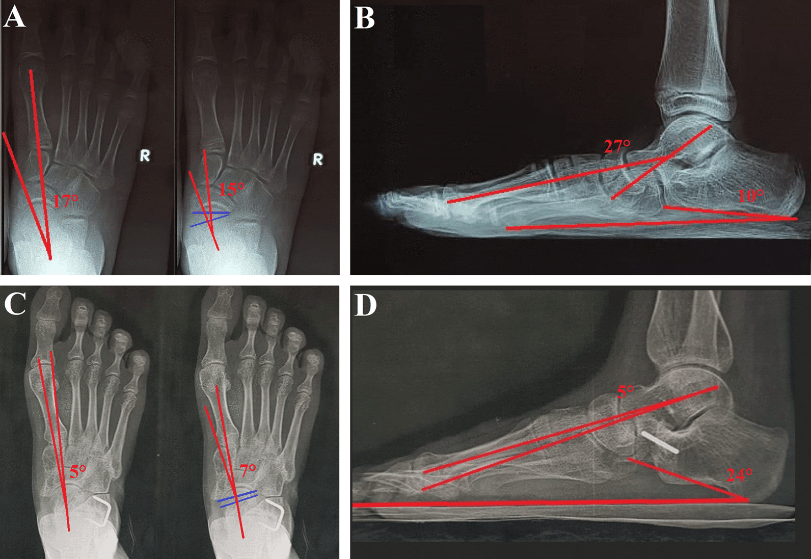
Measuring the Z. No single angle defines skewfoot, so the limbs are measured separately and read together:
- AP talocalcaneal angle - reflects talar-calcaneal divergence; an increased value supports hindfoot valgus but is not diagnostic by itself
- Sgarlato metatarsus adductus angle - defines the lesser-tarsal axis and relates it to the second metatarsal, quantifying the forefoot adduction, the distal limb of the Z
- Talonavicular incongruency angle - quantifies lateral joint mismatch rather than relying on a visual Z
- Talonavicular uncoverage - quantifies lateral navicular displacement over the talar head
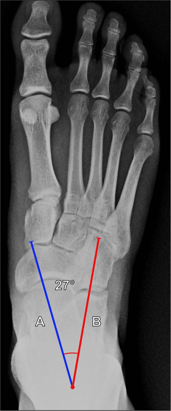
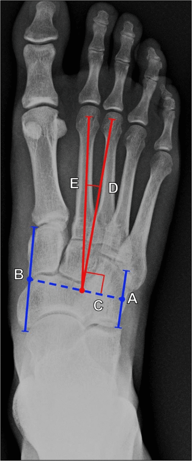
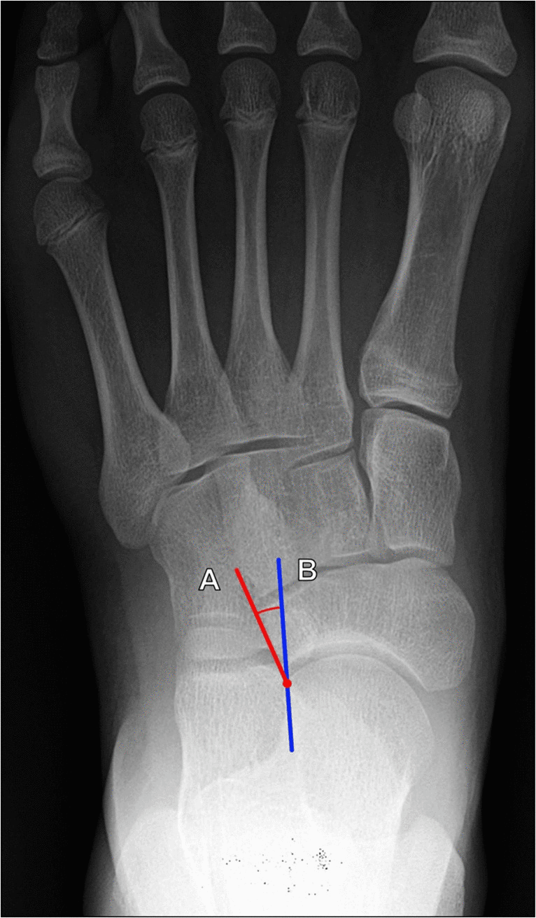
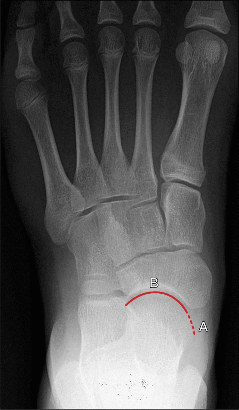
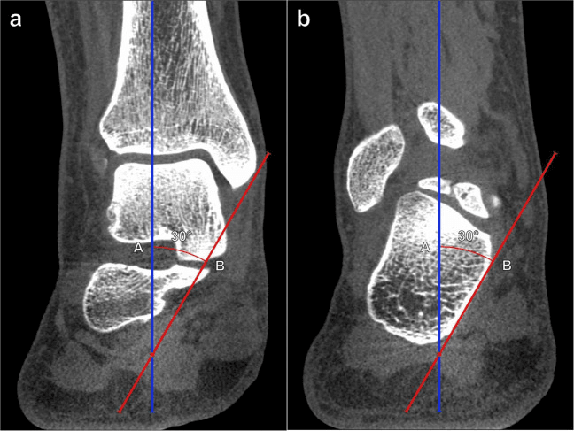
CT. Useful for surgical planning in adolescents, including exclusion of a tarsal coalition, and it can characterise the specific deformity of the medial cuneiform. On coronal cone-beam CT the tibiocalcaneal angle measures hindfoot valgus independently of the forefoot adduction, so the two components can be separated before an osteotomy is chosen.
Detailed Differential Diagnosis
Do not confuse skewfoot with clubfoot or metatarsus adductus: the forefoot is adducted in all three, so check the heel. "If the heel is in valgus and the toes point in, think SKEW."
- Forefoot
- Adducted
- Midfoot (Navicular)
- Medial / Central
- Hindfoot (Calcaneus)
- Neutral / Mild Valgus
- Ankle
- Normal
- Key Differentiator
- Flexible hindfoot; kidney-bean shape
- Forefoot
- Adducted
- Midfoot (Navicular)
- Lateral Subluxation
- Hindfoot (Calcaneus)
- Valgus (fixed, severe); plantarflexed talus
- Ankle
- Equinus often present
- Key Differentiator
- Z-deformity (serpentine)
- Forefoot
- Adducted
- Midfoot (Navicular)
- Medial Dislocation
- Hindfoot (Calcaneus)
- Varus (Fixed)
- Ankle
- Equinus (Rigid)
- Key Differentiator
- Hindfoot varus; small calf, stiff
- Forefoot
- Abducted (dorsiflexed)
- Midfoot (Navicular)
- Dorsolateral Dislocation
- Hindfoot (Calcaneus)
- Valgus (Severe)
- Ankle
- Equinus (Rigid)
- Key Differentiator
- Rocker bottom / vertical talus
- Forefoot
- Abducted
- Midfoot (Navicular)
- Sags Plantar
- Hindfoot (Calcaneus)
- Valgus
- Ankle
- Normal / Equinus
- Key Differentiator
- Forefoot abduction
Why the distinction matters. Treating skewfoot like metatarsus adductus, with casting, causes iatrogenic worsening. Treating it like clubfoot is ineffective, because the hindfoot is already in valgus and Ponseti corrects varus. The Dobbs approach to vertical talus is closer to the mark, but the forefoot deformity is the opposite.
Coalition. A coalition can create rigid hindfoot valgus that mimics one component of skewfoot. Cross-sectional imaging is useful when subtalar stiffness or an atypical clinical course suggests it.
Management Algorithm
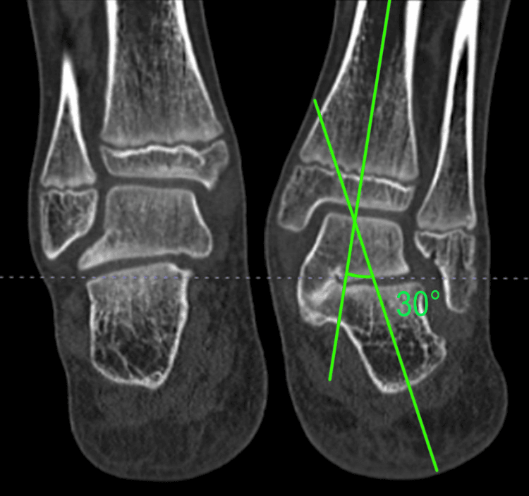
- Confirm the geometry on weight-bearing AP/lateral radiographs when possible, documenting forefoot adduction, talonavicular/midfoot abduction, hindfoot valgus and the apex of each deformity.
- Exclude mimics and associations: metatarsus adductus, clubfoot, vertical talus, planovalgus, coalition and neuromuscular/syndromic disease.
- Assess symptoms and flexibility: shoe conflict, callosity, pain, gait, subtalar motion, Achilles/gastrocnemius tightness and passive correction.
- Observe when appropriate. An asymptomatic flexible foot does not require surgery for appearance alone.
- Use orthoses or carefully moulded casting selectively. Symptom relief and flexibility may improve, but poorly controlled forefoot correction can worsen valgus, and evidence for altering the natural history is limited.
- Reconstruct only the demonstrated deformity. Hindfoot valgus, midfoot/forefoot adduction, equinus and coalition each require their own decision.
- Reassess on the table. Avoid stacking Evans, MDCO, Cotton and metatarsal osteotomies as a routine bundle; each additional procedure must correct a persistent measured component.
There is no validated “older than six years” trigger and no requirement to perform both ends of the Z in every case. Symptoms, rigidity, apex, residual alignment and family goals determine whether and how to reconstruct.
Choosing the Hindfoot Procedure: Lengthening vs Medial Displacement
The management menu offers an Evans calcaneal lengthening and a medial displacement calcaneal osteotomy (MDCO) as if they were interchangeable, but in skewfoot they correct different things, and choosing wrongly leaves residual deformity. The skewfoot hindfoot is a valgus heel plus midfoot abduction, with lateral subluxation of the navicular and uncoverage of the talar head.
Lateral column lengthening. An Evans-type calcaneal lengthening lengthens the anterior calcaneus. That pushes the navicular medially, swinging the abducted midfoot back into adduction, re-covers the talar head and corrects the heel valgus, so one cut addresses all three of these hindfoot and midfoot elements. It is the workhorse hindfoot procedure for skewfoot, and in Mosca's work (Evidence Base) calcaneal lengthening combined with a medial cuneiform osteotomy corrected all components while preserving subtalar motion.
Medial displacement calcaneal osteotomy. Translating the tuberosity medially shifts the Achilles and the ground-reaction-force line medial to the subtalar axis and neutralises the valgus moment. It does not lengthen the lateral column, so it does not correct the midfoot abduction or re-cover the talar head. In skewfoot it is inadequate on its own; it is reserved for a predominantly valgus heel without significant abduction, or as an adjunct when heel valgus persists after a lateral column lengthening.
- Mechanism
- Lengthens anterior calcaneus
- Corrects midfoot abduction / talar uncoverage?
- Yes — re-covers the talar head
- Role in skewfoot
- Workhorse; corrects midfoot abduction, talar uncoverage and heel valgus
- Mechanism
- Translates tuberosity medially
- Corrects midfoot abduction / talar uncoverage?
- No — only neutralises the valgus moment
- Role in skewfoot
- Adjunct, or isolated valgus without abduction
Tailoring the Medial-Column Procedure to the Apex of the Adduction
The forefoot limb of the Z is corrected at whichever level the adduction is maximal. Cotton osteotomy, metatarsal osteotomies and abductor hallucis release form a menu, and the choice is driven by the apex of the deformity and by skeletal maturity.
- Procedure
- Opening-wedge medial cuneiform (Cotton)
- Note
- Motion-preserving; titratable; first choice
- Procedure
- Multiple metatarsal base osteotomies
- Note
- Correct the adduction where it lies
- Procedure
- First tarsometatarsal (Lisfranc) arthrodesis
- Note
- Salvage when a motion-preserving osteotomy will not hold; sacrifices first-ray motion
- Procedure
- Abductor hallucis release
- Note
- Soft-tissue adjunct; most useful in the younger, more flexible forefoot
The Cotton osteotomy. An opening wedge in the medial cuneiform lengthens, realigns and plantarflexes the medial column, restoring talo-first-metatarsal alignment and first-ray support. In Hirose and Johnson's series (Evidence Base) it improved talonavicular coverage by roughly 15 degrees and the lateral talo-first-metatarsal angle by roughly 14 degrees, with no nonunions or malunions. Its advantages over a first-tarsometatarsal fusion are predictable union, preserved first-ray mobility and easily titratable correction.
Never the forefoot alone. Whatever the apex, the forefoot is never corrected in isolation; it is always paired with hindfoot correction. The reverse is not a rule, since there is no requirement to perform both ends of the Z in every case (Management Algorithm): after the hindfoot correction, confirm whether the forefoot adduction apex has also corrected before stopping or adding a medial-column cut.
Surgical Technique
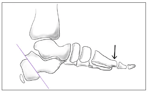
Evans Calcaneal Lengthening Osteotomy
- Lateral incision over the calcaneal neck, 1.5 cm proximal to the calcaneocuboid joint.
- Identify and protect the sural nerve.
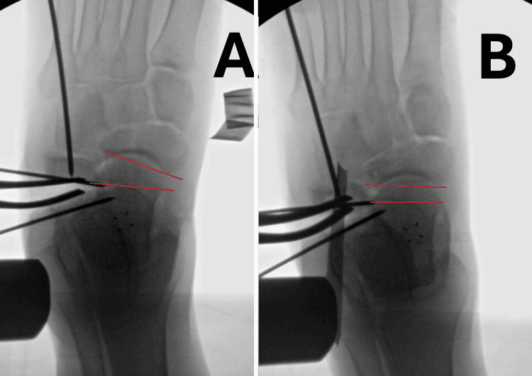
- Transverse osteotomy through the anterior calcaneus, passing between the anterior and middle facets so that it does not enter the posterior facet and stiffen the subtalar joint.
- Distract with a laminar spreader.
- Insert a tricortical iliac crest allograft wedge.
- Fixation: plates or K-wires are not always needed if the graft is tight, but a plate is usually used for stability.
Over-stuffing. Too large a graft lengthens the lateral column beyond the medial and overloads or subluxates the calcaneocuboid joint, giving a stiff, laterally overloaded foot with calcaneocuboid pain; this is the basis of the lateral column overload and calcaneocuboid arthritis complications. Size the graft so the lateral column is not longer than the medial, protect the calcaneocuboid joint, and judge the correction clinically on the table rather than by graft size alone.
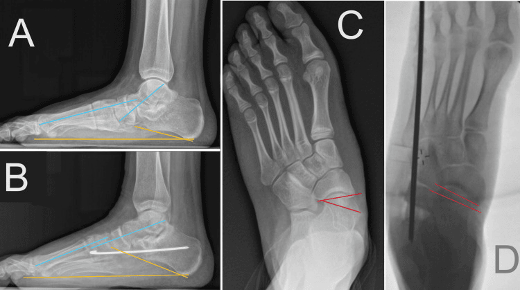
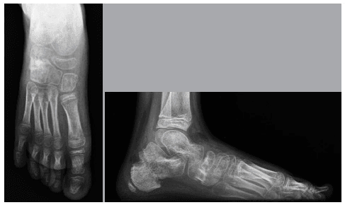
Borrowed evidence. The figures below come from flexible flatfoot and planovalgus reconstruction, one series coalition-associated, and are not skewfoot-specific proof. They teach measurement-driven correction and transferable measurement principles, with the endpoint set by talonavicular coverage and global alignment rather than graft size. In skewfoot, use such procedures only for a matching residual apex, and extrapolate cautiously.
Complications
- Risk Factors
- Addressing only one component (e.g. only the forefoot)
- Prevention/Management
- Address both the hindfoot valgus and the forefoot adduction
- Risk Factors
- Excessive Evans graft size
- Prevention/Management
- Intraoperative sizing; the lateral column should not be longer than the medial
- Risk Factors
- Graft failure; smoking (parents/adolescent)
- Prevention/Management
- Rigid fixation; non-weight-bearing period
- Risk Factors
- Evans osteotomy entering the joint
- Prevention/Management
- Fluoroscopic guidance; stay 1.5 cm proximal to the joint
- Risk Factors
- Lateral approach
- Prevention/Management
- Identify and retract
- Risk Factors
- Too large an Evans graft
- Prevention/Management
- Careful preoperative planning and intraoperative assessment
Postoperative Care
The protocol after a double osteotomy (Evans plus Cotton) runs in five stages:
- 0-2 weeks - splint or backslab in neutral, strict elevation, non-weight-bearing
- 2-6 weeks - wound check, then a short-leg fibreglass cast moulded to hold the hindfoot neutral and the forefoot abducted; still non-weight-bearing
- 6-8 weeks - radiographs for graft union; partial weight bearing if union is evident
- 8-12 weeks - walking cast or CAM boot, full weight bearing
- 3-6 months - shoes with arch support, and physiotherapy for ankle and subtalar motion
Outcomes/Prognosis
Without surgery. The natural history is poorly defined, and Peterson found pain infrequent (Evidence Base). For rigid skewfoot, though, the non-operative outlook is generally poor: pain and footwear difficulties persist, and the deformity tends to progress with growth.
With surgery. Double osteotomy techniques (Evans plus a medial-column procedure) give good functional results, and Mosca (1995) reported satisfactory results using the Evans procedure for valgus deformities including skewfoot. Patient and parent satisfaction with foot shape and shoe fit is high, and most children return to full sports activities.
Long term. Deformity that remains uncorrected carries a risk of early triple arthrodesis because of joint incongruity and degenerative change. Adult skewfoot is notoriously difficult to reconstruct and often requires fusion.
Recurrence. It can occur if the Evans graft resorbs or if the medial column was under-corrected. Monitoring until skeletal maturity is advised.
Guidelines, Registries & Global Practice
Global epidemiology
- Skewfoot is rare; fewer than ~50 cases were reported in the English literature at the time of Peterson's 1986 series, and most modern data remain single-centre case series.
- No dedicated registry exists for paediatric foot deformities; unlike arthroplasty, there is no NJR/AJRR/AOANJRR equivalent capturing skewfoot outcomes, which is itself a recognised evidence gap.
- Where the deformity follows treatment of metatarsus adductus, true incidence is unknown and almost certainly under-reported.
Society guidance and practice positions (no single-country framing)
- Position relevant to skewfoot
- No formal clinical practice guideline; teaching follows the Mosca motion-preserving philosophy (calcaneal lengthening plus medial-column osteotomy, avoid early arthrodesis).
- Position relevant to skewfoot
- Emphasis on accurate differentiation from metatarsus adductus and clubfoot, weight-bearing imaging, and reserving surgery for symptomatic rigid deformity.
- Position relevant to skewfoot
- Reconstruction-principle teaching: correct all components of the deformity, preserve growth and motion, fixation and graft tailored to age.
- Position relevant to skewfoot
- Often frame severe complex metatarsus adductus and skewfoot as a continuum; selective tendon transfer (e.g. tibialis anterior) and soft-tissue balancing feature in the European literature.
High- versus limited-resource practice variation
- High-resource settings: weight-bearing radiographs (or simulated weight-bearing/standing CT in adolescents), allograft availability, image intensifier for the calcaneal osteotomy, and access to specialist paediatric orthopaedic foot services.
- Limited-resource settings: reliance on clinical examination and a single plain film; autograft (iliac crest) preferred over allograft for cost and availability; greater use of soft-tissue and simpler single-osteotomy strategies; later presentation of neglected/rigid deformity is more common.
- Universal principles: differentiate the deformity correctly before treating, never cast/brace a foot in a way that drives the hindfoot into valgus, and address both the forefoot adduction and the hindfoot valgus when operating.
Controversies & Areas of Uncertainty
Skewfoot has one of the weakest evidence bases in paediatric foot surgery — almost all literature is Level IV/V case series. Examiners reward candidates who can articulate the genuine uncertainty rather than overstate dogma.
- Does the iatrogenic mechanism truly exist? The "failed metatarsus adductus casting" story is widely taught and biomechanically plausible (forefoot abduction levering an unstabilised hindfoot into valgus), but it rests on small series and is hard to separate from a primary congenital deformity that was simply misdiagnosed as metatarsus adductus. Many authors regard most cases as primary.
- Is skewfoot a distinct entity or the severe end of a spectrum? Some authors (including the Tönnis tradition) view "skewfoot" and "serpentine foot" as simply the severe end of complex metatarsus adductus rather than a separate disease, which has implications for how aggressively to treat.
- Single- versus double-osteotomy. Whether every operative skewfoot needs both a lateral-column lengthening and a medial-column procedure, or whether selected feet can be corrected with one, is unresolved; the principle of "address both ends of the Z" is consensus-based, not trial-proven.
- Subtalar arthroereisis. Promoted by some as a less invasive option for the valgus component, but evidence in skewfoot specifically is minimal and complication/implant-removal rates are a concern; most paediatric foot surgeons regard it as adjunctive at best.
- Allograft versus autograft and the role of fixation. Both wedge graft choices have advocates; rigid internal fixation versus reliance on a tightly impacted graft is surgeon-dependent.
- Asymptomatic skewfoot. Because long-term natural history is poorly defined and many feet remain pain-free, the threshold for operating on an asymptomatic deformity is genuinely contested.
MCQ Practice Points
Q: What is the hallmark radiographic finding in Skewfoot? A: Increased Talocalcaneal Angle (Valgus) + Adducted Metatarsals. This creates the Z-shape.
Q: Which intervention is a known risk factor for iatrogenic skewfoot? A: Serial Casting for Metatarsus Adductus without stabilizing the hindfoot.
Q: The Evans procedure corrects deformities in which plane? A: Triplanar. It corrects valgus (coronal), abduction (transverse), and dorsiflexion (sagittal).
Q: What is the classic position of the navicular in Skewfoot? A: Dorsolateral subluxation on the talar head. This distinguishes it from MA where the navicular is medial or central.
Q: In the Berg classification, which type represents true Skewfoot requiring surgical treatment? A: Type III - rigid forefoot adduction with fixed hindfoot valgus. Type I is simple metatarsus adductus. Type II is complex metatarsus adductus with lateral midfoot shift.
Q: What is the key clinical finding that distinguishes Skewfoot from Metatarsus Adductus? A: Fixed hindfoot valgus. In Metatarsus Adductus, the hindfoot is neutral or only mildly valgus. The 'Z-shape' appearance on weight-bearing radiographs confirms Skewfoot.
Additional Quiz Questions
Viva Scenarios
Practise clinical reasoning and management decisions out loud
“A 2-year-old child was treated for Metatarsus Adductus with casting. Parents say the foot looks 'flatter and worse'. What happened?”
“8-year-old with painful rigid Skewfoot. Plan surgery.”
“Explain the difference between Clubfoot, Metatarsus Adductus, and Skewfoot to a junior registrar.”
CATCHPHRASE
- Serpentine Foot
- Z-Deformity
- Failed MA Casting
- Idiopathic, Iatrogenic, Inherited
- Rarely resolves spontaneously
TRIAD
- Hindfoot Valgus
- Midfoot Abduction
- Forefoot Adduction
- Plantarflexed talus
- Lateral navicular subluxation
RADIOGRAPHS
- Increased TC Angle (Valgus)
- Adducted Metatarsals
- Lateral Navicular
- Talar head uncoverage
- Weight-bearing views essential
MANAGEMENT
- Observe (if flexible)
- Surgery: Evans + Medial Column Osteotomy
- Avoid isolated MT osteotomy
- Must address both ends
- Casting often fails
KEY TRAP
- Confusing with Clubfoot (Varus)
- Confusing with MA (Neutral Hindfoot)
- Missing the hindfoot valgus
- Incomplete surgical correction
- Not using WB X-rays
Evidence Base
Mosca — Calcaneal Lengthening for Hindfoot Valgus
- 31 severe symptomatic valgus hindfoot deformities in 20 children: 25 flatfeet and 6 skewfeet
- Calcaneal (Evans-type) lengthening combined with opening-wedge medial cuneiform osteotomy to correct both hindfoot and forefoot in the skewfeet
- Satisfactory clinical and radiographic correction in all but the 2 most severely deformed feet; subtalar motion preserved
- Resolved pain and plantar-talar-head callus while avoiding arthrodesis
Hirose & Johnson — Cotton (Medial Cuneiform) Osteotomy
- 16 feet (15 patients) including a 15-year-old skewfoot treated with plantarflexion opening-wedge medial cuneiform osteotomy for fixed forefoot varus
- Talonavicular coverage angle improved a mean of 15 degrees and lateral talo-first metatarsal angle a mean of 14 degrees; no nonunions or malunions
- Preserves first-ray mobility versus first tarsometatarsal arthrodesis, with easily titratable correction
Mosca & Bevan — Calcaneal Lengthening for Talocalcaneal Coalition
- 8 patients (13 feet) showing that correcting the valgus deformity is as important as addressing the coalition
- Calcaneal lengthening with gastrocnemius/Achilles lengthening relieved pain and preserved talonavicular and calcaneocuboid motion
- Supports treating excessive hindfoot valgus rather than defaulting to triple arthrodesis