Identify the dangerous lump before anyone cuts it
- The safe default is: image and refer before cutting. A suspected sarcoma should not be drained, shelled out or excised as a lump.
- Deep, enlarging, painful, recurrent or greater than 5 cm masses need sarcoma-pathway thinking. Small superficial lesions can still be malignant when behaviour is atypical.
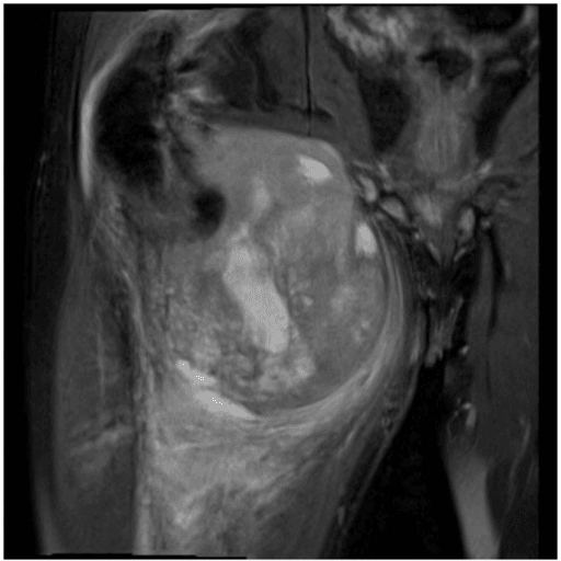
- MRI is obtained before biopsy. It defines compartment, neurovascular relationships, bone involvement, viable biopsy target and the future resection field.
- Biopsy route is part of the operation. The tract must be planned so it can be removed with the tumour.
- Unplanned excision changes prognosis and reconstruction. The tumour bed is contaminated until proven otherwise and usually needs specialist re-excision planning.
- “Do not reassure a patient because the mass is painless; many sarcomas are painless.
- “Ultrasound can confirm a simple cyst or superficial lipoma, but MRI is the key test for an indeterminate or deep mass.
- “Needle biopsy should target viable enhancing tissue, not central necrosis.
- “The first operation is often the best chance to preserve margins, function and reconstructive options.
An excisional biopsy of a suspected sarcoma contaminates the tumour bed, alters MRI interpretation, may force wider re-excision and can compromise limb-sparing reconstruction. If the diagnosis is not confidently benign, stop and refer.

DEEPSarcoma Red Flags
Hook:DEEP masses need deeper thinking.
IMAGEInitial Safety Pathway
Hook:IMAGE before incision.
RESCUEUnplanned Excision Response
Hook:RESCUE the pathway after a whoops procedure.
Overview and Epidemiology
Most soft tissue lumps are benign, but the clinical cost of missing sarcoma is high. The common error is not ignorance of sarcoma subtypes; it is treating an indeterminate lump as a harmless lump before appropriate imaging and referral.
Soft tissue sarcoma is uncommon compared with benign lipoma, cyst, haematoma and muscle injury. That rarity creates danger: a surgeon or general clinician may see many benign lumps for every sarcoma and become overconfident. The safer approach is to recognise behaviour that is not benign.
- Reassuring Pattern
- Small and stable.
- Concerning Pattern
- Greater than 5 cm or increasing over time.
- Reassuring Pattern
- Clearly superficial and mobile above fascia.
- Concerning Pattern
- Deep to fascia, intramuscular or fixed to deeper tissues.
- Reassuring Pattern
- Pain linked to trauma and resolving.
- Concerning Pattern
- Persistent, night, progressive or unexplained pain.
- Reassuring Pattern
- No recurrence after clearly benign treatment.
- Concerning Pattern
- Recurrent after aspiration, drainage or excision.
- Reassuring Pattern
- Classic cyst or lipoma on appropriate imaging.
- Concerning Pattern
- Heterogeneous, infiltrative, vascular, necrotic or deep lesion.
Pathophysiology
Soft tissue sarcomas arise from mesenchymal tissues such as muscle, fat, fibrous tissue, vessels, peripheral nerve sheath or undifferentiated soft tissue. The clinical pathway is similar across subtypes because the first decisions are anatomical and oncological: where is the mass, what does it involve, how should it be sampled, and can it be removed with a planned margin?
Important biological concepts:
- Pseudocapsule: many sarcomas compress adjacent tissue but are not truly encapsulated. Shelling out the mass leaves microscopic disease.
- Reactive zone: oedema, haematoma and post-biopsy change can contain tumour cells and may need inclusion in the resection field.
- Compartment anatomy: deep masses can involve muscle compartments, neurovascular bundles, periosteum, joint capsule or bone.
- Necrosis: large high-grade sarcomas may contain necrotic areas; biopsy should target viable enhancing tissue.
- Skip and metastatic risk: staging is subtype- and grade-dependent, but chest imaging is central for many extremity sarcomas because lung metastasis is common.
The biopsy tract, drain site, haematoma cavity, contaminated planes and previous scar may become part of the definitive resection. That is why the first procedure must be planned.
Classification
Classification in the initial clinic is not about memorising every histological subtype. It is about sorting the mass into a safe management pathway.
- Typical Features
- Small, superficial, soft, mobile, stable and imaging-classic benign.
- Action
- Observe, ultrasound or local treatment when diagnosis is secure.
- Typical Features
- Unclear diagnosis, atypical features or discordant clinical/imaging findings.
- Action
- MRI and specialist review before excision.
- Typical Features
- Deep, enlarging, painful, recurrent or greater than 5 cm.
- Action
- MRI with contrast and sarcoma referral.
- Typical Features
- Sarcoma diagnosed after unplanned excision.
- Action
- Urgent sarcoma MDT, staging, tumour-bed MRI and re-excision planning.
Grading and Staging
Once tissue is obtained, the sarcoma is graded and the disease is staged - the two facts that drive prognosis and the multimodal plan, and the basis of the "low-grade" versus "high-grade" language used above.
Grade (FNCLCC system). The Federation Nationale des Centres de Lutte Contre le Cancer (FNCLCC) grade sums three scored parameters and is the strongest predictor of metastasis.
- Scoring
- Score 1 to 3 (1 resembling normal tissue, 3 poorly differentiated)
- Note
- Subtype-dependent
- Scoring
- Score 1 (under 10 per 10 HPF), 2 (10 to 19), 3 (20 or more)
- Note
- Per 10 high-power fields
- Scoring
- Score 0 (none), 1 (under 50%), 2 (50% or more)
- Note
- On histology
- Scoring
- Grade 1 = 2 to 3; Grade 2 = 4 to 5; Grade 3 = 6 to 8
- Note
- Grade 3 is high-grade
Stage (AJCC TNM). Staging combines tumour size (T), nodes (N), metastasis (M) and grade (G). For extremity and trunk sarcoma the size strata are T1 (5 cm or less), T2 (over 5 to 10 cm), T3 (over 10 to 15 cm) and T4 (over 15 cm); nodal spread (N1) is uncommon but upstages; the commonest distant site is the lung (M1). Crucially, grade drives the stage - a small low-grade tumour is early-stage, while a high-grade tumour is advanced even when small.
The FNCLCC grade (differentiation plus mitoses plus necrosis) is the single best predictor of metastasis. AJCC staging then layers tumour size, nodes, metastasis and grade together - which is why a high-grade sarcoma is never "reassuringly small".
Clinical Presentation
History
Ask precise questions that decide risk and pathway:
- When was the mass first noticed?
- Is it enlarging, stable or fluctuating?
- Was there real trauma, or did trauma simply draw attention to the mass?
- Is there pain, night pain, neurological symptom, vascular symptom or functional loss?
- Has it been aspirated, drained, injected or excised before?
- Is there a history of cancer, radiotherapy, genetic syndrome or immunosuppression?
- Are there systemic features such as weight loss, fever or multiple masses?
Examination
Examine the mass as a surgical field, not only as a lump.
- How To Examine
- Expose the whole limb and compare sides. Inspect scars, swelling, skin tethering, venous prominence, ulceration and previous drain sites.
- What It Means
- Scars and drain sites may define contaminated tissue if sarcoma is confirmed.
- How To Examine
- Define size in centimetres, depth, temperature, tenderness, consistency, fluctuation and relation to fascia.
- What It Means
- Deep or fixed masses are higher risk than mobile superficial lesions.
- How To Examine
- Move adjacent joints and contract nearby muscles while palpating the mass.
- What It Means
- A mass that moves with muscle may be intramuscular; joint restriction may indicate periarticular involvement.
- How To Examine
- Document pulses, capillary refill, sensation, motor power and Tinel sign when near major nerves.
- What It Means
- Nerve or vessel involvement changes biopsy route, resection plan and counselling.
- How To Examine
- Check regional nodes for selected subtypes and examine chest/abdomen only as clinically indicated.
- What It Means
- Most extremity sarcomas metastasise haematogenously, but some subtypes involve nodes.
Differential Diagnosis
Most masses are benign, but the role of the first clinician is to separate the confidently benign from the indeterminate. The table below contrasts the common benign mimics with the malignant lesions they hide, and the feature that should prompt imaging and referral.
- Typical Picture
- Soft, mobile, superficial, slow-growing fatty mass.
- Why It Can Mislead
- Atypical lipomatous tumour / well-differentiated liposarcoma looks almost identical and can be deep.
- Discriminator
- Deep location, size over 5 cm, thick or nodular septa, or non-fatty enhancing areas on MRI warrant referral.
- Typical Picture
- Cystic, transilluminable, near joint or tendon sheath.
- Why It Can Mislead
- Myxoid sarcoma can appear cystic on ultrasound.
- Discriminator
- True simple cysts have no internal enhancement; solid or enhancing 'cyst' needs MRI.
- Typical Picture
- Painful swelling after trauma, should resolve over weeks.
- Why It Can Mislead
- A sarcoma may bleed or simply be noticed after minor trauma.
- Discriminator
- Re-image any haematoma that fails to resolve, enlarges or recurs.
- Typical Picture
- Hot, tender, systemic features, raised inflammatory markers.
- Why It Can Mislead
- Some sarcomas are warm and tender and may be mislabelled infective.
- Discriminator
- If pus is not obtained or the cavity does not settle, treat as a tumour and image.
- Typical Picture
- Mass on a nerve line with Tinel sign or radiating paraesthesia.
- Why It Can Mislead
- Malignant peripheral nerve sheath tumour, especially in NF1.
- Discriminator
- Rapid growth, pain or neurological deficit in a known neurofibroma signals malignant change.
- Typical Picture
- Firm infiltrative mass, locally aggressive but non-metastasising.
- Why It Can Mislead
- Mimics sarcoma clinically and radiologically.
- Discriminator
- Managed by sarcoma MDT; biopsy and imaging still required to confirm.
Investigations

- How To Request It
- Use for clearly superficial lesion or to confirm cystic versus solid nature.
- What It Answers
- May identify simple cyst or lipoma, but cannot safely stage a deep indeterminate mass.
- How To Request It
- MRI of the whole involved anatomical compartment with skin markers over the mass.
- What It Answers
- Depth, size, compartment, margins, neurovascular/bone/joint relationship, necrosis and biopsy route.
- How To Request It
- Request regional x-rays if calcification, bone pain, periosteal reaction or deep mass near bone.
- What It Answers
- Mineralisation, bone erosion, periosteal reaction or alternate diagnosis.
- How To Request It
- Sarcoma staging after suspicion or diagnosis, according to sarcoma team pathway.
- What It Answers
- Pulmonary metastases, especially in high-grade extremity sarcoma.
- How To Request It
- Image-guided core biopsy after MRI, planned with treating sarcoma team.
- What It Answers
- Histology, grade, molecular tests and treatment planning.

Management
Management is pathway-based. The most important decision is often what not to do: do not incise, drain, aspirate repeatedly or excise an indeterminate mass outside a sarcoma plan.
- Safe Action
- Treat locally only when diagnosis is secure and behaviour is benign.
- Avoid
- Ignoring growth or atypical imaging.
- Safe Action
- Ultrasound or MRI depending clinical risk; refer if not confidently benign.
- Avoid
- Office excision to find out what it is.
- Safe Action
- MRI with contrast and sarcoma referral before biopsy.
- Avoid
- Incisional biopsy, drainage or excision outside the resection plan.
- Safe Action
- Urgent imaging and specialist review.
- Avoid
- Delay because the skin looks normal.

Surgical Margins
The topic repeatedly demands a "planned wide excision" and warns about "involved margins"; the underlying language is the margin classification.
Enneking (musculoskeletal) margins describe the plane of the excision relative to the tumour and its reactive zone.
- Plane of dissection
- Through the tumour (debulking, shelling out)
- Residual disease risk
- Gross tumour left behind
- Plane of dissection
- Through the reactive zone or pseudocapsule
- Residual disease risk
- Microscopic satellites or skip lesions left
- Plane of dissection
- Through normal tissue, taking a cuff of normal tissue with the tumour and its reactive zone
- Residual disease risk
- Goal for most sarcomas
- Plane of dissection
- Removal of the entire involved compartment
- Residual disease risk
- Lowest local recurrence but highest morbidity
The pathologist reports the oncological R (residual tumour) status: R0 no residual tumour (margins clear), R1 microscopic residual (margin involved), R2 macroscopic residual. A wide / R0 margin is the surgical aim; an R1 margin - as typically left after an unplanned excision - raises local recurrence and usually drives re-excision and/or radiotherapy.
Aim for a wide Enneking margin and an R0 (microscopically clear) resection. "Shelling out" a sarcoma is an intralesional or marginal margin that leaves disease behind - the reason an unplanned excision (an R1 bed) needs planned re-excision rather than observation.
Complications and Pitfalls
- Why It Matters
- Leaves residual microscopic disease and contaminates the surgical bed.
- Corrective Action
- Refer urgently, restage, MRI tumour bed and plan re-excision.
- Why It Matters
- Forces wider resection or risks local recurrence.
- Corrective Action
- Discuss route with sarcoma surgeon before biopsy.
- Why It Matters
- Distorts anatomy with bleeding and post-procedure change.
- Corrective Action
- MRI first unless an emergency diagnosis changes priorities.
- Why It Matters
- A sarcoma may bleed or be noticed after trauma.
- Corrective Action
- Re-image persistent or enlarging haematoma-like lesions.
- Why It Matters
- Margins may require skin, muscle, nerve, vessel, bone or joint reconstruction.
- Corrective Action
- Plan MDT reconstruction before excision.
Explain that referral does not mean the lump is definitely cancer. It means the lump has features that make specialist imaging and biopsy safer. The goal is to avoid the wrong first operation.
Guidelines, Registries & Global Practice
Soft tissue sarcoma is rare everywhere, which makes centralised, MDT-led care the single most consistent recommendation across regions. The framing below is global: regional guidance is cited as evidence contributing to one shared standard, not as country-specific practice.
Global Epidemiology
- Soft tissue sarcomas account for roughly 1% of adult malignancies and are far outnumbered by benign soft tissue lesions, which are at least 100 times more common.
- They can arise at any site; the extremities (especially the thigh) are the commonest location, followed by trunk and retroperitoneum.
- Incidence rises with age but the disease occurs across all age groups, and over 50 to 70 histological subtypes are recognised in the current WHO classification.
- Lung is the dominant site of distant metastasis for extremity sarcoma, which is why chest imaging anchors staging.
Side-by-Side Guideline Comparison
- Referral and Imaging
- Urgent referral for any unexplained soft tissue lump that is enlarging, deep to fascia or greater than 5 cm; imaging-led diagnosis at a sarcoma centre.
- Biopsy and Treatment
- Core biopsy at the specialist centre; surgery by a sarcoma surgeon with radiotherapy for higher-risk tumours.
- Referral and Imaging
- Refer suspected sarcoma to a reference centre before biopsy; MRI is the mainstay of local imaging.
- Biopsy and Treatment
- Core-needle biopsy as standard, tract planned for excision; multimodal MDT treatment.
- Referral and Imaging
- Imaging then biopsy for masses that are symptomatic, enlarging or greater than 5 cm; MRI for extremity and trunk lesions.
- Biopsy and Treatment
- Core or incisional biopsy through a planned approach; wide excision plus radiotherapy by risk.
- Referral and Imaging
- Echoes Mankin principles: biopsy and definitive surgery at the same specialist centre.
- Biopsy and Treatment
- Biopsy planned by the treating surgeon; poorly planned biopsy is a recognised avoidable harm.
The recommendations converge: image before biopsy, biopsy before excision, and concentrate care in specialist units. Differences are mainly in wording of thresholds rather than principle.
Registries and Reference Data
- National sarcoma registries and reference networks (for example the UK BSG/National Cancer Registration data, the European EURACAN reference network and the US National Cancer Database) consistently show better margins, lower local recurrence and improved survival when patients are treated at high-volume centres.
- Reference-network data also document the persistent cost of unplanned excision, supporting the centralisation message rather than any single national policy.
High- versus Limited-Resource Practice
- Typical Reality
- MRI, image-guided core biopsy and a formal sarcoma MDT are routinely available.
- Pragmatic Adaptation
- Follow the full pathway: MRI, planned core biopsy, MDT, multimodal treatment.
- Typical Reality
- MRI access, specialist pathology and radiotherapy may be delayed or distant.
- Pragmatic Adaptation
- The non-negotiable step is to avoid unplanned excision: confirm depth and size clinically and by ultrasound, then refer the patient (not just the specimen) to the nearest sarcoma capability.
Wherever you practise, the universal rule is the same: do not excise an indeterminate deep or large mass to make the diagnosis. Refer the patient before the tissue is disturbed.
Controversies and Areas of Uncertainty
The referral pathway itself is well established, but several points remain debated and are favourite viva ground.
- Arguments For
- Simple, sensitive and easy to teach; captures most clinically significant sarcomas.
- Counterpoint
- Small superficial sarcomas exist; size must be combined with depth, growth and imaging, not used alone.
- Arguments For
- Ultrasound is cheap, fast and can confirm a simple cyst or superficial lipoma.
- Counterpoint
- It is operator-dependent and cannot stage a deep mass; an indeterminate result still mandates MRI, so some pathways go straight to MRI for deep lesions.
- Arguments For
- Traditional teaching includes the tract in the resection to remove seeded cells.
- Counterpoint
- Modern series with image-guided core biopsy suggest needle-tract seeding is rare; some units no longer routinely excise core-biopsy tracts, though most still mark and consider them.
- Arguments For
- Pre-operative RT uses a smaller field and lower dose with better long-term function.
- Counterpoint
- It roughly doubles major wound-healing complications; timing is an MDT decision balancing wound risk against late fibrosis.
- Arguments For
- Often treated with marginal excision given low metastatic potential.
- Counterpoint
- Local recurrence and rare dedifferentiation mean follow-up and MDT input are still required, especially for deep or retroperitoneal sites.
Clinical Scenarios
Practise clinical reasoning and management decisions out loud
“A 48-year-old patient has a painless enlarging thigh mass. It is about 8 cm, firm and deep to fascia. The skin is normal.”
“A patient is referred after local excision of a presumed lipoma. Final histology shows high-grade soft tissue sarcoma with involved margins.”
“A general practitioner refers a 35-year-old with a soft, mobile, 3 cm subcutaneous mass on the forearm that has been stable for two years. The clinical impression is a lipoma.”
Red Flags
- Deep to fascia
- Greater than 5 cm
- Enlarging
- Painful or persistent
- Recurrent after treatment
Safe Workup
- Document size, depth and neurovascular status
- MRI with contrast before biopsy
- Chest staging after suspicion or diagnosis
- Sarcoma unit referral
- Core biopsy planned in resection field
Do Not Do
- Do not drain an indeterminate mass
- Do not shell out a deep lump
- Do not biopsy before MRI
- Do not place a transverse biopsy tract
- Do not observe unexpected sarcoma histology
“Deep, enlarging, painful, recurrent or greater than 5 cm masses need MRI and sarcoma referral before biopsy or excision.”
Evidence Signals
UK soft tissue sarcoma guideline
- British Sarcoma Group guidance: any patient with a suspected soft tissue sarcoma should be referred to a specialist regional sarcoma service and managed by a sarcoma MDT.
- Diagnosis should be confirmed with appropriate imaging plus biopsy before the main modality of treatment, which is usually surgical excision by a specialist surgeon.
- Pre- or post-operative radiotherapy is considered for tumours at higher risk of recurrence; systemic therapy is reserved for more chemosensitive subtypes.
ESMO-EURACAN-GENTURIS guideline
- Diagnosis and treatment of soft tissue and visceral sarcoma should be planned within specialist multidisciplinary teams.
- MRI is the mainstay of local imaging for extremity, trunk-wall and head-and-neck soft tissue sarcoma.
- Core-needle biopsy is the standard diagnostic approach, planned so the tract can be removed at definitive surgery.
References
- Dangoor A, Seddon B, Gerrand C, Grimer R, Whelan J, Judson I. UK guidelines for the management of soft tissue sarcomas. Clin Sarcoma Res. 2016;6:20. doi:10.1186/s13569-016-0060-4.
- Casali PG, Abecassis N, Aro HT, et al. Soft tissue and visceral sarcomas: ESMO-EURACAN-GENTURIS Clinical Practice Guidelines for diagnosis, treatment and follow-up. Ann Oncol. 2021;32(11):1348-1365. doi:10.1016/j.annonc.2021.07.006.
- Mankin HJ, Lange TA, Spanier SS. The hazards of biopsy in patients with malignant primary bone and soft-tissue tumors. J Bone Joint Surg Am. 1982;64(8):1121-1127.
- Noria S, Davis A, Kandel R, et al. Residual disease following unplanned excision of soft-tissue sarcoma of an extremity. J Bone Joint Surg Am. 1996;78(5):650-655.
- Grimer RJ, Carter SR, Tillman RM, et al. Unplanned excision of soft tissue sarcoma. J Bone Joint Surg Br. 2001;83(2):203-206.
- Venkatesan M, Richards CJ, McCulloch TA, Perks AGB, Raurell A, Ashford RU. Inadvertent surgical resection of soft tissue sarcomas. Eur J Surg Oncol. 2012;38(4):346-351. doi:10.1016/j.ejso.2011.12.011.
- Toki S, Sone M, Yoshida A, et al. Image-guided core needle biopsy for musculoskeletal lesions. J Orthop Sci. 2022;27(2):448-455. doi:10.1016/j.jos.2020.12.017.