SCC in Chronic Wound or Scar | Latency Decades | Biopsy Every Non-Healing Lesion
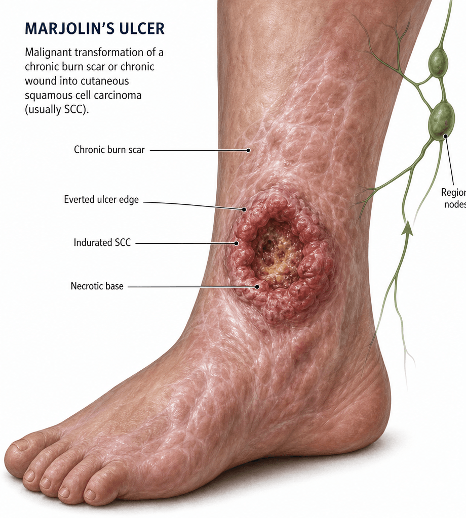
- Marjolin's ulcer is squamous cell carcinoma (SCC) arising in a chronic wound, scar, sinus or ulcer (most commonly old burn)
- Latency in burn scars averages about 31 years in the largest pooled series (mean burn age 20, mean diagnosis 50); the common claim that osteomyelitis-sinus latency is shorter is poorly sourced
- Any chronic wound that fails to heal, develops raised everted edges, foul discharge, bleeding, or pain after long quiescence needs biopsy
- Treatment is wide local excision (2-4 cm margins) or amputation for deep bone / joint involvement, plus nodal staging
- Nodal metastasis is more common than in de novo SCC (20-40 percent at diagnosis) - sentinel node biopsy is increasingly used
- Five-year survival is worse than de novo SCC and depends on stage, grade, depth, and nodal status
- “The ulcer that has been stable for 20 years and suddenly changes is a Marjolin's until proven otherwise
- “Biopsy the edge, not the floor, of a chronic ulcer - the floor is granulation tissue and gives false negatives
- “Osteomyelitis sinus with a Marjolin is a classic viva - squamous epithelium grows down the sinus tract
- “Lower limb is the single most common site because burns and chronic osteomyelitis are concentrated there
- “Always check the regional lymph nodes clinically and radiologically at presentation
Overview and Epidemiology
Definition. A Marjolin's ulcer is a malignancy arising in a chronic wound, scar, sinus or ulcer, most commonly an old burn. Between 75 and 95 percent are squamous cell carcinoma; the remainder include basal cell carcinoma, melanoma, sarcoma and Merkel cell carcinoma. Marjolin described it in 1828, observing tumour change in old burn scars, and the term now covers every aetiology.
Why it matters. Chronic wounds can undergo malignant change, so any non-healing ulcer needs a biopsy, and the tumour that results behaves more aggressively than a de novo SCC, so the treatment is wider. Lower-limb burns, chronic osteomyelitis sinuses and venous ulcers are the typical sources seen in fracture-clinic and sarcoma-unit practice worldwide.
Who. Burn survivors in low- and middle-income countries where childhood burns were managed conservatively; patients with chronic osteomyelitis and a long-standing draining sinus, often for decades; elderly or immobile patients with long-standing venous, pressure or diabetic ulcers of the leg and foot; patients with previous radiotherapy and radiation-induced chronic skin change; and patients with chronic fistulae or surgical scars that have never epithelialised properly.
Where. The lower limb is the single most common site, 40-60 percent of cases, because burns and chronic osteomyelitis are concentrated there. Long-standing leg ulcers are the second most common source after burn scars. Rarer sources are chronic scarring dermatoses (lupus, dystrophic epidermolysis) and vaccination scars.
Time is the risk factor. The single biggest risk factor is time since the burn. Patients burned in childhood, especially in regions with limited acute burn care, are now reaching the latency window in middle age, and the incidence of Marjolin's is expected to rise in these populations. Vigilance in this group is cost-effective: a single targeted biopsy can change management.
Latency. Typically two to four decades, with a mean close to 31 years. Two independent series converge: Kowal-Vern's 412 pooled burn-scar neoplasms gave a mean latency of 31 years (mean age 20 at the burn, 50 at diagnosis), and Hahn's clinical series gave 31.5 years in a cohort that was 32 percent chronic osteomyelitis fistula. The longer a scar has been stable, the more sinister a new change.
The osteomyelitis-sinus caveat. The common teaching that latency is shorter after an osteomyelitis sinus is widely repeated but poorly sourced, and Hahn's osteomyelitis-heavy cohort showed no such shortening. Treat latency as long whatever the aetiology. Acute Marjolin's, under one year from the injury, exists but is rare and behaves more aggressively.
Pathophysiology
Two pathways to malignant change. The first is the chronic-inflammation, or scar-niche, model: persistent wound healing, repeated epithelial injury, free-radical damage, local immunosuppression and bacterial colonisation produce cumulative DNA damage in a setting of impaired immune surveillance. This is the dominant model for burn-scar and osteomyelitis-sinus Marjolin's. The second is direct malignant transformation of unstable scar epithelium, supported histologically by dysplasia at the wound edge adjacent to frank carcinoma in many specimens.
The chronic wound environment. The cellular events described are:
- Repeated epithelial injury and repair
- Sustained cytokine release (TNF-alpha, IL-6)
- Local immunosuppression by regulatory T cells
- Free-radical and bacterial toxin exposure
- Hypoxia and lactate accumulation
The genetic events reported:
- p53 mutation in a high proportion of cases
- Loss of p16 and other cell-cycle regulators
- UV-signature mutations in sun-exposed sites
- Field change: dysplasia extends beyond the visible tumour
Histology. The tumour is most often well- to moderately-differentiated SCC, with keratin pearls, intercellular bridges and invasive nests. The diagnosis is made when SCC is identified arising from, or directly adjacent to, the chronic wound epithelium; the dysplasia-carcinoma sequence is often visible at the wound edge, with adjacent carcinoma in situ.
Why it behaves worse than de novo SCC. Part of the answer is the tumour: often of higher grade than a de novo SCC, deep invasion by the time the diagnosis is made, a high rate of perineural and lymphovascular invasion, and a deep margin that is frequently poorly circumscribed. Part is the patient: older, with more comorbidities, often poorly nourished and chronically unwell, slow to present because the wound was long-standing, and with reconstructive options limited by the surrounding scar. The net effect is lower survival than de novo SCC of equivalent stage.
Classification and Types
Three classifications are in use, by timing of the malignant change, by histology and by the lesion it arose in. The histology is the one that sets the operation.
Classification by Timing of Malignant Change
- Latency
- Less than 1 year from original wound
- Behaviour
- Aggressive, often high grade, deep invasion
- Prognosis
- Worse than chronic form
- Latency
- Over 1 year - usually decades after burn
- Behaviour
- Variable grade, often well-differentiated
- Prognosis
- Guarded - depends on stage, depth, nodal status
- Latency
- Arises in long-standing wound, no preceding scar
- Behaviour
- Behaviour similar to de novo SCC
- Prognosis
- Better than typical Marjolin's
Acute Marjolin's is uncommon and is typically described in acute burns that fail to heal and then turn malignant within months. Chronic Marjolin's is the textbook variant.
The histology table lists angiosarcoma "in chronic lymphoedematous tissue" - this is Stewart-Treves syndrome, the classic exam counterpart to Marjolin's ulcer and a favourite "compare and contrast" question:
- What it is: a cutaneous angiosarcoma arising in chronically lymphoedematous tissue, not an SCC. The classic setting is the arm years after axillary dissection and radiotherapy for breast cancer (post-mastectomy lymphoedema), but it also occurs in chronic primary or filarial lymphoedema of the limb.
- Latency: typically about 10 years of chronic lymphoedema before malignant change.
- Appearance: multiple violaceous/red-blue macules, nodules or plaques in the swollen limb that coalesce - quite unlike the rolled-edge ulcer of Marjolin's; biopsy shows angiosarcoma (CD31/CD34/ERG positive endothelial markers).
- Behaviour and treatment: aggressive, early haematogenous (lung) metastasis and a poor prognosis; treatment is radical excision/amputation often with chemotherapy/radiotherapy, but outcomes are worse than Marjolin's SCC.
Clinical Assessment
History. Establish the original injury (mechanism, age at injury, healing course, treatments received) and the time since the wound in years, decades being typical for a burn scar. Then what has changed: new bleeding, pain, discharge, mass, foul smell or failure to heal. Ask about previous treatment (skin grafts, dressings, surgery, compliance with dressings), past radiotherapy, immune suppression and smoking, and about weight loss, night sweats, anorexia or bone pain, which suggest metastasis.
Examination. Look at the wound and everything around it:
- The wound: size, depth, edges (raised, everted, rolled) and floor (granulation, necrotic, fungating)
- Surrounding skin: satellite nodules, dysplastic change, the quality of the old scar
- Palpation: induration, fixation to deep structures, bone involvement
- Regional lymph nodes: inguinal, axillary, popliteal, epitrochlear or cervical depending on the site
- Distal neurovascular status, especially before any amputation decision
- Distant sites: lung auscultation, abdominal examination for hepatomegaly
The nodes at the first visit. Compare the contralateral limb and assess the regional nodes at the same consultation. A Marjolin's on the lower leg that has travelled to the popliteal or inguinal nodes is a different operation from one that is node-negative. Document the nodal status and use ultrasound or cross-sectional imaging if the nodes are clinically equivocal.
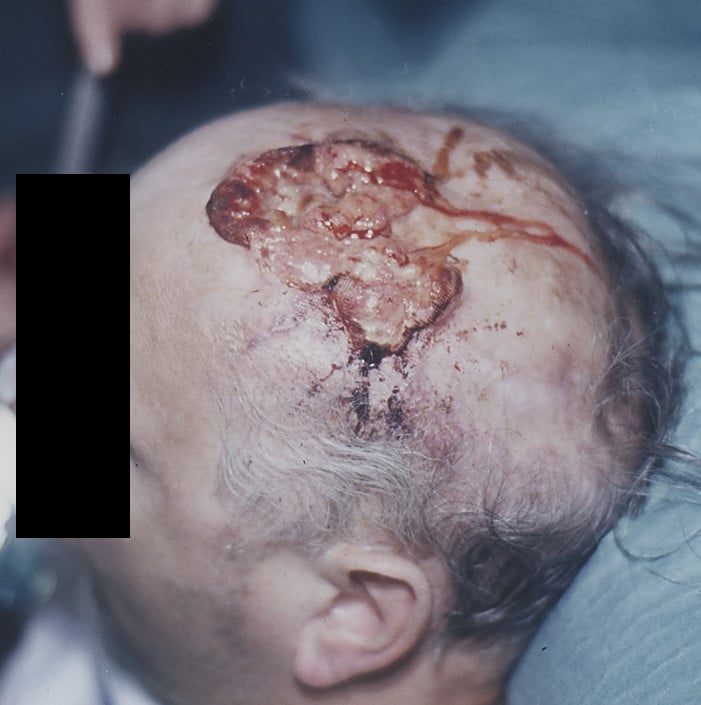
What needs a biopsy. Any chronic wound that fails to heal, or any stable scar that ulcerates, bleeds, becomes painful or develops a mass. A chronic osteomyelitis sinus with increased discharge or a fungating mass at its mouth is biopsied along with the tract, and a venous or pressure ulcer that has outlasted multiple skin grafts is in the same category. The technique is set out under Investigations.
CHANGEMarjolin's - the warning signs in a chronic wound
Hook:Any CHANGE in a chronic wound is a Marjolin's until biopsy proves otherwise!
The differential. A non-healing ulcer in an old scar has other explanations, and the table separates them; the biopsy is what settles it.
- Discriminating feature
- Raised everted edges, new pain, bleeding, mass in old burn scar
- Key test
- Edge biopsy showing SCC with adjacent dysplasia
- Discriminating feature
- Discharge, sinus, no mass; improves with antibiotics
- Key test
- CRP, ESR, MRI, deep swab; biopsy if no improvement
- Discriminating feature
- No ulceration, pruritic, raised but soft; responds to steroid
- Key test
- Clinical diagnosis; biopsy if atypical
- Discriminating feature
- Violaceous undermined edge, often in IBD / arthritis
- Key test
- Biopsy (exclude malignancy), clinical pattern
- Discriminating feature
- Multiple ulcers, systemic features, abnormal serology
- Key test
- ANA, ANCA, biopsy of ulcer edge including vessel
- Discriminating feature
- Classical site (medial gaiter area for venous), Doppler changes
- Key test
- ABI, venous Doppler, biopsy if non-healing after revascularisation
Investigations
Biopsy first. Take a 4 mm punch or wedge biopsy from the edge of the lesion, including both abnormal and adjacent normal-appearing tissue. The floor is granulation tissue, slough or necrotic debris without diagnostic epithelium, so floor biopsies are often false negative. Large or heterogeneous lesions need multiple samples, at least two or three from the most suspicious (raised, indurated, painful) areas, and a benign result from a lesion that still looks suspicious is grounds for a second opinion.
The pathology request. Give the clinical context (Marjolin's suspected), ask for SCC versus another tumour type, and request depth of invasion, grade, lymphovascular and perineural invasion, and margins if the specimen is excisional. Discuss the suspicion explicitly with the pathologist so that multiple levels and immunohistochemistry are considered; immunohistochemistry is useful when the tumour is poorly differentiated (consider p16, p53 and Merkel cell polyomavirus).
Local staging. Plain radiographs of the limb are often obtained to look for bony involvement (chronic osteomyelitis, cortical breach, pathological fracture) and as a baseline for future comparison, but they do not exclude early marrow involvement. MRI of the region is the local staging study of choice for every confirmed case before excision: it shows the enhancing soft-tissue mass, any bone marrow oedema, cortical breach or sinus tract, and it guides the margin and the reconstruction by identifying invasion of the neurovascular bundle or joint.
Nodal staging. Regional nodes are involved in 20-40 percent at presentation, against under 5 percent for de novo cutaneous SCC. The direction is well accepted, but note the provenance: the largest pooled series (Kowal-Vern, 412 cases) found 22 percent regional nodal involvement, the bottom of that range, and made no direct comparison with de novo SCC. No study has matched the two for size, depth and grade, so part of the excess may simply be that these tumours are large and deep by the time anyone biopsies them.
- Ultrasound of the regional nodes is first line in every confirmed case and guides fine-needle aspiration of suspicious nodes
- CT or MRI of the nodal basin for deep nodes (pelvis, retroperitoneum) or when ultrasound is equivocal
- Sentinel lymph node biopsy is increasingly used because of the high rate of microscopic nodal disease, but the evidence is mixed for SCC; consider it in high-grade, deep, large lesions, in conjunction with plastic surgery or nuclear medicine
Distant staging. CT of the chest and abdomen for high-grade lesions, a large primary, clinically positive nodes, recurrent disease or systemic symptoms; CT chest is the standard distant staging study for any high-risk tumour. The lung is the most common distant site, then liver and bone. PET-CT is increasingly used in advanced or recurrent disease, or when cross-sectional imaging is equivocal, to identify occult metastatic disease.
Management Algorithm
Wide Local Excision (2-4 cm Margin) for Localised Soft-Tissue Disease
The goal is complete oncological resection with adequate margins, then reconstruction of the defect.
Before the operation. The diagnosis is confirmed by edge biopsy, with grade and depth on MRI, and the case is reviewed by a multidisciplinary team of oncology, plastic surgery, radiology and pathology. Settle the reconstruction plan (skin graft, local flap, free flap or amputation) and take consent that covers recurrence, nodal disease, amputation and flap failure.
The margin. Excise 2-4 cm of clinically normal tissue around the lesion, though some centres accept 2 cm in low-grade small lesions, down to and including the deep fascia, or to bone if it is involved, taking periosteum or paratenon if the lesion is close to bone or tendon. The width is dictated by field change: dysplasia often extends well beyond the visible tumour, and margins greater than 1 cm give significantly better local control than narrower margins. Orient the specimen with sutures or ink, and use intra-operative frozen section of the peripheral and deep margins if available.
Reconstruction. Split-skin graft for shallow defects, fasciocutaneous flap for deep defects, free flap for extensive defects around joints.
After the operation. Check a graft at day 5 and monitor a flap for 48-72 hours; mobilisation depends on the reconstruction, with limb elevation and a splint as needed. Review the histology to confirm clear margins: a close or involved margin mandates re-excision or amputation, depending on the residual anatomy.
Surveillance runs for five years:
- Wound check at 1, 3 and 6 months, then 6-monthly to 5 years
- Nodal ultrasound at each visit for 2-5 years if SLNB was negative or not done
- Annual CT chest for high-grade or node-positive disease
- Teach the patient to report new bleeding, mass, pain or lymph node swelling urgently
Complications
The complications are those of the tumour, of the reconstruction and of the amputation, and the table sets out the rates and the risk factors.
- Incidence
- 20-50 percent at 5 years
- Risk Factors
- Close / positive margin, deep invasion, high grade
- Management
- Re-excision or amputation; adjuvant radiotherapy
- Incidence
- 20-40 percent at diagnosis, additional 10-20 percent at follow-up
- Risk Factors
- Tumour depth greater than 4-5 mm, high grade, lymphovascular invasion
- Management
- Therapeutic dissection; adjuvant radiotherapy
- Incidence
- 10-30 percent overall, more with positive nodes
- Risk Factors
- Node-positive disease, high grade, recurrent primary
- Management
- Systemic therapy; palliative radiotherapy; surgical resection of isolated metastasis
- Incidence
- 10-30 percent after wide excision with reconstruction
- Risk Factors
- Poor surrounding skin, infection, irradiated field
- Management
- Debridement, regraft, salvage flap, or amputation
- Incidence
- Common - phantom pain, skin breakdown, neuroma
- Risk Factors
- Poor soft-tissue cover, neuroma at nerve end, diabetes
- Management
- Stump revision, neuroma excision, prosthetic adjustment
- Incidence
- 10-20 percent over lifetime
- Risk Factors
- Field change in chronic wound skin, sun exposure, immune suppression
- Management
- Lifelong skin surveillance, biopsy any new lesion
Late recurrence. Marjolin's ulcer is one of the few cutaneous malignancies that can recur 10-20 years after apparently successful treatment, which is why follow-up is lifelong. A healed scar that changes again is re-biopsied and treated as a new Marjolin's until proven otherwise, and the patient is told at discharge to seek urgent review for any new bleeding, mass, pain, nodal swelling or non-healing in the scar, even decades later.
Outcomes and Prognosis
Prognosis in a Marjolin's ulcer is quoted by stage and by matched stage, so the staging framework itself is worth setting out - and it is the one examiners expect:
- AJCC 8th edition T-stage (head and neck cutaneous SCC): T1 tumour under 2 cm; T2 2 cm up to under 4 cm; T3 4 cm or more, OR minor bone erosion, OR perineural invasion, OR deep invasion (beyond subcutaneous fat or over 6 mm deep); T4 gross cortical bone/marrow or skull-base invasion. N and M follow standard TNM (nodal disease being the dominant driver in Marjolin's).
- The Brigham and Women's Hospital (BWH) alternative T-stage was designed because AJCC poorly separates high-risk cutaneous SCC. It counts four high-risk factors: tumour 2 cm or more, poorly differentiated, perineural invasion 0.1 mm or larger, and invasion beyond the subcutaneous fat (excluding bone, which upstages to T3). T1 = 0 factors, T2a = 1, T2b = 2-3, T3 = 4 factors or bone invasion. BWH T2b/T3 stratify the patients who actually metastasise and die far better than AJCC - most poor outcomes cluster there.
- Why it matters in Marjolin's: these tumours are typically deep, often of higher grade and large at diagnosis, so they cluster in the high-risk (BWH T2b/T3) groups - which is precisely the population in whom SLNB, adjuvant radiotherapy and close surveillance are justified.
Nodal status is the single most important prognostic factor, so document it carefully. The survival figures by stage:
- 5-year survival
- 70-90 percent
- Notes
- Wide excision with clear margins - best outcome
- 5-year survival
- 40-60 percent
- Notes
- Therapeutic dissection plus adjuvant radiotherapy
- 5-year survival
- Less than 20 percent
- Notes
- Systemic therapy; palliative radiotherapy
- 5-year survival
- 30-50 percent
- Notes
- Amputation often required; aggressive multimodality therapy
Favourable factors are a small, well-differentiated, node-negative tumour without lymphovascular or perineural invasion or bone involvement, excised with clear margins from an immunocompetent host.
Unfavourable factors. Any of the following worsens the outlook:
- Deep invasion (greater than 4-5 mm)
- High grade
- Lymphovascular invasion
- Perineural invasion
- Positive nodes
- Bone or joint involvement
- Recurrent tumour
- Immunocompromised host
- Latent period under 1 year (acute Marjolin)
- Older age and comorbidities
Against de novo SCC. A de novo cutaneous SCC follows years of cumulative sun damage, is usually superficial at diagnosis and is variable in grade, often well-differentiated. A Marjolin's follows decades of latency, is often deep by the time of biopsy, is often of higher grade, and carries the nodal rate set out under Investigations. Even stage for stage, Marjolin's carries a lower survival than primary cutaneous SCC because of higher grade, deeper invasion at presentation and more frequent nodal disease.
Guidelines, Registries & Global Practice
- Lower-limb burn scars in patients from low- and middle-income countries are the dominant source; incidence reflects regional burn-care history
- Chronic osteomyelitis sinus is a consistent source in all regions, particularly post-traumatic tibial disease
- Venous and pressure ulcers are an increasing source in ageing populations globally
- Radiation-induced chronic skin change is concentrated in patients treated for head and neck, breast, and pelvic malignancy
- High-resource centres: MRI, sentinel node biopsy with dual tracer, free-flap reconstruction routinely available
- Limited-resource settings: wide local excision with split-skin graft remains the workhorse; SLNB is not universally available
- Burnscar surveillance programmes: established in some high-income systems; opportunistic in many regions - any clinic visit is a chance to inspect an old burn scar
- Reconstructive options: free flap reconstructive surgery for extensive defects is concentrated in tertiary centres
- Diagnosis emphasis
- Edge biopsy for any non-healing ulcer or changing scar; MRI for deep lesions
- Treatment emphasis
- Wide local excision 2 cm minimum margin for low-risk; 4 cm for high-risk; consider SLNB
- Follow-up
- 3-6 monthly for 2 years, 6-12 monthly to 5 years, then annual lifelong
- Diagnosis emphasis
- Biopsies of any lesion that has changed within a scar; multidisciplinary review for high-risk
- Treatment emphasis
- Complete excision with 2-4 cm margin; nodal staging with ultrasound and SLNB for high-risk
- Follow-up
- Regular review for 5 years minimum; patient education on self-examination
- Diagnosis emphasis
- Biopsy confirmation essential; cross-sectional imaging for advanced disease
- Treatment emphasis
- Wide excision with adequate margins; nodal dissection for positive nodes
- Follow-up
- Multidisciplinary follow-up; adjuvant radiotherapy for high-risk nodal disease
- Diagnosis emphasis
- High index of suspicion in chronic burn and osteomyelitis wounds
- Treatment emphasis
- Wide excision and reconstructive ladder; amputation for unresectable disease
- Follow-up
- Lifelong scar surveillance and patient education
There is no dedicated Marjolin's ulcer registry. Evidence comes from single-centre retrospective series, narrative reviews, and case reports rather than randomised trials, because the disease is uncommon and clinically heterogeneous. The dominant national registry evidence in adjacent areas - cutaneous SCC registries, burn registries, and sarcoma registries - supports the principles of biopsy-driven diagnosis, wide margin excision, and nodal staging, but does not provide high-level evidence for specific surgical techniques.
Record in every suspected case:
- Date and mechanism of original wound
- Latency since original wound in years
- Description of recent change (bleeding, pain, discharge, mass)
- Site, size, edge characteristics of the lesion
- Palpation of regional lymph nodes with size and character
- Biopsy site, type, and pathology result
- Multidisciplinary team discussion outcome and treatment plan
A missed or delayed diagnosis of Marjolin's ulcer is a recognised source of complaints and medico-legal claims. Always document the biopsy site and result for any non-healing wound that you have seen in clinic.
Controversies & Areas of Uncertainty
Sentinel node biopsy is widely used in melanoma and increasingly used in high-risk cutaneous SCC, including Marjolin's. However, no randomised trial has yet shown a survival benefit of SLNB-guided management in cutaneous SCC. The pragmatic view: offer SLNB in high-risk lesions (deep, high grade, large, lymphovascular invasion) and act on the result within a multidisciplinary framework. The clinical question is unresolved.
The 2-4 cm margin range reflects consensus rather than randomised comparison. Smaller margins (1 cm) have been used for low-risk cutaneous SCC, but in Marjolin's the field-change effect argues for wider margins where reconstructive anatomy allows. The exact minimum is unknown.
For advanced disease with bone or joint involvement, the question of when to amputate is judgemental. Modern free-flap techniques can extend limb-sparing options, but at the cost of long surgery, prolonged rehabilitation, and risk of flap failure. The decision is shared with the patient and is not protocol-driven.
Adjuvant radiotherapy is accepted for node-positive disease and for close or involved surgical margins. Its role in node-negative, margin-negative, high-grade primaries is debated. Emerging evidence suggests benefit in selected high-risk features (perineural invasion, lymphovascular invasion, depth greater than 4-5 mm), but this is not yet a universal recommendation.
Historically, systemic therapy for advanced SCC had limited efficacy. Immune checkpoint inhibitors (PD-1 inhibitors) and EGFR inhibitors (cetuximab) have shown activity in advanced cutaneous SCC, but their role in Marjolin's ulcer specifically is not yet defined. Trials are ongoing.
Most recurrences occur in the first 2-3 years, but late recurrence (over 10 years) is well described. Lifelong annual review with low threshold for re-biopsy is the prudent approach, although this is not universally implemented and is a topic of resource-allocation debate.
MCQ Practice Points
Q: What is Marjolin's ulcer? A: Squamous cell carcinoma (most common) or other malignancy arising within a chronic wound, scar, sinus or ulcer, typically decades after the original injury. Originally described by Marjolin in 1828 from malignant change in old burn scars.
Q: What is the typical latency between the original wound and malignant change in burn-scar Marjolin's? A: Decades - typically 20 to 40 years, with a mean close to 31. Two independent series agree: Kowal-Vern's 412 pooled burn-scar neoplasms gave a mean of 31 years, and Hahn's series 31.5 years. Acute Marjolin's (latency under 1 year) is rare and more aggressive. Do not repeat the common claim that osteomyelitis-sinus latency is markedly shorter - it is unsourced, Hahn's 31.5-year mean came from a cohort that was 32 percent osteomyelitis fistula, and the ranges in print for sinus latency disagree with each other (10-20, 10-25 and 20-30 years all appear).
Q: Where should the biopsy be taken from in a suspected Marjolin's ulcer? A: The edge of the ulcer, including the transition between abnormal and adjacent normal-appearing skin. Floor biopsies are unreliable because the floor is granulation tissue. Multiple biopsies should be taken for large or heterogeneous lesions.
Q: What is the most common histological type of Marjolin's ulcer? A: Squamous cell carcinoma, accounting for 75-95 percent of cases. Less common types include basal cell carcinoma, melanoma, sarcoma, and Merkel cell carcinoma.
Q: What is the recommended surgical margin for wide local excision of a Marjolin's ulcer? A: 2-4 cm of clinically normal skin and underlying deep fascia. Margins less than 2 cm are associated with higher local recurrence because of the field-change effect. Amputation is indicated for deep bone or joint involvement.
Q: What is the rate of regional nodal metastasis at presentation in Marjolin's ulcer compared with de novo cutaneous SCC? A: Up to 20-40 percent in Marjolin's ulcer, against under 5 percent for de novo cutaneous SCC - and the direction is what examiners want. Be able to source it: the largest pooled series (Kowal-Vern, 412 burn-scar neoplasms) found 22 percent regional nodal involvement with 14 percent distant metastasis and 21 percent mortality, and it made no head-to-head comparison with de novo SCC. This drives ultrasound of the nodal basin and consideration of sentinel node biopsy. The threshold worth knowing for SLNB: in Schmitt's pooled cutaneous SCC data every positive sentinel node occurred in a lesion over 2 cm, with positivity rising from 7.1 percent in BWH T2a to 29.4 percent in T2b - and a Marjolin's ulcer is almost always over 2 cm by the time it is diagnosed.
Q: What is the most important prognostic factor in Marjolin's ulcer? A: Nodal status at diagnosis. Other adverse factors include deep invasion (greater than 4-5 mm), high grade, lymphovascular and perineural invasion, recurrent tumour, bone involvement, and immunocompromise.
Exam Viva Scenarios
Practise clinical reasoning and management decisions out loud
“A 58-year-old man presents with a 6-month history of increasing pain, malodorous discharge and intermittent bleeding from a chronic ulcer on the anterolateral aspect of his right lower leg. The ulcer sits within an extensive burn scar he sustained at age 6 in a house fire. Examination shows a 4 cm ulcer with heaped-up everted edges and a friable floor; the surrounding scar is unstable. There is palpable ipsilateral inguinal lymphadenopathy. An X-ray of the tibia is normal. What is your diagnostic and management approach?”
“A 64-year-old man with a 22-year history of post-traumatic chronic osteomyelitis of the left tibia presents with a 4-month history of increased purulent discharge from his medial sinus and a 2 cm fungating mass at the sinus mouth. He has had no fevers and his inflammatory markers are normal. X-rays show a chronic sclerotic tibia with a thick corticated cloaca. MRI demonstrates a soft-tissue mass continuous with the sinus tract, no abscess, and no marrow oedema. What is your differential, and how do you confirm and manage this?”
Definition and Histology
- Malignant transformation in a chronic wound, scar, sinus or ulcer (most often SCC, 75-95 percent)
- Originally described by Marjolin in 1828 in old burn scars; term now covers all aetiologies
- Less common types: basal cell carcinoma, melanoma, sarcoma, Merkel cell carcinoma
- Field change - dysplasia often extends beyond the visible tumour
Aetiology - BURNSS Mnemonic
- Burn scars (classic, latency 20-40 years)
- Ulcers (venous, pressure, diabetic) - common in elderly
- Radiation-induced chronic dermatitis (10-30 years latency)
- Non-healing traumatic wounds, fistulae, surgical scars
- Sinus of chronic osteomyelitis (epithelium grows down the tract)
Warning Signs in a Chronic Wound - CHANGE
- Crusting or bleeding from a previously stable scar
- Heaped-up everted edges
- Atypical new pain in a previously painless scar
- New foul or bloody discharge
- Growth or mass within the wound
Diagnosis
- Biopsy the EDGE of the ulcer (punch or wedge), not the floor - floor is granulation tissue and gives false negatives
- Multiple biopsies for large or heterogeneous lesions
- MRI of the limb for local staging (depth, bone, sinus)
- Ultrasound of regional nodes with FNA of suspicious nodes
- CT chest for distant staging in high-grade or node-positive disease
Treatment
- Wide local excision with 2-4 cm margins down to deep fascia
- Amputation for bone, joint or extensive deep disease - it is a legitimate curative option, not a failure
- Sentinel node biopsy for high-risk lesions (deep, high grade, large)
- Therapeutic lymph node dissection for clinically positive nodes
- Adjuvant radiotherapy for close / involved margins or high-risk nodal disease
Prognosis
- Worse than de novo cutaneous SCC at matched stage
- Nodal status is the dominant prognostic factor
- 5-year survival: 70-90 percent node-negative, 40-60 percent node-positive, under 20 percent distant metastasis
- Local recurrence 20-50 percent at 5 years; late recurrence (over 10 years) is well recognised
- Second primary SCC in 10-20 percent - lifelong skin surveillance required
Evidence Base and Key Trials
Burn Scar Neoplasms: A Literature Review and Statistical Analysis
- 412 burn-scar neoplasms pooled from 146 articles published between 1923 and 2004 - still the largest aggregated dataset in this disease
- SCC accounted for 71 percent (293 cases), but nearly three in ten were NOT squamous: BCC 12 percent (48), melanoma 6 percent (23), sarcoma 5 percent (21), other 4 percent (16)
- Mean age at burn 20 years, mean age at tumour diagnosis 50 years, MEAN LATENCY 31 YEARS
- Counterintuitively, BCC arose at a significantly LATER age than SCC and sarcoma (p less than 0.02) yet after a SHORTER latency (p less than 0.004)
- Local recurrence 16 percent, regional nodal involvement 22 percent, distant metastasis 14 percent, and overall MORTALITY 21 percent
- Only 5 percent of these burns had been excised and grafted at the time of injury (p less than 0.001) - the paper's prevention message
Marjolin Ulcer: A Comprehensive Review
- Comprehensive review confirming that the lower extremity is the most common site because of the concentration of burn and chronic-wound pathology there
- Biopsy of any non-healing wound, including the edge of long-standing burn-scar ulceration, is the diagnostic step that cannot be omitted
- Wide local excision with adequate margins is the mainstay; sentinel lymph node biopsy is suggested as a staging adjunct in high-risk lesions, though level of evidence remains lower than for melanoma
- Recurrence and nodal failure after apparently curative surgery can occur more than 5 years out - justifying prolonged surveillance