Benign Fibrous Metaphyseal Lesion | Fibrous Cortical Defect | Paediatric Incidental Finding
- NOF is a very common benign developmental fibrous lesion in children and adolescents
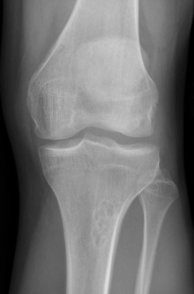
- Classic X-ray: eccentric, cortically based, well-defined metaphyseal lucency with a scalloped sclerotic rim
- Most are incidental, asymptomatic, and ossify or disappear with skeletal maturity
- Fracture risk increases when the lesion occupies greater than 50 percent cortical width or is longer than 33 mm
- Differentiate from fibrous dysplasia: central intramedullary lesion with ground-glass matrix
- “Do not biopsy a classic incidental NOF in the right age group
- “Large distal tibial lesions are the fracture-risk scenario examiners like
- “Pain at the lesion, cortical thinning, or a limp should trigger fracture-risk imaging review
- “Multiple NOFs plus cafe-au-lait macules should prompt syndromic assessment
Most NOFs need no treatment. A classic incidental lesion in a child or adolescent is benign, self-limiting, and usually reossifies with skeletal maturity.
Large lesions matter. Concern rises when the lesion occupies greater than 50 percent of bone diameter or is longer than 33 mm, especially in weight-bearing bones.
X-ray is usually enough. Biopsy is not indicated when age, location, margins, and cortically based metaphyseal pattern are classic.
Fibrous dysplasia is central and ground-glass. NOF is eccentric, cortical, lytic, and rimmed by sclerosis.
- Diagnosis
- Classic eccentric NOF on plain radiographs
- Treatment
- Reassure and observe
- Key Pearl
- No biopsy for a textbook lesion
- Diagnosis
- Greater than 50 percent width or longer than 33 mm
- Treatment
- Activity advice, surveillance, consider prophylactic curettage if high risk
- Key Pearl
- Assess true cortical support, not just lesion length
- Diagnosis
- Pathological fracture through NOF
- Treatment
- Treat fracture first, curettage only if lesion remains high risk
- Key Pearl
- Most fractures heal with standard paediatric fracture care
CORTEXRadiographic Pattern
Hook:NOF lives in the CORTEX: cortically based, eccentric, rimmed, and diagnosed on X-ray.
BIGFracture-Risk Screen
Hook:A BIG NOF is the one that breaks: width, length, and symptoms decide risk.
EDGENOF versus Fibrous Dysplasia
Hook:NOF is at the EDGE; fibrous dysplasia sits centrally with ground-glass bone.
Overview and Epidemiology
Non-ossifying fibroma is one of the commonest benign bone lesions in the growing skeleton. The clinical skill is not making an exotic diagnosis; it is recognising the classic benign pattern, avoiding unnecessary biopsy, identifying the small subgroup at fracture risk, and knowing when multiple lesions imply a syndromic diagnosis.
- Age: child or adolescent with open physes
- Symptoms: usually none; found after unrelated trauma or knee pain imaging
- Sites: distal femur, proximal tibia, distal tibia, proximal humerus, and other long-bone metaphyses
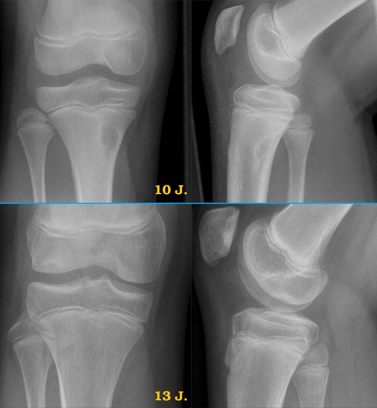
- Natural history: migrates away from the physis with growth, becomes sclerotic, and usually disappears or leaves a healed cortical scar
- Reassurance: most lesions require explanation rather than intervention
- Fracture risk: large weight-bearing lesions can weaken cortex
- Avoid harm: unnecessary biopsy can create anxiety and morbidity
- Syndromic clue: multiple lesions may be associated with Jaffe-Campanacci phenotype or neurofibromatosis type 1 features
Pathophysiology
NOF is best understood clinically as a benign developmental fibrous defect of metaphyseal cortex. Histology shows fibroblastic spindle cells, collagen, foamy histiocytes, and osteoclast-like giant cells, but biopsy is rarely required because the radiographic pattern is usually diagnostic. The lesion is active during growth, then ossifies as skeletal maturity approaches.
- Mechanism
- Small focus of fibrous tissue in cortex
- Clinical effect
- Incidental and mechanically insignificant
- When it matters
- Usually less than 2 cm and self-resolving
- Mechanism
- Larger eccentric fibrous metaphyseal lesion
- Clinical effect
- Can thin and expand cortex
- When it matters
- Assess size and cortical support in weight-bearing bones
- Mechanism
- Loss of cortical cross-sectional strength
- Clinical effect
- Pain, limp, deformity, or acute fracture
- When it matters
- Risk rises with large diameter and length
Slow growth lets the surrounding bone form a thin sclerotic margin. The lobulated fibrous tissue produces a scalloped or multiloculated contour rather than a permeative aggressive pattern.
With physeal closure and remodelling, the fibrous focus progressively ossifies. The lesion may become densely sclerotic, shrink, or become invisible on later radiographs.
Classification and Types
Classification by Radiographic Maturation
- Radiographic Features
- Eccentric lytic metaphyseal defect with thin rim
- Fracture Relevance
- Risk depends on size and cortex
- Management
- Observe if small and asymptomatic
- Radiographic Features
- Increasing peripheral and internal sclerosis
- Fracture Relevance
- Mechanical risk falling as bone fills in
- Management
- Reassure and reduce follow-up if stable
- Radiographic Features
- Dense cortical or metaphyseal sclerosis
- Fracture Relevance
- Low risk
- Management
- No treatment
Staging is useful because the active lucent stage is the period when fracture risk is most relevant.
Ritschl radiographic staging is the named NOF-specific scheme that formalises this natural history into four stages:
- Stage A — a small, round/oval cortical lucency lying close to the physis (the fibrous cortical defect end of the spectrum).
- Stage B — a larger, polycyclic/multilocular well-marginated lesion that may extend into the medulla as the bone grows.
- Stage C — sclerosis begins (typically from the diaphyseal margin) as the lesion migrates away from the physis and starts to heal.
- Stage D — complete sclerosis/ossification: the healed, often invisible residual lesion.
When multiple (multifocal/polyostotic) non-ossifying fibromas occur together with café-au-lait macules and extraskeletal anomalies, consider Jaffe-Campanacci syndrome. The recognised features are: multiple NOFs (causing bone fragility and recurrent pathological fractures), café-au-lait spots, and a variable combination of intellectual disability, hypogonadism or cryptorchidism, ocular anomalies, and cardiovascular malformations. It overlaps clinically with neurofibromatosis type 1 (and both involve RAS-pathway/mosaic mutations — see the KRAS evidence), so look specifically for neurofibromas and Lisch nodules and involve genetics/paediatrics. The orthopaedic priority is whole-skeleton fracture-risk assessment because multiple large lesions weaken several bones at once.
Clinical Assessment
- Why imaging was done: trauma, knee pain, limp, deformity, or incidental radiograph
- Pain pattern: true lesion pain is focal and activity-related; many children have unrelated anterior knee pain
- Fracture symptoms: sudden pain, inability to weight bear, swelling after low-energy trauma
- Syndrome screen: cafe-au-lait macules, neurofibromas, developmental delay, jaw swelling, endocrine history, family history of NF1
- Look: limb alignment, swelling, deformity, gait, skin stigmata of NF1
- Feel: focal tenderness over the lesion; diffuse joint-line pain suggests another diagnosis
- Move: adjacent joint range, effusion, mechanical symptoms, hip and spine screen if pain is poorly localised
- Function: single-leg hop, squat, sport tolerance, and weight-bearing ability when safe
Most NOFs are incidental. If the lesion is small and painless to direct palpation, search for common causes of pain: trauma, osteochondritis dissecans, stress injury, infection, inflammatory arthropathy, hip pathology referred to the knee, or sporting overuse. A classic NOF explains pain mainly when it is large, fractured, or directly tender.
- Typical Imaging
- Eccentric cortical metaphyseal lucency with sclerotic rim
- Clinical Clue
- Usually asymptomatic
- Discriminating Feature
- Classic scalloped rim, narrow transition zone
- Typical Imaging
- Central intramedullary ground-glass lesion
- Clinical Clue
- May cause deformity or pain
- Discriminating Feature
- Central, expansile, ground-glass matrix rather than cortical NOF
- Typical Imaging
- Expansile eccentric lytic lesion, often blow-out cortex
- Clinical Clue
- Painful, enlarging, sometimes after minor trauma
- Discriminating Feature
- MRI fluid-fluid levels and expansile behaviour
- Typical Imaging
- Central metaphyseal lucency, often proximal humerus or femur
- Clinical Clue
- Fracture presentation common
- Discriminating Feature
- Central lesion; fallen fragment sign after fracture
- Typical Imaging
- Wide transition zone, cortical destruction, periosteal reaction, soft tissue mass
- Clinical Clue
- Night pain, systemic symptoms, progressive pain
- Discriminating Feature
- Aggressive radiographic features; urgent sarcoma referral
NOF is eccentric and cortical with a sclerotic scalloped rim. Fibrous dysplasia is central and intramedullary with a ground-glass matrix and may produce expansile deformity. This distinction is a common exam trap because both occur in young patients and both are benign fibrous bone lesions.
Investigations
Imaging Protocol
Views: orthogonal AP and lateral radiographs of the involved bone including adjacent joints.
Look for: eccentric metaphyseal lucency, sclerotic scalloped rim, cortical thinning, fracture line, lesion length, and percentage bone width occupied.
Clinical correlation: if the child is pain-free and the radiographic pattern is classic, plain films are diagnostic.
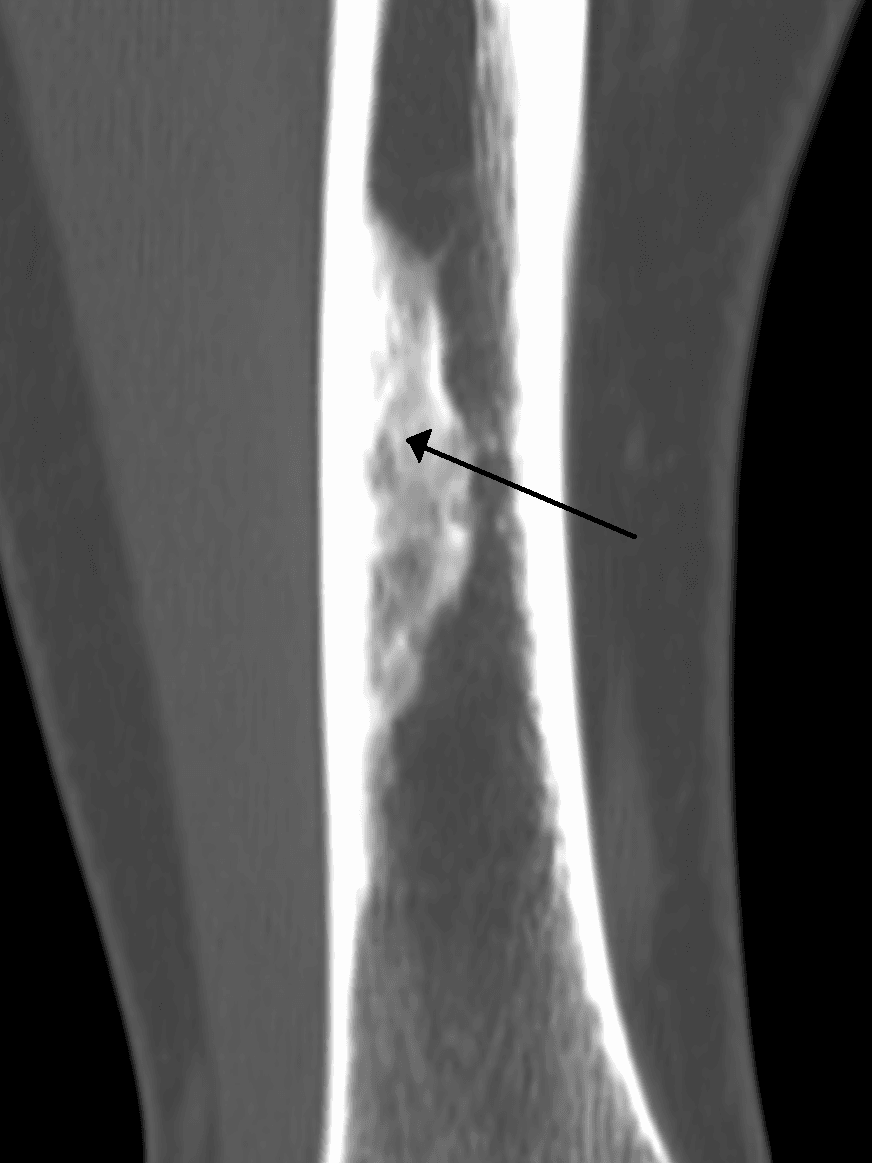
Indication: selected large lesions where cortical support or fracture risk cannot be judged on plain radiographs.
Use: define cortical breach, residual cortical shell, and geometry before prophylactic curettage or fixation.
Caution: avoid routine CT in classic small lesions because radiation rarely changes management.
Indication: atypical margins, aggressive features, persistent unexplained pain, possible infection, ABC, or malignant mimic.
Findings: NOF can show variable signal and enhancement; MRI is most useful to exclude alternative diagnoses, not to prove a classic NOF.
Referral trigger: soft tissue mass, marrow oedema out of proportion, cortical destruction, or wide zone of transition.
Measure the lesion in two ways: maximum length and maximum transverse diameter as a proportion of the bone diameter on AP and lateral views. The classic Drennan / Arata risk flags are length greater than 33 mm and occupation of greater than 50 percent of the bone diameter.
- What to Record
- Age, metaphyseal location, eccentric cortical position, narrow transition zone
- Management Impact
- Classic lesion avoids biopsy and extensive workup
- What to Record
- Length in millimetres and percentage bone width on both views
- Management Impact
- Large lesions need fracture-risk counselling and surveillance
- What to Record
- Subtle cortical break, periosteal reaction, angulation, or displacement
- Management Impact
- Treat fracture according to paediatric fracture principles
- What to Record
- Poorly defined margin, soft tissue mass, aggressive periosteal reaction, epiphyseal extension
- Management Impact
- Urgent tumour referral and advanced imaging
Management Algorithm
Incidental Classic NOF
Goal: reassure the family, avoid unnecessary biopsy, and confirm the lesion is mechanically safe.
Observation Protocol
Confirm pattern: child or adolescent, metaphyseal, eccentric, cortically based, well-defined sclerotic rim.
Explain: benign developmental lesion, not cancer, no malignant transformation expected, usually resolves with maturity.
Assess risk: measure width and length; examine for focal tenderness and gait change.
Criteria: small lesion, asymptomatic, no cortical breach, less than fracture-risk thresholds.
Treatment: normal activities, reassurance, no biopsy, no immobilisation.
Follow-up: discharge or one interval radiograph if diagnostic confidence or family anxiety requires it.
Criteria: approaching greater than 50 percent bone width or greater than 33 mm, particularly in tibia or femur.
Treatment: activity modification for high-impact sport if symptomatic or cortex very thin.
Follow-up: repeat radiographs until sclerosis or reduced mechanical concern.
The family needs two clear messages: the lesion is benign and usually self-resolving, but a large lesion can behave like a stress riser. That framing prevents both over-treatment and unsafe dismissal.
Complications
- Incidence
- Uncommon overall, concentrated in large lesions
- Risk Factors
- Greater than 50 percent bone width, greater than 33 mm length, lower limb, active lucent stage
- Management
- Immobilise or fix fracture; reassess lesion after union
- Incidence
- Avoidable when imaging is classic
- Risk Factors
- Failure to recognise benign radiographic pattern
- Management
- Correlate age, location, margin, and cortical position before referral
- Incidence
- Rare but high consequence
- Risk Factors
- Painful atypical lesion, wide transition zone, soft tissue mass, periosteal reaction
- Management
- Urgent MRI and sarcoma pathway referral
- Incidence
- Rare
- Risk Factors
- Multiple lesions, cafe-au-lait macules, jaw giant-cell lesions, endocrine or developmental features
- Management
- Genetics / paediatric assessment and whole-skeleton risk review
NOF does not become malignant. The reason to follow or treat a large lesion is mechanical weakness, especially in a weight-bearing bone. Explain this clearly: a child may need surveillance or curettage because the cortex is thin, not because the lesion is biologically dangerous.
Outcomes and Prognosis
- Treatment
- Reassurance, no intervention
- Expected Outcome
- Spontaneous sclerosis or resolution with maturity
- Long-term Function
- Normal function and unrestricted activity
- Treatment
- Observation or selective curettage
- Expected Outcome
- Usually stabilises as it ossifies; surgery prevents selected fractures
- Long-term Function
- Excellent if fracture is avoided or healed
- Treatment
- Standard fracture care with or without grafting / fixation
- Expected Outcome
- Union expected in most paediatric patients
- Long-term Function
- Return to sport after union and cortical restoration
Best prognosis: classic incidental lesion, small size, no pain, increasing sclerosis, and approaching skeletal maturity.
Higher-risk prognosis: large active lucent lesion, thin cortex in the distal tibia or femur, focal pain, or a previous pathological fracture.
Key threshold: large size changes fracture risk, not tumour biology.
Guidelines, Registries & Global Practice
- Worldwide lesion: NOF is common in children and adolescents across all health systems
- Detection pattern: most are found incidentally on radiographs taken for trauma or sports symptoms
- Clinical burden: low oncological burden, but large lower-limb lesions create fracture-risk decisions
- Syndromic cases: multiple lesions are uncommon and should prompt a broader clinical examination
- High-resource: MRI or CT may be used for atypical lesions or pre-operative mechanical assessment
- Limited-resource: plain radiographs are usually sufficient for classic lesions and fracture follow-up
- Universal principle: classic, small, asymptomatic NOF needs reassurance, not biopsy
- Referral: atypical imaging or aggressive features should enter a bone-tumour pathway wherever the child is treated
- Diagnosis emphasis
- Benign fibroblastic / histiocytic lesion of growing skeleton
- Observation / follow-up
- Clinical-radiographic diagnosis when classic
- Surgery / referral
- Histology reserved for atypical or uncertain cases
- Diagnosis emphasis
- Do not biopsy classic benign latent lesions unnecessarily
- Observation / follow-up
- Follow only if lesion is symptomatic, large, or atypical
- Surgery / referral
- Refer aggressive features to specialist tumour care
- Diagnosis emphasis
- Assess size, cortex, symptoms, and weight-bearing location
- Observation / follow-up
- Serial radiographs for large active lesions until sclerosis
- Surgery / referral
- Selective curettage / grafting for impending or recurrent fracture risk
- Diagnosis emphasis
- Treat pathological fracture by stability, alignment, age, and bone involved
- Observation / follow-up
- Observe lesion after union if cortical support restores
- Surgery / referral
- Fix unstable fractures and fill defects when structural support is inadequate
There is no implant or arthroplasty registry equivalent for NOF because most lesions are non-operative developmental findings. Evidence is mainly historical descriptions, retrospective fracture series, and observational natural-history studies. The practical global standard is therefore principle-based: recognise the classic benign lesion, measure mechanical risk, and refer atypical or aggressive imaging.
Record in every NOF assessment:
- Why the radiographic appearance is classic or atypical
- Lesion length and percentage bone diameter on AP and lateral views
- Whether there is focal tenderness, limp, fracture line, or cortical breach
- Family counselling: benign natural history, fracture-risk advice if large, and return precautions
Clear documentation prevents two opposite errors: unnecessary tumour anxiety in small classic lesions and unsafe reassurance in large lower-limb lesions.
Controversies & Areas of Uncertainty
Greater than 50 percent bone diameter and greater than 33 mm length are useful classic risk flags, but they are not absolute rules. Symptoms, site, cortical thickness, lesion stage, and child activity level all modify risk.
There is no universally mandated follow-up schedule. Many units discharge small classic lesions, while large active lesions are followed until sclerosis or skeletal maturity reduces risk.
Surgery can prevent fracture in selected large symptomatic lesions, but many large lesions heal without operation. The decision balances fracture probability, sport exposure, lesion site, and family preference.
Multiple NOFs overlap clinically with NF1-spectrum findings. Whether a child has a distinct syndrome or NF1-related manifestation may require genetics and paediatric assessment.
MCQ Practice Points
Q: What is the classic radiographic appearance of a non-ossifying fibroma? A: Eccentric, cortically based, metaphyseal, well-defined lytic lesion with a scalloped sclerotic rim in a child or adolescent. The long axis often parallels the bone, and the transition zone is narrow.
Q: Which NOFs are at risk of pathological fracture? A: Large active lesions in weight-bearing bones, especially those occupying greater than 50 percent of the bone diameter or measuring greater than 33 mm in length. Symptoms, cortical thinning, and site matter as much as a single measurement.
Q: How do you distinguish NOF from fibrous dysplasia? A: NOF is eccentric and cortical; fibrous dysplasia is central and ground-glass. Fibrous dysplasia may expand the medullary canal and produce deformity, whereas NOF has a thin sclerotic rim and cortical metaphyseal origin.
Q: What is the treatment for an incidental small NOF? A: Reassurance and observation. No biopsy, curettage, or activity restriction is required when the lesion is classic, asymptomatic, and mechanically low risk.
Q: What should multiple NOFs make you consider? A: Jaffe-Campanacci syndrome or NF1-spectrum features. Look for cafe-au-lait macules, neurofibromas, jaw giant-cell lesions, developmental or endocrine abnormalities, and family history.
Clinical Imaging
Radiographic Pattern of Non-Ossifying Fibroma
The classic lesion is eccentric, cortically based, metaphyseal, and well circumscribed with a thin scalloped sclerotic rim. It is usually discovered incidentally on radiographs taken for trauma, limp, alignment assessment, or knee pain unrelated to the lesion.
- Typical NOF
- Eccentric metaphysis of a long bone, often around the knee
- Why it matters
- A classic location in a child supports diagnosis without biopsy
- Typical NOF
- Narrow zone of transition with a scalloped sclerotic rim
- Why it matters
- A sharp rim indicates slow benign growth and host response
- Typical NOF
- Cortically based lucency with expansion or thinning if large
- Why it matters
- Cortical involvement determines fracture risk
- Typical NOF
- Lytic and multiloculated without aggressive mineralised matrix
- Why it matters
- Ground-glass central matrix suggests fibrous dysplasia instead
Exam Viva Scenarios
Practise clinical reasoning and management decisions out loud
“A 12-year-old footballer has knee radiographs after a twisting injury. The knee injury is improving, but the AP film shows a 22 mm eccentric lytic metaphyseal lesion in the distal femur with a thin scalloped sclerotic rim. He has no focal tenderness over the lesion and no systemic symptoms. How do you manage this?”
“A 10-year-old presents with activity-related distal tibial pain. X-rays show a 45 mm eccentric cortically based metaphyseal lesion with a sclerotic rim. It occupies greater than 50 percent of the tibial diameter on the lateral view, with marked cortical thinning but no fracture line. What is your assessment and management?”
Key Imaging
- Child or adolescent with eccentric cortically based metaphyseal lucency
- Thin scalloped sclerotic rim and narrow zone of transition
- Usually around the knee or distal tibia, but can occur in other long bones
- Fibrous dysplasia is central and ground-glass, not eccentric cortical
Diagnosis
- Plain radiographs diagnose a classic NOF
- No biopsy for an asymptomatic textbook lesion
- MRI only for atypical features, persistent unexplained pain, or aggressive mimic
- Multiple lesions require skin, jaw, endocrine, developmental, and family-history assessment
Fracture Risk
- Large lesion: greater than 50 percent bone diameter or greater than 33 mm length
- Risk highest in lower-limb weight-bearing bones with cortical thinning
- Pain, limp, and active lucent stage increase concern
- Size thresholds are risk flags, not automatic surgery indications
Treatment Algorithm
- Small incidental NOF = reassure, observe, no activity restriction
- Large asymptomatic NOF = measure, counsel, consider surveillance and activity advice
- Symptomatic high-risk NOF = consider curettage and graft / substitute
- Fractured NOF = treat fracture first, then reassess residual lesion
Exam Traps
- Do not call NOF malignant; it is benign and self-limiting
- Do not confuse NOF with central ground-glass fibrous dysplasia
- Do not ignore a large distal tibial lesion because the diagnosis is benign
- Do not miss Jaffe-Campanacci / NF1 features when lesions are multiple
Evidence Base and Key Trials
Diagnostic and treatment strategies for adult non-ossifying fibromas: a case report and literature review
- NOF is the commonest benign bone tumour of childhood and usually regresses spontaneously; in skeletally mature patients it can persist and mimic GCT or aneurysmal bone cyst
- Surgical indication is based on lesion size (over 33 mm), cortical integrity and fracture risk (over 50 percent of bone diameter) rather than symptoms alone
- Large cortically-compromised lesions were managed with extended curettage, PMMA cement and prophylactic fixation; biopsy is mandatory for radiologically indeterminate lesions
Trametinib for multiple non-ossifying fibromas due to KRAS mosaic mutations: two case reports
- KRAS mosaic activating variants are the main molecular cause of non-ossifying fibromas
- Multifocal (polyostotic) NOFs cause bone fragility and recurrent fractures (e.g. Jaffe-Campanacci spectrum)
- The MEK inhibitor trametinib produced radiological reossification and stopped fractures, with relapse after cessation