Prevention | Measurement | Intraoperative Tools | Medicolegal Risk
- Document preoperative LLD - medicolegal essential, compare to contralateral
- 10mm threshold - patient dissatisfaction increases dramatically over 10mm
- Lengthening safer than shortening - nerve palsy risk with overlengthening over 4cm
- Multiple measurement methods - intraoperative verification with at least 2 techniques
- Offset restoration - LLD often related to femoral offset reconstruction errors
- “LLD is a leading source of negligence complaints after THA
- “3D CT planning predicts combined cup and stem size in 96% vs 16% for 2D templating
- “Navigation's pooled benefit for LLD is modest; clearest gain is cup orientation
- “Shoe lift usually needed only once LLD approaches 20mm
Among the highest litigation risks in THA. Document preoperative LLD, templating plan, intraoperative measurements, and postoperative counseling. Informed consent must include LLD possibility (even with perfect technique).
Multiple techniques required. Never rely on single method. Use combination of: Shuck test, direct measurement from fixed points, calibrated pins, intraoperative fluoroscopy, and surgical navigation.
LLD often secondary to offset error. Restoring femoral offset is key - inadequate offset leads to compensatory lengthening. Template femoral offset carefully; measure both length and offset intraoperatively.
Preoperative counseling essential. Some patients tolerate LLD better than others. High-demand patients, fixed spinal deformity, and bilateral disease require extra caution. Document baseline gait and spine pathology.
- Acceptable LLD
- Under 5mm
- Management
- Strict intraoperative measurement, both offset and length
- Key Pearl
- Computer navigation or robotics strongly recommended
- Acceptable LLD
- 5-10mm
- Management
- Template carefully, use 2+ measurement methods
- Key Pearl
- Most patients tolerate this range well
- Acceptable LLD
- 10-15mm
- Management
- Accept lengthening for stability if needed
- Key Pearl
- Document trade-off: stability vs LLD
- Acceptable LLD
- 15-20mm acceptable
- Management
- Nerve monitoring, gradual lengthening protocol
- Key Pearl
- Over 4cm = high sciatic nerve palsy risk
OFFSETCauses of Iatrogenic LLD in THA
Hook:OFFSET errors cause most LLD - restore anatomy before adjusting length!
Overview and Epidemiology
Leg length discrepancy (LLD) after total hip arthroplasty is the most common reason for patient dissatisfaction and litigation despite being a known complication of the procedure. Even with modern techniques, some degree of LLD is almost universal.
LLD affects gait mechanics, patient satisfaction, and medicolegal risk. Small discrepancies (under 10mm) are usually well-tolerated, but patient perception often exceeds actual measured LLD. The key is documentation - proving preoperative assessment and intraoperative diligence protects against litigation even if LLD occurs.
- Most primary THAs achieve residual LLD under 10mm with careful technique
- Up to 10mm natural LLD exists in 60-95% of the population (O'Brien 2010)
- Larger discrepancies (over 15mm) less common but cause functional gait changes
- Revision and dysplasia carry higher rates of significant LLD
- Developmental dysplasia (high hip center)
- Revision surgery (bone loss, soft tissue laxity)
- Severe deformity (Crowe III-IV DDH)
- Inadequate templating (most preventable cause)
- ~97% perceive a 10mm imposed discrepancy (O'Brien 2010)
- No subject perceived a 5mm increase as uncomfortable
- Psychological factors influence perception
- Body image and expectations play major role
- Leading source of THA negligence complaints
- Uncemented stems less forgiving for LLD (Whittingham-Jones 2012)
- Preoperative planning critical with abnormal anatomy
- Documentation is defense - proves due diligence
Anatomy and Biomechanics
Anatomical Determinants of Leg Length
True leg length is ASIS to medial malleolus. Apparent length is umbilicus to medial malleolus and reflects pelvic obliquity. THA aims to restore true length while accounting for fixed pelvic tilt and spinal deformity.
- Landmarks
- ASIS to medial malleolus
- What It Assesses
- Actual skeletal length
- Clinical Use
- Primary measurement for LLD
- Landmarks
- Umbilicus to medial malleolus
- What It Assesses
- Functional length with pelvic tilt
- Clinical Use
- Screening for pelvic obliquity
- Landmarks
- Lesser trochanter to acetabular teardrops
- What It Assesses
- Hip offset and neck length
- Clinical Use
- Templating and intraoperative check
Femoral Offset and Length Relationship
Femoral offset is the perpendicular distance from the center of rotation of the femoral head to the long axis of the femur. Restoring offset is essential - inadequate offset forces the surgeon to lengthen the leg to achieve stability and abductor tension.
- Abductor moment arm restored (reduces limp)
- Stability without excessive lengthening
- Range of motion improved
- Gait efficiency normalized
- Under-restore offset → compensatory lengthening for stability
- Over-restore offset → potential impingement
- Offset error of 5mm can translate to 10mm LLD
- Template offset first then adjust length
Acetabular Offset, Hip Centre and Global Offset
Teaching on length restoration concentrates on the femoral side, but the acetabular component is an equal determinant of both length and offset - a point the warnings above about a "high, medial" cup and the Weber trial's use of global offset make explicit.
- Centre of rotation (hip centre). The vertical position of the cup sets the acetabular contribution to leg length. A cup placed superiorly (a high hip centre) shortens the limb relative to anatomic; restoring the centre of rotation inferiorly to its anatomic level lengthens it. This is why a high, medialised cup tempts the surgeon into compensatory femoral overlengthening to regain stability.
- Global (total) offset = acetabular offset + femoral offset. Acetabular offset is the horizontal distance from the cup centre of rotation to the pelvic (teardrop) reference; femoral offset is the distance from the head centre to the femoral axis. Medialising the cup reduces acetabular offset and therefore global offset, weakening the abductor lever arm exactly as an under-restored femoral offset does. Restoring the anatomic centre of rotation preserves global offset and avoids the trap of lengthening the leg to chase abductor tension.
Leg length and offset are restored on both sides of the joint. A perfectly templated femoral stem cannot rescue a cup that is too high (shortens) or too medial (loses global offset). Set the acetabular centre of rotation anatomically first, then template the femoral side to it - this is the logic behind "restore the hip centre, then restore offset, then fine-tune length."
Soft Tissue Tension and Length
The soft tissue envelope determines acceptable leg length changes. Overlengthening stretches neurovascular structures; excessive shortening creates laxity and instability.
- Effect of Lengthening
- Traction neuropraxia
- Critical Threshold
- Over 4cm lengthening
- Clinical Consequence
- Foot drop, sciatic palsy
- Effect of Lengthening
- Excessive tension
- Critical Threshold
- Over 2cm lengthening
- Clinical Consequence
- Pain, Trendelenburg gait
- Effect of Lengthening
- Laxity if shortened
- Critical Threshold
- Over 1cm shortening
- Clinical Consequence
- Instability, dislocation risk
Classification Systems
LLD Classification by Etiology
Pre-existing Leg Length Discrepancy
Discrepancy present before THA due to underlying hip disease or developmental abnormalities.
- Typical LLD
- 5-15mm shortening
- Clinical Features
- Joint space loss, femoral head collapse
- Surgical Consideration
- Document baseline, can often equalize safely
- Typical LLD
- 10-25mm shortening
- Clinical Features
- High hip center, shallow acetabulum
- Surgical Consideration
- Gradual correction, may need soft tissue releases
- Typical LLD
- Over 40mm shortening
- Clinical Features
- High dislocation, severe bone loss
- Surgical Consideration
- Accept residual LLD - nerve palsy risk if fully equalized
- Typical LLD
- Variable (10-30mm)
- Clinical Features
- Malunion, bone loss, deformity
- Surgical Consideration
- Complex reconstruction, may need femoral osteotomy
Clinical Assessment
History and Physical Examination
- Baseline gait - did patient limp before surgery, use walking aids
- Shoe modifications - prior shoe lift use indicates tolerance
- Spine pathology - scoliosis, spinal fusion, low back pain
- Contralateral hip - arthritis or prior THA affects comparison
- Patient expectations - some patients fixate on perfect symmetry
- Functional goals - high-level athletics vs basic mobility
- Gait analysis - Trendelenburg, limp, compensatory trunk lean
- True leg length - ASIS to medial malleolus with tape measure
- Apparent leg length - umbilicus to medial malleolus
- Block test - place blocks under short leg to level pelvis
- Spine exam - fixed scoliosis vs compensatory curve
- Contralateral comparison - measure both sides identically
Preoperative LLD Measurement Technique
Clinical Measurement Protocol
Supine on firm surface (exam table, not soft bed). Both ASIS and pubic symphysis must be palpable. Ensure pelvis is level - check that both ASIS are at same height.
Align both limbs identically in neutral rotation and extension. Draw line from umbilicus through pubic symphysis to ensure midline reference. Correct any pelvic obliquity or rotation.
Palpate and mark ASIS bilaterally with pen. Palpate medial malleolus bilaterally and mark. Use consistent technique for both sides (same fingertip, same bony point).
Tape measure from ASIS to medial malleolus on each side. Record in centimeters. Repeat measurement twice to confirm. Document which side is longer/shorter and by how much.
Place measured blocks (5mm increments) under short leg. Progressive stacking until pelvis is level (check ASIS heights). Final block height confirms functional LLD magnitude.
Document every measurement in clinic notes and operative planning: (1) True leg length bilaterally, (2) Apparent leg length, (3) Direction and magnitude of LLD, (4) Whether patient uses shoe lift currently, (5) Discussion with patient about LLD risk and management options. This documentation defends against litigation even if postoperative LLD occurs.
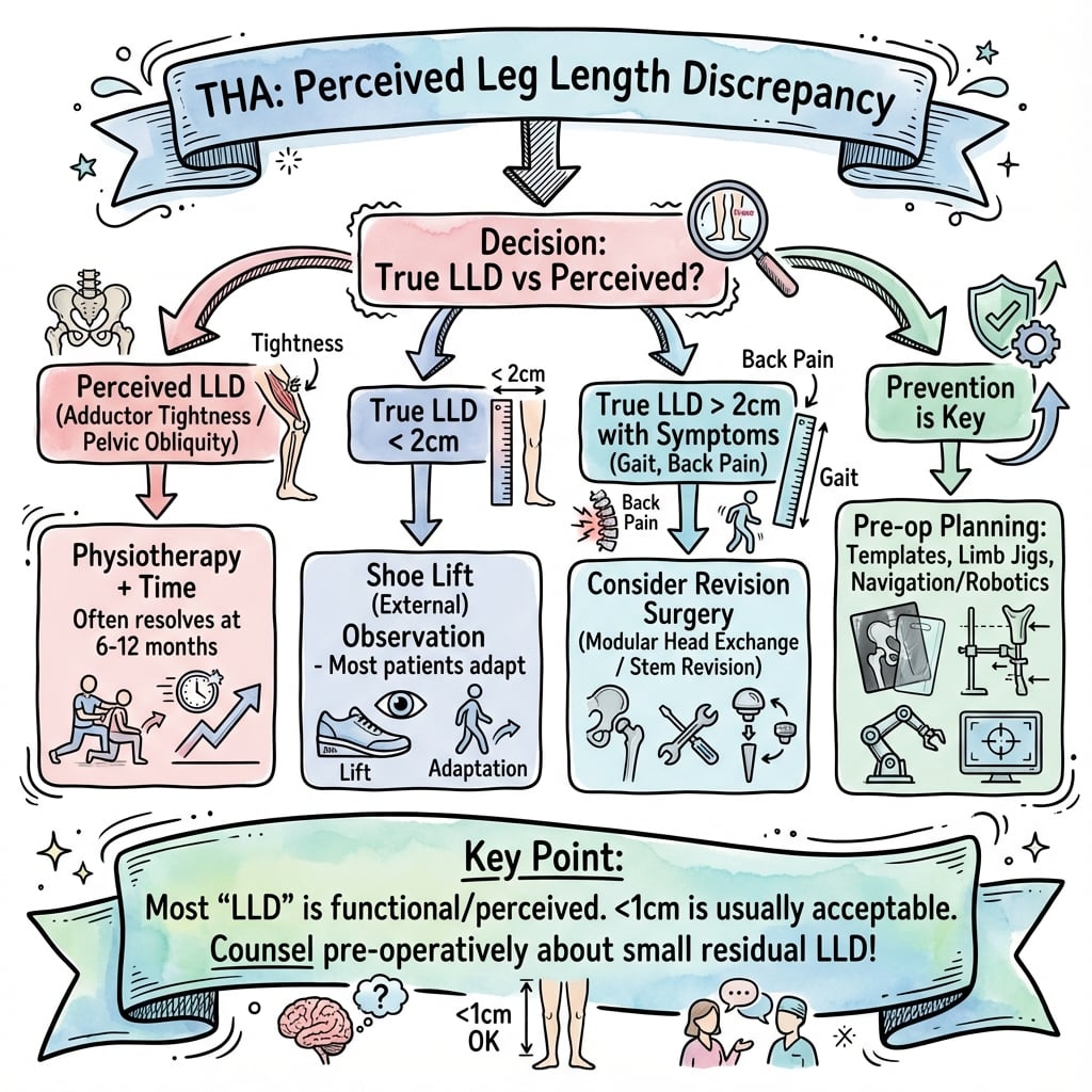
Differential Diagnosis of Perceived Limb Length Inequality After THA
Not every patient who reports a "long" or "short" leg has a true skeletal LLD. The key clinical task is to separate true (skeletal) LLD from functional/apparent LLD and from other causes of an asymmetric-feeling gait.
- Mechanism
- Real difference in ASIS-to-medial-malleolus length
- Distinguishing Feature
- Block test levels pelvis; teardrop-to-lesser-trochanter differs on AP pelvis
- Management
- Shoe lift if symptomatic; address cause
- Mechanism
- Fixed pelvic obliquity, hip adduction/flexion contracture or scoliosis
- Distinguishing Feature
- True lengths equal but apparent (umbilicus-malleolus) lengths differ
- Management
- Treat contracture/spine; lift rarely helps
- Mechanism
- Under-restored offset or abductor insufficiency
- Distinguishing Feature
- Trendelenburg gait, weak abduction; offset reduced on radiograph
- Management
- Strengthening; revise offset if structural
- Mechanism
- Fixed lumbar deformity or prior fusion altering pelvic tilt
- Distinguishing Feature
- Compensatory trunk lean, persists when supine pelvis levelled
- Management
- Spine assessment; cautious lift trial
- Mechanism
- Altered proprioception and heightened expectation post-THA
- Distinguishing Feature
- Measured LLD minimal; complaint disproportionate; often improves over months
- Management
- Reassurance, education, show radiographs
Pelvic Obliquity and Contracture: True versus Functional Length
The discrepancy the surgeon actually equalises at THA is the true (skeletal) length; what the patient feels is the functional length, which a fixed pelvic obliquity or a hip contracture can distort even when the skeletal lengths are equal. The TEMPLATE checklist's "fixed vs compensatory pelvic obliquity" step and the apparent-length measurement exist precisely to separate these.
Classify the source of a pelvic obliquity - it determines whether THA (or a lift) can help:
- Suprapelvic - driven from above by a fixed structural scoliosis or sagittal imbalance. A fixed suprapelvic obliquity persists after a perfectly balanced THA, so the patient may still perceive inequality; the spinopelvic and component-positioning consequences are addressed in the hip-spine relationship and spinopelvic-parameter topics.
- Infrapelvic - driven from below by a hip adduction or flexion contracture (or a knee/ankle deformity). A fixed adduction contracture makes that leg sit functionally short (apparent shortening); an abduction contracture makes it sit functionally long. Releasing the contracture at THA corrects this functional component without changing skeletal length.
- Intrapelvic - asymmetry within the pelvic ring itself (post-traumatic, post-radiation, congenital).
Why it matters. A flexible/compensatory obliquity corrects once the hip is balanced, so aim for true skeletal equality. A fixed obliquity can leave the patient feeling unequal even after full skeletal equalisation - so document it, set expectations before surgery, and base any shoe-lift trial on the functional (block-test) length rather than the radiographic skeletal difference.
You can only restore skeletal length at THA. Where a fixed pelvic obliquity or contracture drives functional LLD, equalising the bones will not satisfy the patient - identify it preoperatively (block test, standing films, spine exam), document it, and make it part of the consent conversation. This is a common reason a "radiographically perfect" THA still generates an LLD complaint.
Investigations
Radiographic Assessment Protocol
Imaging Sequence for LLD Assessment
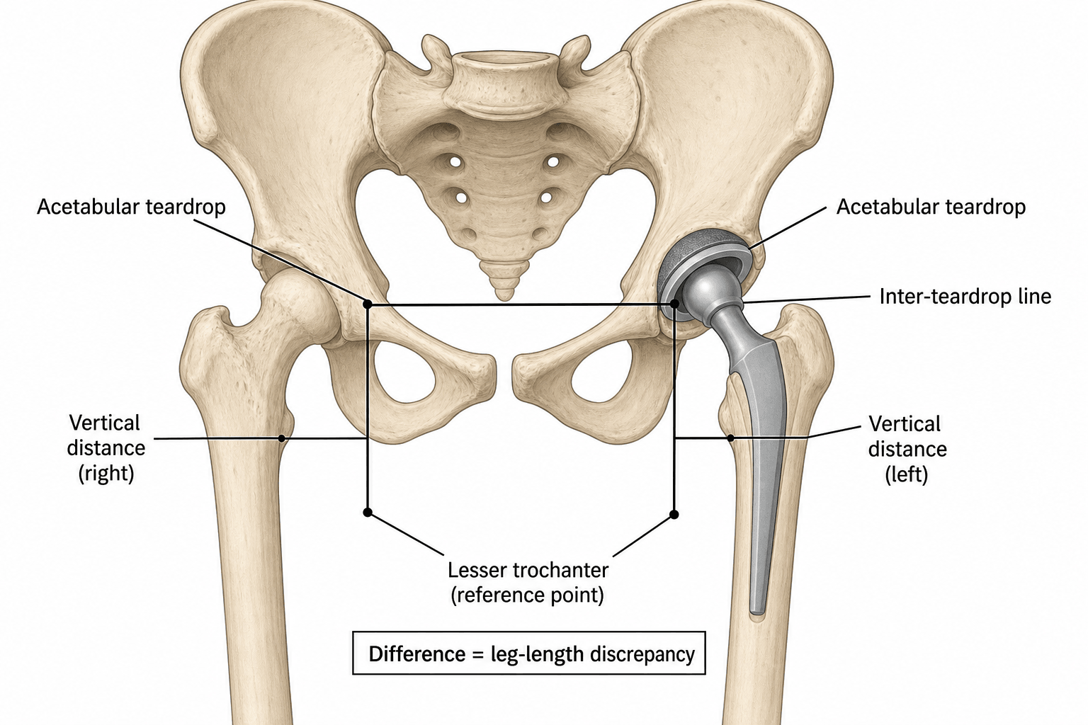
Gold standard for LLD measurement. Patient standing, weight-bearing bilateral. Ensure pelvis is level - check obturator foramina symmetry. Measure from acetabular teardrops (or inter-teardrop line) to lesser trochanters bilaterally. Calculate difference.
Assess femoral offset and neck length. Measure perpendicular distance from femoral axis to center of femoral head (offset). Assess femoral bow and canal width for stem selection. Identify anatomic landmarks for neck cut level.
For preoperative LLD over 2cm, DDH, or bilateral disease. Measure mechanical axis and true leg length on calibrated images. Assess compensatory knee or ankle changes. Plan for gradual correction if large LLD.
For severe DDH, post-traumatic deformity, or revision with bone loss. Assess bone stock and 3D anatomy. Plan for custom implants or osteotomy. Required for robotic-assisted surgery planning.
Radiographic Measurement Techniques
Inter-Teardrop Line Method (Most Common)
Draw horizontal line connecting medial acetabular teardrops. Measure vertical distance from this line to the tip of each lesser trochanter. Calculate difference between sides.
- Standardized reproducible method
- Not affected by pelvic tilt
- Used in most published studies
- Easy to measure on PACS systems
- Assumes teardrops are symmetric (may not be in DDH)
- Obscured teardrops in severe arthritis
- Small measurement errors magnified
- Requires good quality AP pelvis
Management Algorithm
Treatment Decision Algorithm
Preoperative and Intraoperative Prevention
Best approach is prevention - careful planning and intraoperative measurement substantially reduce the incidence of clinically significant LLD.
Prevention Protocol
Document baseline LLD in clinic notes. Digital templating on calibrated AP pelvis - measure hip center, femoral offset, neck length targets. Informed consent discussion including LLD risk. Equipment check - ensure measurement tools available (calipers, navigation if planned).
Review template with surgical team. Confirm component sizes in stock including backup sizes. Position patient carefully with pelvis level and secured. Baseline clinical measurement before prepping.
Restore hip center anatomically (not high-medial). Restore femoral offset first using templated size and offset option. Neck cut per template (measure from lesser trochanter). Trial reduction with multiple measurements - shuck test, direct measurement, fluoroscopy. Adjust components systematically if LLD detected. Final verification before closure.
Record measurement methods used. Document trial reduction findings. State final leg length relative to contralateral and preoperative baseline. This protects against litigation.
Preoperative Assessment and Templating
Clinical Examination
- Baseline gait - preexisting limp or shoe lift
- Spine pathology - fixed scoliosis or kyphosis
- Contralateral hip - arthritic or operated
- Patient expectations - tolerance for minor LLD
- True leg length - ASIS to medial malleolus bilaterally
- Apparent leg length - umbilicus to malleolus
- Pelvic obliquity - assess for fixed vs compensatory
- Block test - place blocks under short leg to level pelvis
Essential medicolegal step: Measure and document preoperative leg lengths in medical record and operative note. State magnitude, direction, and whether patient currently uses shoe lift. This protects against litigation if postoperative LLD occurs.
Radiographic Assessment
Imaging Protocol for LLD Assessment
Essential view for bilateral comparison. Ensure pelvis is level (check obturator foramina symmetry). Measure from acetabular teardrops to lesser trochanters bilaterally. Assess hip center height and pelvic obliquity.
Femoral offset assessment. Measure offset from femoral axis to center of femoral head. Plan stem size and neck length. Assess anterior femoral bow (affects stem seating depth).
For preoperative LLD over 2cm or DDH. Assess overall limb alignment and compensatory knee changes. Measure mechanical axis. Check for fixed deformities.
Digital Templating
Preoperative templating improves the accuracy of restoring length and offset and is now standard of care; 3D CT-based planning predicts combined cup and stem size in 96% of cases versus 16% for conventional 2D templating (Sariali 2012).
- Calibrate images to known object (femoral head or marker)
- Template acetabulum first - position, size, inclination
- Template femoral component - size, neck length, offset
- Measure both length and offset on template
- Hip center height - superior/inferior relative to teardrops
- Femoral offset - medial/lateral from femoral axis
- Neck length - distance from stem shoulder to head center
- Target LLD - document planned length change
Template the contralateral normal hip if unilateral disease. This gives target values for offset and neck length. If bilateral disease, template worst hip first and use it as reference for second side to minimize bilateral LLD.
TEMPLATEPreoperative LLD Assessment
Hook:TEMPLATE your hips before surgery - proper planning prevents LLD litigation!
Intraoperative LLD Measurement Techniques
Measurement Methods Overview
No single measurement technique is 100% reliable. Best practice is to use at least two different methods and cross-check results.
Shuck Test (Ligamentous Laxity Assessment)
Most common bedside technique - assess relative laxity compared to contralateral hip.
Shuck Test Technique
Patient supine. Contralateral hip flexed and externally rotated (removes it from pelvis). Operative hip extended in neutral rotation.
Grasp operative limb at ankle. Apply axial traction and compression. Note excursion distance (typically 5-10mm normal laxity).
Reduce trial components. Repeat traction-compression. Compare excursion to baseline and to contralateral side.
Equal laxity = good match. Tight (less laxity) = overlengthened. Loose (more laxity) = underlengthened or unstable.
- Quick and simple
- No equipment needed
- Assesses soft tissue tension
- Compares to contralateral
- Subjective feel
- Unreliable in bilateral disease
- Affected by muscle relaxation
- Does not give absolute measurement
SHUCKIntraoperative LLD Measurement Techniques
Hook:SHUCK the hip multiple ways - never rely on one measurement method alone!
Postoperative Assessment and Management
Postoperative LLD Measurement
Postoperative Assessment Protocol
Clinical examination: Measure apparent and true leg lengths. Compare to preoperative baseline. Document in progress notes. Discuss findings with patient and family.
Standing AP pelvis radiograph: Measure from teardrops to lesser trochanters bilaterally. Assess hip center position and offset. Compare to templated plan. Measure actual LLD.
Shoe lift trial: Start with half the measured LLD (if 10mm LLD, try 5mm lift). Assess gait and comfort. Increase incrementally if needed. Consider full-length films if bilateral disease or spine pathology.
Management of Postoperative LLD
Non-Operative Management of LLD
Most LLD under 20mm is managed non-operatively with shoe modifications and physiotherapy.
- Start low: Begin with 50% of measured LLD
- External lift: Easier to adjust and remove if uncomfortable
- Internal lift: More cosmetic but limited to 10mm
- Full-length insole: Better than heel lift alone
- Gait training: Teach energy-efficient gait patterns
- Core strengthening: Address compensatory trunk lean
- Hip abductor exercises: Restore normal biomechanics
- Flexibility: Address soft tissue contractures
Shoe lifts are needed for LLD over 20mm in most patients. Between 10-20mm, it is patient preference. Under 10mm, lifts often make symptoms worse due to altered biomechanics. Trial before permanent prescription.
Patient perception often exceeds actual LLD. Some patients with 5mm actual LLD report feeling 2cm discrepancy. Reassurance and education are critical. Show them the radiographs. Explain that some LLD is expected even with perfect technique. Empathy and communication prevent escalation to litigation.
Surgical Technique for LLD Prevention
Preoperative Planning Checklist
- Informed consent - include LLD possibility
- Baseline documentation - preoperative leg lengths
- Expectations - acceptable range discussion
- Shoe lift - mention as option if needed
- Digital template both hips (compare to normal)
- Document target values - offset, neck length, cup position
- Plan component sizes - have backup sizes available
- Measure preoperative LLD on images
Planning Sequence
Measure and document true and apparent leg lengths. Assess spine and contralateral hip. Discuss LLD risk and management options with patient.
Obtain quality AP pelvis and lateral hip. Perform digital templating. Calculate target offset and neck length. Plan acetabular position.
Ensure measurement tools available (calipers, pins, navigation if planned). Confirm component sizes in stock. Review template with surgical team.
Complications of LLD
- Threshold
- Over 10mm perceived LLD
- Clinical Features
- Complaints of limp, uneven shoe wear, low back pain
- Management
- Reassurance, PT, shoe lift trial, revision if over 20mm
- Threshold
- LLD over 15mm
- Clinical Features
- Trendelenburg gait, increased energy expenditure, limp
- Management
- Gait training, shoe lift, consider revision
- Threshold
- LLD over 10mm with scoliosis
- Clinical Features
- Compensatory lumbar curve, muscle spasm, radiculopathy
- Management
- PT, NSAIDs, shoe lift, rarely spine surgery
- Threshold
- Overlengthening over 4cm
- Clinical Features
- Foot drop, sensory loss, pain posterior leg
- Management
- Decompress acutely, nerve monitoring, revision to shorten
- Threshold
- Any LLD if not documented
- Clinical Features
- Patient files lawsuit, claims negligence
- Management
- Defense requires preop documentation and informed consent
Sciatic nerve traction injury occurs with acute lengthening over 4cm (40mm). Risk higher in DDH and revision surgery. Prevention: Gradual lengthening protocol if needed, nerve monitoring, accept some residual LLD rather than risk palsy. Treatment: If palsy occurs, decompress immediately - revise to shorten limb.
- Nerve palsy - sciatic or femoral traction
- Wound breakdown - tension from overlengthening
- Dislocation - inadequate soft tissue tension from shortening
- Patient dissatisfaction - immediate perception of LLD
- Chronic back pain - compensatory spinal changes
- Hip abductor weakness - altered biomechanics from offset error
- Knee pain - contralateral knee overload
- Litigation - delayed lawsuit filing (up to 3 years)
Outcomes and Prognosis
Patient Outcomes After LLD
Prognosis for LLD after THA depends on magnitude, patient factors, and management. Most patients with minor LLD (under 10mm) adapt well and are satisfied with their outcomes. Larger discrepancies often require intervention but can be successfully managed non-operatively in majority of cases.
- Patient Satisfaction
- 95% satisfied
- Functional Outcome
- Normal gait and function
- Long-term Prognosis
- Excellent - no intervention needed
- Patient Satisfaction
- 85-90% satisfied
- Functional Outcome
- Minimal gait changes, most adapt
- Long-term Prognosis
- Good - temporary shoe lift in 20%, most discontinue
- Patient Satisfaction
- 60-70% satisfied
- Functional Outcome
- Visible limp, compensatory trunk lean
- Long-term Prognosis
- Fair - 60% need permanent shoe lift, 10% consider revision
- Patient Satisfaction
- 40-50% satisfied
- Functional Outcome
- Significant gait abnormality, increased energy
- Long-term Prognosis
- Poor - permanent lift required, 20-30% request revision
Predictors of Patient Satisfaction
- Preoperative counseling - expectations set realistically
- Small magnitude - LLD under 10mm
- Gradual onset - body adapts over months
- Restored offset - biomechanics optimized
- Good communication - surgeon responsive to concerns
- No spine pathology - flexible compensation
- No preoperative discussion - feels blindsided
- Large magnitude - LLD over 15mm
- High patient expectations - perfectionist personality
- Inadequate offset - biomechanics suboptimal
- Fixed spine deformity - cannot compensate
- Bilateral hip disease - comparison to other THA
Functional Outcomes by Management Strategy
- Gait Improvement
- Gradual improvement over 6-12 months
- Energy Expenditure
- Normalized in most with LLD under 10mm
- Return to Activities
- 95% return to pre-disease activity level
- Gait Improvement
- Immediate gait improvement with lift
- Energy Expenditure
- Reduced by 30-50% with properly fitted lift
- Return to Activities
- 80% return to activities with lift use
- Gait Improvement
- Variable - 60-70% gait improvement
- Energy Expenditure
- May worsen initially, improve by 6 months
- Return to Activities
- 70% return to activities, 20% new complications
Long-term Complications and Surveillance
Long-term Follow-up Considerations
Gait adaptation period. Most patients show improvement as abductors strengthen and gait pattern normalizes. Reassess LLD clinically and radiographically. Trial shoe lift if symptomatic.
Peak adaptation. Patient should have reached their new baseline gait. If still significantly symptomatic, consider formal gait analysis, full-length imaging, and spine assessment. Refer to orthotics for professional shoe lift if needed.
Monitor for secondary changes. Assess for: (1) Contralateral hip or knee pain from overload, (2) Low back pain or progressive scoliosis, (3) Prosthetic loosening (may cause subsidence and increasing LLD), (4) Patient satisfaction trends. Document for medicolegal protection.
Patient perception of LLD often improves over time even without intervention. Studies show that 40% of patients who initially complain of LLD at 6 weeks no longer report it as problematic at 1 year. Avoid rushing to intervention - give the body time to adapt. Premature revision rarely helps.
Litigation Outcomes and Prevention
LLD is a leading allegation in THA negligence claims but most cases are defensible with proper documentation. Successful defense requires proof of: (1) Preoperative LLD measurement and documentation, (2) Informed consent discussion including LLD risk, (3) Templating with target values documented, (4) Intraoperative measurement attempts, (5) Postoperative follow-up and management. Claims are often filed well after surgery, so maintain excellent records and communication throughout the follow-up period.
- Complete documentation of all measurements
- Informed consent specific to LLD risk
- Templating with documented plan
- Multiple measurement methods used intraoperatively
- Postoperative management attempted before revision
- Outcome within acceptable range (under 15mm)
- No preoperative LLD documentation
- Generic consent without LLD discussion
- No templating or intraoperative measurement
- Component malposition contributing to LLD
- Delayed or inadequate postoperative follow-up
- LLD over 20mm without explanation
Guidelines, Registries & Global Practice
Global Epidemiology
Minor leg length inequality is part of normal human variation: discrepancies of up to 10mm exist in 60-95% of the asymptomatic population and are usually neither symptomatic nor functionally significant (O'Brien 2010). Against this background, even a small change in length after THA can be perceived by the patient. THA volumes are rising worldwide - the major registries (NJR England & Wales, AJRR USA, AOANJRR Australia, Swedish/SHAR and Norwegian registries) each capture hundreds of thousands of procedures - so LLD, although rarely the sole reason for revision, is a high-frequency source of dissatisfaction and complaint across all health systems.
Society Guidance, Side by Side
- Position on LLD
- LLD is a recognised, consentable complication; emphasises preoperative templating
- Practical recommendation
- Routine digital templating and intraoperative leg-length assessment; disclose LLD risk
- Evidence level
- Consensus / expert opinion (Grade C)
- Position on LLD
- No LLD-specific threshold; addressed within primary THA quality and consent standards
- Practical recommendation
- Shared decision-making and documented consent covering length change
- Evidence level
- Guideline-based consensus
- Position on LLD
- Restore hip biomechanics (offset AND length) as a single planning goal
- Practical recommendation
- Template offset first; aim residual LLD under 10mm; accept residual LLD in severe dysplasia
- Evidence level
- Expert consensus, level III-IV supporting data
- Position on LLD
- LLD seldom recorded as an isolated revision indication; tracked via instability/malposition
- Practical recommendation
- Benchmark dislocation and revision-for-instability as surrogates for malposition
- Evidence level
- Registry observational data
Registry Evidence and Practice Variation
- Isolated revision for LLD is rare across AOANJRR, NJR and AJRR
- LLD is captured indirectly through dislocation and revision-for-instability
- Registries benchmark prostheses and surgeons but do not set an LLD threshold
- Offset/length error contributes to instability, the commonest early revision cause
- Target threshold (under 10mm) is broadly consistent internationally
- Verification method varies: caliper/shuck (most centres) to navigation/robotics (selected high-resource units)
- Limited-resource settings rely on clinical measurement and 2D templating
- Dysplasia-endemic regions accept larger residual LLD to avoid nerve palsy
Essential documentation to defend against LLD litigation:
-
Preoperative: Measure and document true and apparent leg lengths bilaterally. State magnitude and direction of any preexisting LLD. Document discussion of LLD risk in clinic notes and consent form.
-
Templating: Save digital templating images with measurements. Document target values for cup position, femoral offset, and neck length.
-
Intraoperative: Record measurement method(s) used. Document trial reduction findings and any adjustments made. State final LLD in operative note.
-
Postoperative: Measure and document leg lengths at first follow-up. Discuss radiographic findings with patient. Document any conservative management instituted (shoe lift, PT).
This documentation proves due diligence even if LLD occurs. Absence of documentation is the primary factor in successful litigation against surgeons.
- Written consent must include LLD risk explicitly
- Verbal discussion should cover: possibility of LLD even with perfect technique, shoe lift as management option, revision rarely needed
- Set expectations - explain acceptable range (under 10mm) and that patient may perceive LLD even if minimal
- Document discussion in clinic notes for medicolegal protection
Common Litigation Scenarios
- Patient Allegation
- Surgeon did not assess baseline LLD
- Surgeon Defense
- Difficult to defend without records
- Prevention Strategy
- Always measure and document preoperative leg lengths
- Patient Allegation
- LLD risk was not disclosed
- Surgeon Defense
- Consent form shows generic risks only
- Prevention Strategy
- Specific discussion of LLD possibility, document in notes
- Patient Allegation
- Surgeon was negligent in technique
- Surgeon Defense
- No record of measurement attempts
- Prevention Strategy
- Use and document multiple measurement methods intraoperatively
- Patient Allegation
- Surgical error caused LLD
- Surgeon Defense
- Radiographs show cup too high/medial or offset error
- Prevention Strategy
- Template carefully, verify component position intraoperatively
MCQ Practice Points
Q: What is the most accurate radiographic method for measuring leg length discrepancy in THA? A: Measuring the vertical distance from the inter-teardrop line to the lesser trochanters bilaterally on a standing AP pelvis radiograph. The pelvis must be level (check obturator foramina symmetry) and magnification must be accounted for.
Q: What is the most accurate intraoperative method for assessing leg length in THA? A: Calibrated pin device with one pin in the pelvis and one in the femur, with a measuring device between them. This is independent of limb position and muscle relaxation. Accuracy within 2mm in most studies.
Q: What is the recommended intraoperative target for residual leg length discrepancy after THA, and what evidence supports it? A: Less than 10mm. O'Brien and Beverland (2010) imposed graded discrepancies in healthy volunteers and found 96.7% perceived a 10mm difference while a 5mm increase went unnoticed, supporting a sub-10mm target. Up to 10mm of natural LLD exists in 60-95% of the population.
Q: What is the critical threshold for sciatic nerve palsy from overlengthening in THA? A: 4cm (40mm) acute lengthening. Risk increases dramatically above this threshold, particularly in developmental dysplasia and revision surgery. Prevention: gradual lengthening, nerve monitoring, accept residual LLD rather than risk palsy.
Q: What is the effect of computer navigation on leg length discrepancy outcomes in THA compared to conventional technique? A: Evidence is mixed. A pooled meta-analysis of RCTs (Jia et al. 2019) found no significant LLD difference between imageless navigation and conventional THA, its clearest benefit being cup orientation. A single RCT (Weber et al. 2014) found navigation matched fluoroscopy for accuracy but improved precision and reduced outliers (93% vs 54% within 5mm). Net effect on LLD is modest; technology is best targeted to complex cases.
Q: Why is leg length discrepancy such a prominent driver of medicolegal claims after THA? A: Medicolegal case-series (e.g. Whittingham-Jones et al. 2012) identify LLD as a leading source of negligence complaints after THA, because patients perceive it readily and it is often attributed to surgical error. Defence depends on documented preoperative LLD, templating, intraoperative measurement and an LLD-specific consent discussion.
Exam Viva Scenarios
Practise clinical reasoning and management decisions out loud
“You are planning a primary THA for a 65-year-old woman with end-stage hip osteoarthritis. She has no significant past medical history. On examination, her true leg length is 85cm on the left (operative side) and 88cm on the right. How do you assess and plan for leg length discrepancy?”
“You are performing a primary THA via posterior approach. After trial reduction with a 28mm +0 head on a size 12 stem and 54mm cup, your shuck test suggests the hip is tight compared to baseline. Your direct measurement from the ASIS to medial malleolus shows the operative leg is 8mm longer than the contralateral side. The hip feels stable. How do you proceed?”
“You see a 58-year-old man in clinic 6 weeks after primary THA. He is very unhappy and complains that his operated leg is significantly longer. He has difficulty walking and feels unbalanced. On examination, true leg length measurement shows 12mm lengthening on the operative side. His radiographs show well-positioned components with good fixation. He demands you fix this immediately. How do you manage this situation?”
Key Thresholds
- 6-9mm = acceptable LLD in most patients
- 10mm = patient dissatisfaction threshold (32% dissatisfied if over 10mm)
- 15mm = functional gait abnormality threshold
- 20mm = shoe lift typically needed
- 40mm (4cm) = sciatic nerve palsy risk with acute lengthening
Preoperative Assessment
- Measure true leg length (ASIS to medial malleolus) - document in notes
- Radiographic measurement (teardrops to lesser trochanter bilaterally)
- Digital templating - hip center, femoral offset, neck length targets
- Informed consent - include LLD risk discussion, document in clinic notes
Measurement Methods
- Shuck test = quick bedside, compare laxity to contralateral
- Direct measurement = caliper from ASIS to femur, needs identical positioning
- Calibrated pins = most accurate (within 2mm), independent of position
- Fluoroscopy = K-wire from sacrum to lesser trochanter bilaterally
- Computer navigation = real-time tracking, reduces outliers by 50%
Intraoperative Prevention
- Restore femoral offset FIRST - inadequate offset forces lengthening
- Multiple measurement methods - never rely on single technique
- Femoral neck cut per template - 1cm above lesser trochanter typically
- Trial reduction checks - adjust head size, stem depth, or offset
- Final verification before closure - document in operative note
Postoperative Management
- LLD under 10mm = reassurance, most patients adapt well
- LLD 10-20mm = shoe lift trial (start at 50% of measured LLD), PT, NSAIDs
- LLD over 20mm = full-length shoe lift, gait training, consider revision if conservative fails
- Nerve palsy from overlengthening = urgent revision to decompress and shorten
Medicolegal Essentials
- LLD is a leading allegation in THA negligence claims (Whittingham-Jones 2012)
- Document preoperative LLD, templating, intraoperative measurements, consent
- Uncemented stems are less forgiving - plan carefully in abnormal anatomy
- Defense requires proof of due diligence - documentation is key
- Navigation/fluoroscopy may reduce outliers but pooled LLD benefit is modest
Evidence Base and Key Studies
Imageless Navigation vs Intraoperative Fluoroscopy for Leg Length and Offset in THA (RCT)
- RCT of 125 patients randomised to imageless navigation or intraoperative fluoroscopy during minimally invasive THA
- Relative accuracy of leg length restoration was equivalent (mean difference 0.2mm between methods, p=0.729)
- Reconstruction within 5mm of the contralateral leg length and global offset succeeded in 93% with navigation vs 54% with fluoroscopy
- Navigation improved absolute precision of length and offset by a mean of 1.7mm (p less than 0.001)
Perception Threshold for Imposed Leg Length Inequality (Landmark)
- Experimental study imposing 5-25mm leg length inequality with calibrated blocks in 30 healthy young adults
- No subject perceived a 5mm increase as uncomfortable, but 29 of 30 (96.7%) detected a 10mm discrepancy
- All subjects were aware of discrepancies of 20mm and 25mm
- Authors conclude the surgeon should aim for a residual LLD of less than 10mm at THA
Negligence Claims in UK Total Hip Arthroplasty
- Review of negligence reports detailing 227 complaints from the practices of two orthopaedic surgeons (167 consecutive cases)
- Leg length discrepancy was a leading source of complaints, alongside femoral fracture and cup malposition
- Complaint patterns differed between cemented and uncemented implants, with uncemented stems being less forgiving
- Preoperative planning, particularly in abnormal anatomy, was emphasised as central to avoiding claims
Australian Orthopaedic Association National Joint Replacement Registry (AOANJRR)
- Australia's near-complete national registry capturing essentially all primary and revision hip arthroplasties
- Revision specifically and solely for leg length discrepancy is uncommon; most revisions are for loosening, dislocation, infection and fracture
- Component malposition (which contributes to LLD and offset error) is captured indirectly through dislocation and revision-for-instability rates
- Registry data are used to benchmark surgeon and prosthesis performance nationally
3D CT-Based Planning vs Conventional 2D Templating for THA (RCT)
- Randomised comparison of CT-based 3D planning vs calibrated 2D templating in 60 cementless THAs (direct anterior approach)
- Leg length prediction accuracy improved with 3D planning (mean error -1.8 plus or minus 3.6mm vs +1.4 plus or minus 6.4mm for 2D, p less than 0.0001)
- Femoral offset prediction also tighter with 3D (-0.07 plus or minus 2.7mm vs +0.33 plus or minus 5.7mm)
- Combined stem and cup size prediction was 96% with 3D vs 16% with 2D templating
Neurologic Injury and Limb Lengthening After THA
- Narrative review of neurologic injury after THA: reported incidence ranges from 0.08% to 7.6% overall
- Primary THA incidence 0.09-3.7%; revision THA 0-7.6%
- Significant limb lengthening is a recognised cause alongside direct injury, retractor placement, cement and haematoma
- Risk factors include developmental dysplasia, female sex, post-traumatic arthritis and revision surgery
Navigation vs Conventional THA: Systematic Review and Meta-Analysis
- Meta-analysis of 5 RCTs comparing imageless navigation (OrthoPilot) with conventional manual THA
- No significant difference in postoperative leg length discrepancy between navigation and conventional technique
- Navigation altered cup orientation (inclination and anteversion) and showed differences in restored femoral offset
- Overall complication rates and functional improvement were similar between groups