Toddler's Fracture | Cozen's Phenomenon | Tibial Spine
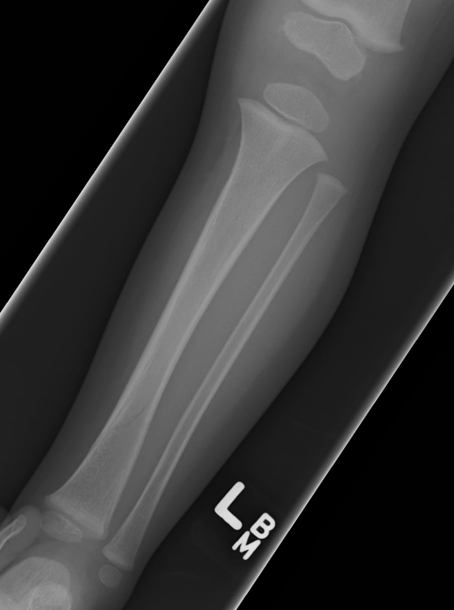
- Toddler's fracture: spiral tibia in 9 months to 3 years, limping child, X-ray often normal initially
- Cozen's phenomenon: progressive valgus after proximal tibial metaphyseal fracture, self-corrects
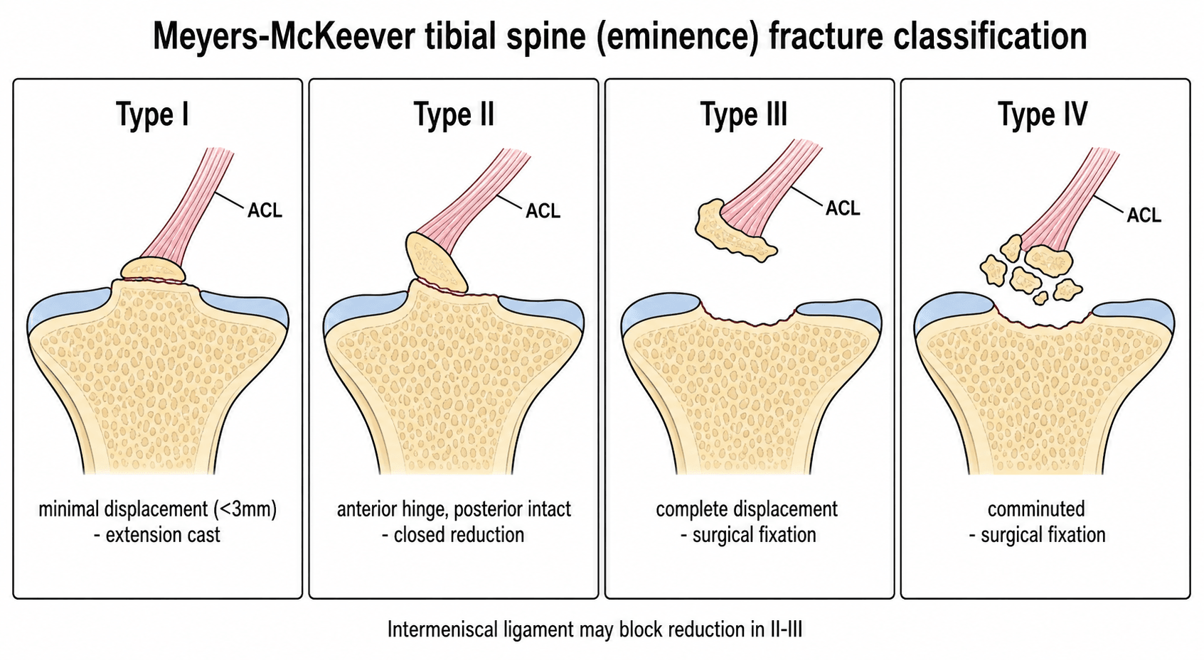
- Tibial spine fracture = pediatric ACL injury - treat based on displacement
- Floating knee in children: ipsilateral femur and tibia fractures - high energy
- Intact fibula may cause valgus deformity in tibial shaft fractures
- “Negative X-ray does not rule out toddler's fracture - treat clinically if suspected
- “Cozen's valgus peaks at 12-18 months then spontaneously corrects - observe
- “Tibial spine Type III = surgical (ORIF or arthroscopic reduction)
- “Age over 10 years: consider flexible IM nails for tibial shaft fractures
Non-displaced spiral tibia in walking child 9 months to 3 years. X-ray often negative initially. Clinical diagnosis - limp, refuse to bear weight. Cast 3-4 weeks even if X-ray negative.
Progressive valgus after healed proximal tibial metaphyseal fracture. Develops 6-12 months post-injury, peaks at 18 months. Self-corrects by skeletal maturity. DO NOT overcorrect initially.
Pediatric ACL equivalent. Meyers-McKeever classification. Type I/II = non-operative (extension cast). Type III/IV = surgical reduction and fixation.
Ipsilateral femur and tibia fractures = high energy. Screen for other injuries. May need surgical stabilization of both levels. Higher complication rate.
- Age
- 9 months - 3 years
- Key Feature
- Spiral tibia, often occult
- Treatment
- Cast 3-4 weeks
- Age
- 3-10 years
- Key Feature
- Risk of Cozen's valgus
- Treatment
- Cast, observe for valgus
- Age
- 8-14 years
- Key Feature
- ACL equivalent
- Treatment
- Type III = surgical
- Age
- All ages
- Key Feature
- High remodeling
- Treatment
- Cast or flexible nails
- Age
- Any age
- Key Feature
- High energy
- Treatment
- Surgical stabilization
VALCozen's Phenomenon
Hook:VAL-gus develops then VAL-ishes (vanishes)!
Overview and Epidemiology
Different tibial fracture patterns occur at different ages. Toddler's fracture (9 months to 3 years), proximal metaphyseal Cozen's type (3-10 years), tibial spine (8-14 years), shaft fractures (all ages).
- Second most common pediatric long bone fracture
- 15% of all pediatric fractures
- Peak incidence: toddlers and adolescents
- Boys more than girls (2:1)
- Left and right equal
- Toddler's: low energy twist/fall
- Proximal metaphyseal: direct impact
- Tibial spine: hyperextension (bicycle)
- Shaft: direct blow or torsion
- Floating knee: high energy (MVA)
Anatomy and Biomechanics
The proximal tibial physis grows faster than the distal (57% vs 43% of tibial growth). Injuries to the proximal physis have greater potential for growth disturbance.
Tibial Growth Plate Anatomy
Contributes 57% of tibial length. Located 1-2 cm distal to joint line. Protected by tibial tubercle apophysis.
Contributes 43% of tibial length. Asymmetric closure (central, then medial, then lateral).
Secondary ossification center. Vulnerable during adolescence (Osgood-Schlatter).
Classification Systems
Toddler's Fracture (CAST)
Childhood Accidental Spiral Tibial fracture
Characteristics: Age 9 months to 3 years (walking age). Non-displaced spiral or oblique fracture. Distal tibial shaft most common. Often not visible on initial X-ray. Low energy mechanism (twist, fall).
Clinical Assessment
- Refuses to bear weight
- Limping or not walking
- Point tenderness over tibia
- Often no swelling initially
- May have normal X-rays
- History of minor fall/twist
- Acute knee pain after hyperextension
- Knee effusion (hemarthrosis)
- Unable to extend knee fully
- Positive Lachman (if tested)
- Often bicycle handlebar injury
High energy injury. Assess for associated injuries: ipsilateral hip, knee, ankle. Neurovascular exam essential. Screen for head, chest, abdominal trauma. Higher risk of compartment syndrome.
Be vigilant for compartment syndrome especially in floating knee, both bone fractures, and high energy mechanisms. Pain out of proportion, pain with passive stretch, tense compartments.
Investigations
X-ray Protocol
AP and lateral tibia/fibula. Include knee and ankle joints.
May be negative initially. Look for subtle periosteal reaction at 10-14 days. Bone scan or MRI if clinical suspicion high.
AP and lateral knee. CT if surgical planning needed.
Differential Diagnosis
A toddler refusing to weight-bear with a normal radiograph has a broad differential. The job is to separate a benign occult fracture from infection, malignancy and non-accidental injury before reassuring the family.
- Typical age
- 9 months - 3 years
- Key discriminator
- Point tibial tenderness, low-energy twist, periosteal reaction at 10-14 days
- Pitfall to avoid
- Calling it 'normal' on day 1 film
- Typical age
- Any
- Key discriminator
- Fever, raised CRP/ESR, warmth, refusal to move joint
- Pitfall to avoid
- Casting an infected limb
- Typical age
- Pre-ambulatory or inconsistent history
- Key discriminator
- Mechanism-injury mismatch, other injuries on survey
- Pitfall to avoid
- Failing to safeguard a young child
- Typical age
- 3-8 years
- Key discriminator
- Hip-referred pain, recent viral illness, settles quickly
- Pitfall to avoid
- Missing a tibial source of pain
- Typical age
- Any
- Key discriminator
- Pain before injury, lytic lesion, trivial trauma
- Pitfall to avoid
- Treating fracture without imaging the lesion
- Typical age
- 3-10 years
- Key discriminator
- Cozen's appears 8-19 months after a healed metaphyseal fracture
- Pitfall to avoid
- Blaming initial treatment for late valgus
Non-Accidental Injury and the Paediatric Tibial Fracture
A tibial fracture may be the first presentation of non-accidental (inflicted) injury, and recognising the red flags before reassuring or discharging a family is a non-negotiable safeguarding duty. The single most important discriminator is whether the child is developmentally able to have caused the injury.
- Why it is concerning
- A spiral or long-bone tibial fracture needs force a pre-walking baby cannot generate; a toddler's fracture by definition needs a walking child
- Action
- Treat as suspected NAI until explained; escalate to safeguarding
- Why it is concerning
- Mechanism-injury mismatch and inconsistent accounts are classic
- Action
- Document verbatim histories; involve the child-protection team
- Why it is concerning
- Metaphyseal corner and bucket-handle fractures are highly specific for inflicted injury
- Action
- Skeletal survey to find them and other occult fractures
- Why it is concerning
- Healing fractures at varied stages, bruising in a non-mobile baby, torn frenulum or burns
- Action
- Full skeletal survey and multidisciplinary safeguarding assessment
- Why it is concerning
- Late presentation without adequate explanation
- Action
- Consider in the safeguarding picture rather than in isolation
In any young child (generally under 2 years) with a tibial fracture that does not fit the history or who is not yet walking, obtain a skeletal survey (per AAP/NICE guidance) and make a child-protection referral before discharge. Casting the fracture without addressing the safeguarding concern is a serious and recurring error - the orthopaedic injury and the safeguarding pathway must run in parallel.
SPIRALToddler's Fracture Features
Hook:SPIRAL describes both the fracture pattern and key features!
1234Meyers-McKeever Tibial Spine
Hook:Type 3+ = Surgery (the 3 looks like a backwards S for Surgical)
Management
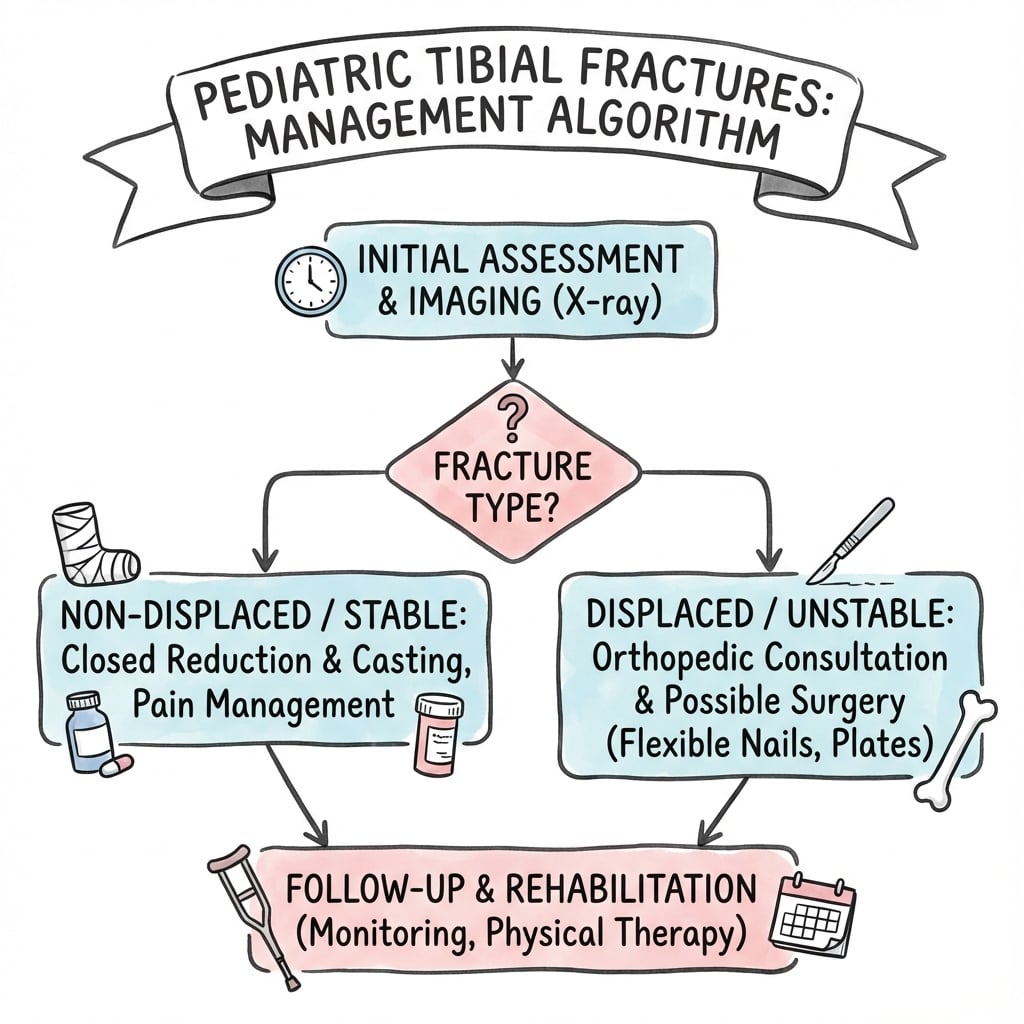
Under 6 years: Cast treatment for most fractures. High remodeling potential. 6-10 years: Cast for stable, operative for unstable or acceptable alignment not achieved. Over 10 years: Consider flexible IM nails for shaft fractures. Lower remodeling potential.
Toddler's Fracture Management
Long leg cast or walking boot for 3-4 weeks.
- Treat clinically even if X-ray negative
- No reduction needed (non-displaced)
- Rapid healing in this age group
- Follow-up X-ray at 2 weeks shows callus
Excellent. Heals rapidly with no long-term sequelae.
Angulation: Up to 10 degrees in sagittal plane, 5 degrees in coronal plane. Shortening: Up to 1-1.5 cm (will remodel with growth). Rotation: Minimal accepted (does not remodel). Younger children tolerate more deformity due to greater remodeling potential.
Principles of Remodelling in the Paediatric Tibia
The acceptable-deformity thresholds above are not arbitrary - they follow the predictable rules of physeal remodelling. Knowing why a deformity will or will not correct is what lets you justify closed treatment versus reduction in the viva.
- Remodels well
- Young child with years of growth ahead
- Remodels poorly / not at all
- Adolescent near skeletal maturity
- Remodels well
- Deformity close to an active physis
- Remodels poorly / not at all
- Deformity at the mid-diaphysis, far from the physis
- Remodels well
- Angulation in the plane of joint motion (sagittal at the knee and ankle)
- Remodels poorly / not at all
- Coronal-plane (varus/valgus) angulation and, above all, ROTATION, which does not remodel
The proximal tibial physis contributes more growth (around 57 percent) than the distal (around 43 percent), but the tibia overall remodels less reliably than the femur, so coronal angulation and rotation are corrected more readily here. This is why the accepted limits tighten with age and why a rotational malalignment must be corrected at the time of treatment rather than left to remodel.
The single rule that catches candidates out: rotational malalignment does NOT remodel at any age, so it must be corrected during reduction. Sagittal angulation near a physis in a young child remodels best; coronal angulation and any deformity in an older child or far from the physis remodel least - which is exactly why the acceptable thresholds are age- and plane-dependent.
Surgical Technique Considerations
Flexible IM Nailing (TENS/ESIN)
Age over 6-10 years, unstable shaft fractures, polytrauma.
Medial and lateral distal metaphysis (avoid physis).
40% of medullary canal at isthmus.
Pre-contour nails for apex anterior angulation. Avoid proximal entry (tibial tubercle physis damage).
Complications
Complications by Fracture Type
- Fracture Type
- Proximal metaphyseal
- Management
- Observe - self-corrects by maturity
- Fracture Type
- Shaft fractures
- Management
- Remodeling or corrective osteotomy if needed
- Fracture Type
- Tibial spine
- Management
- Proper reduction and fixation, ACL rehab
- Fracture Type
- Floating knee, high energy
- Management
- Urgent fasciotomy
- Fracture Type
- Physeal injuries
- Management
- Bar resection or corrective procedures
- Fracture Type
- Rare in children
- Management
- Operative intervention if occurs
Progressive valgus deformity after proximal tibial metaphyseal fracture. Mechanism unclear (asymmetric growth stimulation, tethering by fibula). Develops 6-18 months post-fracture. Spontaneous correction expected by skeletal maturity. Osteotomy rarely indicated before maturity.
Postoperative Care
Post-Treatment Protocol
Cast immobilization. Non-weight bearing. Monitor for compartment syndrome in high-energy injuries.
X-ray at 2-3 weeks to confirm alignment. Toddler's fracture usually healed. Weight bearing as tolerated in cast.
Remove cast when clinically and radiographically healed. Begin weight bearing. Tibial spine: begin ROM.
Follow proximal metaphyseal fractures for Cozen's valgus. Document and reassure. Tibial spine: assess for ACL laxity.
Outcomes and Prognosis
Prognosis by Fracture Type
Excellent prognosis. Complete healing in 3-4 weeks. No long-term sequelae.
Good prognosis despite Cozen's phenomenon. Most remodel by skeletal maturity.
Good outcomes with proper treatment. Residual ACL laxity possible but usually not symptomatic.
Excellent prognosis. High union rates. Good remodeling potential in younger children.
Guidelines, Registries & Global Practice
- Tibia/fibula fractures are among the commonest paediatric long-bone injuries worldwide
- Bimodal pattern: toddlers (low-energy twisting) and adolescents (sport, road traffic)
- Male predominance roughly 2:1 across most published cohorts
- Mechanism shifts by setting: trampolines, scooters and cycling in high-income regions; falls and road traffic dominate elsewhere
- Age-based remodelling logic is shared across all major training systems (FRCS, FRACS, EBOT, ABOS, DNB/MS)
- Toddler's fracture is a clinical diagnosis treated on suspicion regardless of resources
- Displaced tibial spine and floating-knee injuries are operative everywhere when expertise allows
- Non-accidental injury must be considered in any young, pre-ambulatory child with a tibial fracture
- Toddler's & shaft
- Cast for low-energy; flexible nails for unstable adolescents
- Tibial spine
- Arthroscopic suture fixation favoured for displaced
- NAI screening
- Skeletal survey under 2 years per AAP
- Toddler's & shaft
- Closed treatment first; ESIN for length-unstable
- Tibial spine
- Refer to paediatric ortho/sports knee service
- NAI screening
- Follow BOAST safeguarding standard
- Toddler's & shaft
- ESIN entry distal to tubercle apophysis
- Tibial spine
- ORIF/ARIF avoiding physis
- NAI screening
- Document mechanism vs injury concordance
- Toddler's & shaft
- Stable angulation thresholds by age
- Tibial spine
- Suture over screw to avoid removal
- NAI screening
- National safeguarding pathways
- No dedicated paediatric tibial-fracture implant registry exists; evidence is cohort/series level
- Trauma registries (e.g. national paediatric trauma networks) inform floating-knee and polytrauma outcomes
- Flynn criteria remain the common outcome language for flexible-nail series
- High-resource: routine arthroscopy, intra-operative imaging, MRI for occult injury, guided-growth implants
- Limited-resource: closed reduction and casting prioritised; open reduction when arthroscopy unavailable
- Telemedicine and serial radiographs substitute for advanced imaging where access is constrained
- Outcomes for closed-treatable patterns remain excellent regardless of setting
Special Considerations
Floating Knee (Pediatric)
Ipsilateral femur and tibia fractures.
High energy trauma (MVA, fall from height).
Knee ligament injuries (40-80%), vascular injuries, other trauma.
Usually requires surgical stabilization of both levels. Femur typically flexible nails. Tibia cast or nails depending on pattern.
Highest risk of compartment syndrome. LLD possible.
Controversies and Areas of Uncertainty
Age, sex and initial angulation do not reliably predict who develops late valgus, and the deformity usually self-corrects. Routine prophylactic intervention is not justified; the debate is how long to observe before offering guided growth.
Comparative data favour suture-based fixation for function and lower implant-removal rates, but no high-quality randomised trial defines the optimal construct. Open versus arthroscopic reduction also remains debated.
The angulation/shortening a growing tibia will remodel is age-dependent and imprecise. Heavier, older adolescents behave more like adults, narrowing the case for purely closed treatment.
MRI, bone scan and ultrasound can confirm occult fractures, but most authorities treat clinically without advanced imaging. The controversy is cost and radiation versus diagnostic certainty.
MCQ Practice Points
Q: A 2-year-old refuses to walk after a fall. X-ray is normal. What is the management? A: Treat as toddler's fracture with cast 3-4 weeks. Clinical diagnosis is sufficient. X-ray may be negative initially.
Q: What is the management of progressive valgus 12 months after proximal tibial metaphyseal fracture? A: Observation and reassurance. Cozen's phenomenon self-corrects by skeletal maturity. Do not operate early.
Q: Which Meyers-McKeever type requires surgical treatment? A: Type III and IV. Type I and II are typically non-operative. Type III is completely displaced and requires fixation.
Q: What is the main complication risk in floating knee injury? A: Compartment syndrome. Floating knee is high energy with highest compartment syndrome risk. Also screen for other injuries.
Q: How much angulation is acceptable in pediatric tibial shaft fractures? A: 10 degrees sagittal, 5 degrees coronal. Younger children tolerate more. Rotation does not remodel.
Q: What is the risk of tibial shaft fracture with intact fibula? A: Valgus deformity. Intact fibula acts as tether, preventing shortening but may cause progressive valgus.
Exam Viva Scenarios
Practise clinical reasoning and management decisions out loud
“A 2-year-old child is brought to ED by his mother. He has been refusing to bear weight on his left leg since yesterday after a fall from a low chair. On examination, there is no obvious swelling but tenderness over the distal tibia. X-rays appear normal. How would you manage this?”
“You are seeing an 8-year-old boy in clinic 12 months after he sustained a proximal tibial metaphyseal fracture that was treated in a cast. The fracture has healed but the parents are worried because his leg has become progressively bowed outwards. Examination confirms a 12 degree valgus deformity. How would you manage this?”
“A 12-year-old girl fell off her bicycle and presents with a swollen, painful right knee. She cannot fully extend her knee. X-ray shows a tibial spine fracture that appears displaced by about 8mm with complete loss of contact. How would you manage this?”
“A 10-year-old is brought in after being struck by a car. He has ipsilateral closed femoral shaft and tibial shaft fractures. Six hours after admission the nursing staff report escalating pain and increasing analgesia requirement despite the leg being splinted. How do you proceed?”
Toddler's Fracture
- Age 9 months to 3 years
- Spiral tibia, often occult on X-ray
- Clinical diagnosis - treat if suspected
- Cast 3-4 weeks, excellent prognosis
Cozen's Phenomenon
- Progressive valgus after proximal tibial metaphyseal fracture
- Develops 6-18 months post-injury
- Self-corrects by skeletal maturity
- DO NOT operate early
Tibial Spine
- Pediatric ACL equivalent
- Meyers-McKeever I-IV
- Type I-II: non-operative (cast)
- Type III-IV: surgical fixation
Floating Knee
- Ipsilateral femur and tibia fractures
- High energy - look for other injuries
- Highest compartment syndrome risk
- Usually requires surgical stabilization
Acceptable Deformity
- 10 degrees sagittal plane
- 5 degrees coronal plane
- 1-1.5 cm shortening
- Rotation: minimal (doesn't remodel)
Evidence Base and Key Studies
Each card below is anchored to a verified PubMed record (PMID and DOI shown). Use the level of evidence and sample size to judge how heavily to lean on a given statement in a viva.
Cozen's Phenomenon: Natural History and Resolution
- Retrospective series of 33 children (6 months to 14 years), mean follow-up 8.8 years
- 15 of 33 developed late valgus between 8 and 19 months (mean onset 12.5 months)
- 24 of 33 developed tibial elongation; neither finding correlated with age at injury
- Maximum valgus subsequently corrected to near the initial post-treatment angle by final follow-up
Guided Growth for Persistent Cozen's Deformity
- Largest reported series (19 patients, 24 Cozen's phenomena) treated with medial proximal tibial guided growth
- Corrective osteotomy abandoned because of frequent recurrent valgus
- Mechanical axis and medial proximal tibial angle corrected in all but one patient
- Five recurrences after implant removal; managed by repeat or retained metaphyseal screw
Tibial Eminence Fractures: Surgical Outcomes Systematic Review
- Systematic review of 12 studies of operatively treated Meyers-McKeever type II-IV fractures
- Open reduction showed higher Tegner/Lysholm scores and less arthrofibrosis than arthroscopic fixation
- Arthroscopic suture fixation outperformed arthroscopic screw fixation on functional scores
- Screw fixation carried the highest rate of implant removal
Arthroscopic Fixation Techniques for Pediatric Tibial Eminence Fractures
- Narrative review confirming arthroscopic reduction as the contemporary standard for surgical TEFs
- Anchor, suture and screw constructs gave broadly comparable clinical and radiographic results
- Suture/anchor techniques minimise physeal damage and are favoured in skeletally immature knees
- Higher-quality comparative studies are still needed to define the optimal construct
Titanium Elastic Nails for Pediatric Tibial Shaft Fractures
- 19 consecutive children (mean age 12.2 years) with unstable tibial shaft fractures treated with TENs
- All united at a mean of 11 weeks; final mean angulation 2 degrees sagittal, 3 degrees coronal
- No leg-length discrepancy or physeal arrest; entry-site irritation in 26 percent
- 12 excellent, 6 satisfactory and 1 poor result by Flynn criteria
Adolescent Tibial Shaft Fractures: Treatment Outcomes and Risk Factors
- 52 patients aged 10-18 years treated by ESIN, interlocking nail, plate, external fixator or cast
- No significant difference in union rate between fixation methods
- Open fractures had longer time to union and a 5.5-fold higher complication risk
- Heavier body weight correlated with lower radiographic union scores at 12 weeks
Occult Toddler's-Type Tibial Fractures
- Described subtle, easily missed occult tibial fractures in infants and young children
- Radiographic findings parallel the classic spiral toddler's fracture of Dunbar
- Knowledge of mechanism plus focused exam allows confident diagnosis
- Reinforces that initial radiographs are frequently unremarkable
Foundational Classification & Description (Historical)
- Meyers-McKeever (1959/1970) defined the tibial spine fracture classification still used today
- Cozen (1953) first described post-traumatic proximal tibial valgus
- Dunbar et al. (1964) coined the 'toddler's fracture' as a childhood accidental spiral tibial fracture
- These remain the eponymous source descriptions for the entities on this page