Plantarflexion & Sole Sensation
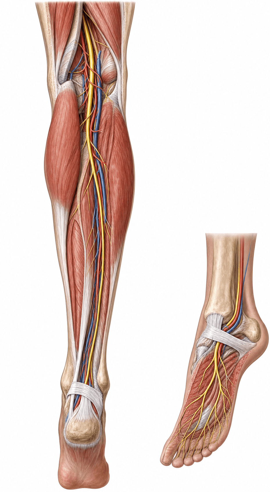
- The tibial nerve is the larger terminal branch of the sciatic nerve (L4-S3), beginning at the apex of the popliteal fossa.
- In the popliteal fossa it lies most superficial of the neurovascular bundle (nerve over vein over artery).
- It supplies the superficial AND deep posterior compartments of the leg (plantarflexors, invertor, toe flexors).
- It passes behind the medial malleolus through the TARSAL TUNNEL (deep to the flexor retinaculum) and divides into the medial and lateral plantar nerves.
- It provides sensation to the SOLE of the foot (via plantar nerves) and the heel (medial calcaneal branch).
- A high tibial nerve injury produces an INSENSATE SOLE plus loss of plantarflexion and toe flexion - relevant to limb-salvage decisions.
- “Tarsal tunnel contents (anterior to posterior): Tibialis posterior, FDL, posterior tibial Artery/vein, tibial Nerve, FHL - 'Tom, Dick, And Very Nervous Harry'.
- “The first branch of the lateral plantar nerve is Baxter's nerve (inferior calcaneal nerve), a cause of heel pain.
- “An insensate sole was historically a relative indication against limb salvage - now interpreted with caution.
Deep to the flexor retinaculum behind the medial malleolus, anterior to posterior: Tibialis posterior, flexor Digitorum longus, posterior tibial Artery and Vein, tibial Nerve, flexor Hallucis longus. The nerve divides here into medial and lateral plantar nerves.
Compression of the tibial nerve (or its plantar branches) in the tunnel causes burning sole pain, paraesthesia and a positive Tinel sign behind the medial malleolus. Causes include space-occupying lesions (ganglion, varicosities), deformity (hindfoot valgus) and trauma.
Origin & Course
Origin
- The tibial nerve is the larger terminal branch of the sciatic nerve, carrying fibres from the anterior (ventral) divisions of L4-S3.
- The sciatic nerve typically divides into the tibial and common peroneal nerves at the apex of the popliteal fossa (though a higher division is a common variant).
Motor & Sensory Supply
The tibial nerve supplies both posterior leg compartments: superficial (gastrocnemius, soleus, plantaris) and deep (tibialis posterior, flexor digitorum longus, flexor hallucis longus, popliteus), plus all the intrinsic plantar muscles via the plantar nerves.
- Plantarflexion (gastrocnemius-soleus - "tiptoe"), inversion (tibialis posterior) and toe flexion (FDL/FHL) all depend on the tibial nerve.
- Sensory: the sole of the foot (medial plantar - medial sole and medial 3.5 toes; lateral plantar - lateral sole and lateral 1.5 toes), the heel (medial calcaneal), and a contribution to the sural nerve (posterolateral leg/lateral foot).
Clinical Correlations
Proximal (High) Tibial Nerve Injury
- Occurs with knee dislocation, proximal tibial trauma, or popliteal injury.
- Deficit: loss of plantarflexion, inversion and toe flexion (a calcaneovalgus foot), loss of intrinsic foot muscles, and crucially an insensate sole.
- The insensate plantar surface historically influenced limb-salvage-versus-amputation decisions, though this is now applied cautiously as outcomes data have evolved.
TDANHTarsal Tunnel Contents (Ant → Post)
Hook:'Tom, Dick, And Very Nervous Harry' - the order through the tarsal tunnel.
Evidence Base
Tibial Nerve Termination & Variation in the Tarsal Tunnel
- The tibial nerve is the larger terminal branch of the sciatic nerve and terminates in the tarsal tunnel as the medial and lateral plantar nerves
- Reports a trifurcation variant of the tibial nerve within the tarsal tunnel
- The variant branch curved laterally and ran deep to quadratus plantae
- The posterior tibial artery showed a corresponding three-branch termination
Lower-Extremity Entrapment Neuropathies (incl. Posterior Tibial)
- Reviews cause, signs, diagnosis and treatment of lower-limb entrapment neuropathies including the posterior tibial nerve (tarsal tunnel syndrome)
- Accurate identification and management prevents pain, sensory loss and weakness affecting mobility
- Diagnosis relies on clinical examination supported by electrodiagnosis and imaging (ultrasound/MRI)
- Considers the tibial nerve alongside common peroneal, femoral and lateral femoral cutaneous neuropathies
Viva Scenarios
Practise clinical reasoning and management decisions out loud
“After a knee dislocation that was reduced, a patient cannot plantarflex the ankle or flex the toes and has a numb sole. What nerve is injured and what are the implications?”
Guidelines, Registries & Global Practice
Global Practice Picture
Tibial nerve anatomy underpins management of tarsal tunnel syndrome, plantar heel pain (Baxter's nerve), posterior leg compartment and popliteal injuries, and the assessment of the insensate foot. The internationally consistent principles are: know the tarsal tunnel contents and plantar division, diagnose tarsal tunnel syndrome clinically with electrodiagnostic/imaging support, and treat the underlying cause.
Side-by-Side Synthesis
- Detail
- Larger terminal branch of sciatic (L4-S3)
- Detail
- Superficial + deep posterior compartments
- Detail
- Tarsal tunnel (behind medial malleolus)
- Detail
- Medial + lateral plantar (+ medial calcaneal)
- Detail
- Sole of foot, heel, (sural contribution)
- Detail
- Tarsal tunnel syndrome; high injury → insensate sole
Anatomy
- Larger terminal branch of sciatic (L4-S3)
- Most superficial in popliteal fossa
- Deep posterior compartment of leg
- Tarsal tunnel → medial + lateral plantar nerves
Supply
- Motor: plantarflexors, invertor, toe flexors, foot intrinsics
- Sensory: sole (plantar nn.) + heel (medial calcaneal)
- Contributes to sural nerve
- Baxter's = 1st branch lateral plantar
Clinical
- Tarsal tunnel syndrome (Tinel behind medial malleolus)
- High injury → insensate sole + lost plantarflexion
- Knee dislocation puts it at risk (+ popliteal artery)
- Contents: Tom, Dick, And Very Nervous Harry