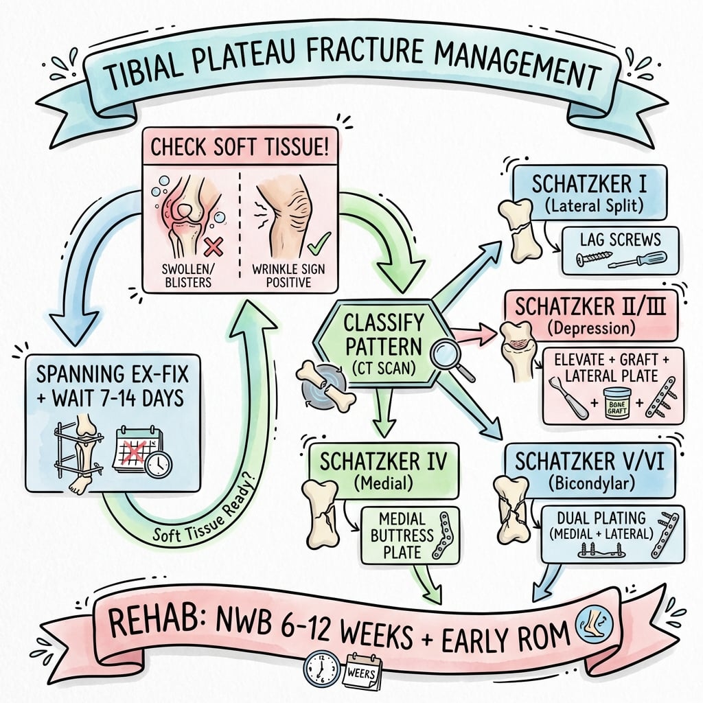
Schatzker Classification | Three-Column Concept | Staged Surgery
- CT is ESSENTIAL for surgical planning - reveals posterior column and true depression
- Wrinkle sign indicates safe soft tissue for ORIF (no wrinkles = no surgery)
- Three-column concept: Lateral, Medial, Posterior - each needs specific approach
- Bicondylar patterns (V-VI) require staged approach and dual plating
- Single lateral plate fails in bicondylar fractures - this is a common exam trap
- “Hoffa fragment = posterior column involvement requiring specific approach
- “Medial plateau fracture = high-energy until proven otherwise
- “Moore classification for fracture-dislocation patterns
- “Rasmussen radiological criteria for articular reduction assessment

Wrinkle test mandatory before ORIF. No wrinkles = spanning ex-fix. High-energy (V-VI) = staged approach.[5] Compartment syndrome in 10-15%.
CT-based surgical planning. Lateral column = anterolateral approach. Medial = anteromedial/posteromedial. Posterior = separate posterior approach.
Under 2mm step-off goal. Reduce depression through cortical window. Raft screws support subchondral bone. Bone graft prevents subsidence.
Schatzker I-III = lateral plating. IV = medial plate essential. V-VI = dual plates staged. Single lateral plate in bicondylar = FAILURE.
- Fracture Pattern
- Schatzker I (lateral split)
- Treatment
- Percutaneous lag screws
- Key Pearl
- Simple pattern = simple fix
- Fracture Pattern
- Schatzker II (split-depression)
- Treatment
- ORIF with elevation + bone graft
- Key Pearl
- Support the subchondral bone
- Fracture Pattern
- Schatzker III
- Treatment
- Arthroscopic-assisted vs open
- Key Pearl
- Elevate via cortical window
- Fracture Pattern
- Schatzker IV (medial)
- Treatment
- Medial buttress plate required
- Key Pearl
- High-energy = ligament injury
- Fracture Pattern
- Schatzker V-VI
- Treatment
- STAGE: Ex-fix → wait → dual ORIF
- Key Pearl
- Wrinkle sign before definitive
SPlIT-D-PURESchatzker Classification
Hook:Split patterns (I-III) are LOW energy, Medial + Bicondylar + Dissociation (IV-VI) are HIGH energy!
WRINKLESoft Tissue Management
Hook:WRINKLE test = No wrinkles on skin, no surgery on fracture!
Overview and Epidemiology
Tibial plateau fractures test your understanding of articular fracture principles, soft tissue management, and staged surgery. Examiners focus on classification, when NOT to operate, approach selection, and complication avoidance.
- Bimodal distribution: Young (high-energy) + Elderly (low-energy)
- Lateral plateau more common (55-70%)
- Male predominance in young, female in elderly
- Sports injuries, MVA, falls from height
- Weight-bearing articular surface injury
- Long-term OA risk (10-30%)[8,9]
- Associated soft tissue injuries common
- Significant rehabilitation required
- Work and function implications
Anatomy and Biomechanics
The popliteal artery lies directly posterior to the tibia, separated by only the popliteus muscle. During posterior approaches, the neurovascular bundle must be carefully protected. Tethering at the soleal arch makes it vulnerable to injury with posterior displacement.
Surface Anatomy
- Convex surface - less congruent with femoral condyle
- Higher position (3mm proximal to medial)
- More prone to depression fractures
- Lateral meniscus covers 80%
- Concave surface - greater congruency
- Larger weight-bearing surface (60%)
- Stronger subchondral bone
- Medial meniscus covers 50%
Tibial Slope: 7-10° posterior slope - must restore during fixation
Intercondylar Eminence: Central, contains ACL and PCL insertions
Classification Systems
Schatzker Classification (1979)[1]
- Pattern
- Lateral split (wedge)
- Mechanism
- Low-energy valgus
- Treatment
- Lag screws ± buttress plate
- Pattern
- Lateral split-depression
- Mechanism
- Low-energy valgus
- Treatment
- ORIF: elevate, graft, buttress plate
- Pattern
- Pure lateral depression
- Mechanism
- Low-energy axial
- Treatment
- Elevation, raft screws, graft
- Pattern
- Medial plateau fracture
- Mechanism
- HIGH-energy varus
- Treatment
- Medial buttress plate essential
- Pattern
- Bicondylar (split)
- Mechanism
- HIGH-energy axial
- Treatment
- Dual plating, staged approach
- Pattern
- Metaphyseal-diaphyseal dissociation
- Mechanism
- HIGH-energy
- Treatment
- Staged, dual/ring fixation
Type II vs Type III: Type II has a split component (cortical break), Type III is pure depression (cortex intact). This changes surgical approach - Type II can be opened like a book to access the depression.
Clinical Assessment
- Mechanism: Axial load + valgus/varus (bumper fracture)
- Energy level: Fall height, MVA speed
- Comorbidities: Osteoporosis, diabetes, smoking
- Anticoagulation status: Affects timing
- Functional demands: Activity level, occupation
- Look: Swelling, bruising, deformity, wounds
- Feel: Tenderness, crepitus, effusion
- Move: Limited by pain, instability
- Special: Neurovascular status MANDATORY
- Compartments: Assess all four leg compartments
Vascular: Palpate dorsalis pedis and posterior tibial pulses. Calculate ABI if ANY concern. Popliteal artery injury in 2-3% of tibial plateau fractures.
Neurological: Common peroneal nerve (foot drop), posterior tibial nerve (sole sensation).
Compartments: Pain with passive stretch of toes, tense compartments, pain out of proportion. Low threshold for fasciotomy.
Critical for Surgical Timing
Soft Tissue Checklist
- Open fracture? (Gustilo classification)
- Fracture blisters? (Clear vs blood-filled)
- Tension? Tenting? Impending necrosis?
- Wrinkle test: Can you see skin wrinkles over anterior tibia?
- No wrinkles = too swollen for ORIF
- Document in notes with photos
- Wrinkle positive + good skin = proceed to ORIF
- Wrinkle negative = spanning external fixator
- Wait 7-14 days for swelling to resolve
Clear blisters = epidermis only, can operate through. Blood-filled (hemorrhagic) blisters = full-thickness injury, AVOID incisions through these areas.
Differential Diagnosis of the Acute Painful Swollen Knee After Injury
- Discriminating Features
- Bony tenderness over the plateau, valgus/varus mechanism, lipohaemarthrosis, axial-load injury
- Key Investigation
- AP/lateral radiographs then CT
- Discriminating Features
- Younger patient, ACL-type mechanism, central bony fragment without articular depression
- Key Investigation
- Radiograph / CT; MRI for ACL
- Discriminating Features
- Tenderness localised to femoral condyle, coronal split on lateral film
- Key Investigation
- CT (coronal plane fracture often missed on X-ray)
- Discriminating Features
- Gross instability, dimple sign, high suspicion for vascular injury
- Key Investigation
- Vascular assessment, ABI, CT angiography
- Discriminating Features
- Anterior tenderness, inability to straight-leg-raise, palpable gap
- Key Investigation
- Radiograph; ultrasound/MRI for tendon
- Discriminating Features
- Effusion without bony tenderness, normal radiographs, mechanism-specific instability
- Key Investigation
- MRI
- Discriminating Features
- Effusion, normal radiographs, focal subchondral signal
- Key Investigation
- MRI
Investigations
Imaging Protocol
Standard knee series: AP, lateral, oblique views
Key measurements:
- Tibial plateau angle (joint line)
- Condylar widening
- Depression depth
- Stress views if ligamentous injury suspected
Mandatory for surgical planning in ALL displaced fractures.
What CT reveals:
- True articular depression (often underestimated on X-ray)
- Posterior column involvement (missed in 30% on X-ray)
- Comminution and fragment size
- Coronal plane fracture lines
Reconstructions: Axial, coronal, sagittal, 3D
Indications:
- Suspected meniscal injury (for surgical planning)
- Ligamentous injury evaluation
- Occult fracture in non-displaced injuries
Timing: Usually done post-operatively or after swelling subsides.
Posterior column involvement (seen on CT) requires ADDITIONAL posterior approach - cannot be addressed through anterolateral approach alone. This is WHY CT is mandatory!
Management Algorithm
Indications for Conservative Management
- Non-displaced or minimally displaced (under 2mm step-off)
- Stable knee on stress testing
- Low functional demand patient
- Severe medical comorbidities precluding surgery
- Long leg cast or hinged knee brace
- Non-weight-bearing 6-8 weeks
- Progressive ROM starting week 2-4
- Serial X-rays to monitor position
- Full weight-bearing by 12 weeks
Weekly X-rays for first 2-3 weeks to monitor for displacement. If any secondary displacement, convert to operative management.
The staged pathway above ends in dual plating, but a viva will expect the alternative definitive strategy for the high-energy bicondylar fracture with a poor soft-tissue envelope: circular (Ilizarov/hybrid) external fixation, which the topic's own COTS randomised trial supports.
- Best indications: Schatzker V-VI (especially VI metaphyseal-diaphyseal dissociation) with compromised soft tissues, open injuries, or where extensive dual-plate dissection would risk the skin.
- The concept - "limited internal fixation plus a ring frame": the articular surface is reduced and held with percutaneous or limited screws, then the metaphyseal-diaphyseal zone is bridged by the ring/hybrid frame - achieving stability without the soft-tissue stripping of open plating.
- Safe-zone rule: periarticular tensioned wires/half-pins should stay roughly at least 14mm below the joint line to remain extracapsular - a wire that enters the capsular reflection can seed a septic arthritis.
- Evidence (COTS RCT): versus dual plating, ring fixation gave less blood loss, shorter hospital stay and fewer/less-severe reoperations, with equivalent articular reduction and 2-year WOMAC - the trade-off is pin-site infection, frame burden, knee stiffness and the need for surgeon expertise.
Exam point: for the high-energy bicondylar/metaphyseal-diaphyseal fracture with a poor soft-tissue envelope, circular (ring) fixation with limited internal fixation is a valid lower-wound-morbidity alternative to dual plating (COTS RCT) - reduce and fix the joint percutaneously, bridge the metaphysis with the frame, and keep periarticular wires extracapsular (about 14mm below the joint) to avoid septic arthritis.
Surgical Technique
Pre-operative Planning
- Infection: 2-5% superficial, 1-2% deep
- Wound complications: 5-10% (higher in Schatzker V-VI)
- Nerve injury: Peroneal nerve at risk (1-2%)
- Stiffness: Common, may need MUA/arthrolysis
- Malunion/nonunion: 5-10%
- Post-traumatic OA: 10-30%
- Hardware removal: May be needed
- Plates: Lateral locking plate, medial buttress plate, posterior plates
- Screws: Lag screws, raft screws, locking screws
- Graft: Bone graft substitute or autograft
- C-arm: Two image intensifiers ideal
- Reduction tools: Femoral distractor, reduction clamps, bone tamp
- Fluoro views: AP, lateral, Schatzker views
For Lateral Column
Position: Supine, bump under hip, slight knee flexion
Step-by-Step
Landmarks: Line from fibular head to Gerdy's tubercle
Curved incision parallel to lateral joint line, centered over lateral plateau
Length: 10-15cm, extensile if needed
- Incise iliotibial band in line with skin incision
- Identify and protect common peroneal nerve at fibular neck
- Retract tibiofemoral joint with Z-retractors
Common peroneal nerve wraps around fibular neck. Flex knee to relax. Never use retractors near fibular head without visualization.
- Submeniscal arthrotomy to visualize articular surface
- Elevate anterior tibialis muscle from lateral tibial surface
- Expose metaphysis for plate application
For Type II (split-depression):
- Open the split fragment like a book
- Visualize depressed articular fragments
- Use bone tamp to elevate depression
- Pack with bone graft
- Reduce split fragment over elevated joint
- Provisional K-wire fixation
- Lag screws for split component
- Raft screws (subchondral) to support elevated segment
- Apply lateral buttress/locking plate
- Final imaging in all planes
Intraoperative Troubleshooting
- Cause
- Inadequate exposure
- Solution
- Use femoral distractor to open joint, extend arthrotomy, consider arthroscopy
- Cause
- Impacted into metaphysis
- Solution
- Use larger tamp, more force, consider wider cortical window
- Cause
- No subchondral support
- Solution
- Add raft screws, more bone graft, check for void
- Cause
- Inadequate reduction of split
- Solution
- Re-apply reduction clamp, add lag screw across condyle
- Cause
- Osteoporosis
- Solution
- Use locking screws, consider cement augmentation, longer construct
- Cause
- Wrong approach
- Solution
- Add posteromedial or posterolateral approach
The quick-decision table mentions "arthroscopic-assisted vs open," but ARIF is worth knowing as a technique in its own right - and it carries a classic examiner trap.
Where it fits: ARIF suits simple low-energy patterns - Schatzker I, II and III (a clean split and/or central depression without metaphyseal comminution). It is not appropriate for Schatzker V-VI bicondylar or metaphyseal-comminuted fractures.
Advantages:
- Direct articular visualisation of the reduction - more accurate than fluoroscopy for confirming a step-off under 2mm.
- Lets you diagnose and treat the associated intra-articular injuries that are so common here (lateral meniscal tears in well over half of displaced lateral fractures, chondral and cruciate injury).
- Less soft-tissue stripping and smaller incisions than open exposure; the depression is elevated through a metaphyseal cortical window/bone tamp under scope control and held with percutaneous raft/lag screws plus or minus a small plate.
The trap - fluid extravasation and compartment syndrome: arthroscopic irrigation fluid can leak through the fracture and capsular tear into the leg compartments and precipitate acute compartment syndrome. Mitigate with low pump/gravity inflow, the shortest possible operative time, and continuous calf monitoring, and avoid ARIF in high-energy/comminuted fractures with capsular disruption where the risk is greatest.
Exam point: ARIF is for Schatzker I-III without metaphyseal comminution - it gives direct articular reduction and treats meniscal/chondral injury, but arthroscopy fluid can extravasate through the fracture and cause compartment syndrome, so use low-pressure inflow, limit time, monitor the calf, and avoid it in high-energy comminuted patterns.
LMPThree-Column Surgical Planning
Hook:Lateral-Medial-Posterior = Every column needs its own approach and plate!
Complications
- Incidence
- 5-10%
- Risk Factors
- Schatzker V-VI, diabetes, smoking, early surgery
- Management
- Early: debridement, ABx. Late: may need flap
- Incidence
- 5-10%
- Risk Factors
- High-energy, polytrauma, prolonged surgery
- Management
- Four-compartment fasciotomy URGENT
- Incidence
- 10-20%
- Risk Factors
- Immobility, no prophylaxis
- Management
- Prophylaxis mandatory, anticoagulation
- Incidence
- 10-30%
- Risk Factors
- Poor reduction, cartilage damage, malalignment
- Management
- Activity modification, eventual arthroplasty
- Incidence
- 5-10%
- Risk Factors
- Poor fixation, single plate for bicondylar
- Management
- High tibial osteotomy if symptomatic
- Incidence
- 15-25%
- Risk Factors
- Delayed mobilization, prolonged immobilization
- Management
- Early physio, may need MUA or arthrolysis
- Incidence
- 10-20%
- Risk Factors
- Thin soft tissue, prominent plate
- Management
- Hardware removal when healed
High-energy tibial plateau fractures have 5-10% compartment syndrome risk. Monitor closely postoperatively. Pain out of proportion, pain with passive toe stretch, tense compartments = fasciotomy. Clinical diagnosis - do not wait for compartment pressure measurements if clinical suspicion.
Varus collapse occurs when medial column is not adequately supported. Single lateral plate in bicondylar fracture allows medial column to collapse. Prevention: dual plating, adequate medial buttress.
Postoperative Care and Rehabilitation
Rehabilitation Protocol
- DVT prophylaxis: Enoxaparin or TED/pneumatic boots
- Pain control: Multimodal, consider nerve block
- Wound care: Monitor for hematoma, infection
- ROM: Start passive ROM in CPM if available
- Weight-bearing: Toe-touch or non-weight-bearing
- Weight-bearing: Non-weight-bearing or touch-weight-bearing
- ROM goals: 0-90° by 6 weeks
- Exercises: Quad sets, straight leg raises, gentle ROM
- X-rays: 2 weeks, 6 weeks for union assessment
- Remove sutures: 2-3 weeks
- Weight-bearing: Progressive based on radiographic healing
- ROM goals: Full ROM by 12 weeks
- Strengthening: Progressive resistance exercises
- X-rays: 12 weeks for union confirmation
- Full weight-bearing: When radiographic union confirmed
- Return to work: Sedentary 6-8 weeks, manual labor 4-6 months
- Return to sport: 6-12 months depending on sport
- Surveillance: Annual X-rays for OA development
- Schatzker I-III: May allow early protected WB
- Schatzker IV-VI: Strict NWB 6-8 weeks minimum
- Osteoporotic bone: Extended protected WB
- Progression based on radiographic healing
- Increasing pain or swelling (infection?)
- Loss of ROM (stiffness, arthrofibrosis)
- Hardware prominence (may need removal)
- Progressive deformity (loss of fixation)
Outcomes and Prognosis
- Good Outcomes
- 80-90%
- Key Factors
- Anatomic reduction, stable fixation
- Good Outcomes
- 75-85%
- Key Factors
- Elevation quality, graft support
- Good Outcomes
- 70-80%
- Key Factors
- Medial buttress, ligament healing
- Good Outcomes
- 60-75%
- Key Factors
- Soft tissue handling, staged approach
- Good Outcomes
- 40-60%
- Key Factors
- High complication rate
Poor prognostic factors:
- High-energy mechanism (Schatzker V-VI)
- Bicondylar involvement
- Greater than 10mm initial depression
- Associated ligament injury
- Post-traumatic OA precursors (cartilage damage)
- Smoking, diabetes, obesity
- Articular step-off greater than 2mm
Guidelines, Registries & Global Practice
Global Epidemiology
Tibial plateau fractures account for roughly 1% of all fractures and about 8% of fractures in the elderly, with a bimodal distribution: high-energy injuries (road traffic and fall-from-height) in younger men, and low-energy fragility fractures in older women. The lateral plateau is involved most often (Schatzker I-III predominate in most series), reflecting the physiological valgus axis of the knee. Incidence is rising in ageing high-income populations driven by osteoporotic and sporting injuries, while motor-vehicle trauma dominates in low- and middle-income settings.
Side-by-Side Guidance
- Region
- Global
- Key Position
- Articular reduction (step 2mm or less), restore mechanical axis and slope, column-specific approaches, staged ORIF for compromised soft tissues
- Evidence Basis
- Expert consensus + cohort evidence
- Region
- UK
- Key Position
- Senior decision-making, ortho-plastic input for high-energy/open patterns, definitive fixation only when soft tissues allow
- Evidence Basis
- Guideline (consensus)
- Region
- UK
- Key Position
- Senior multidisciplinary planning, CT for intra-articular fractures, VTE assessment, regional major-trauma networks
- Evidence Basis
- Guideline (GRADE)
- Region
- Canada
- Key Position
- Ring fixation a valid lower-morbidity alternative to dual plating in bicondylar fractures
- Evidence Basis
- Level 1 RCT [PMID 17142411]
- Region
- USA
- Key Position
- Anatomic articular restoration; individualised VTE prophylaxis after lower-limb trauma
- Evidence Basis
- Guideline / appropriate-use
Registry & Practice Variation
There is no dedicated international tibial plateau fracture registry; most outcome data derive from trauma databases and population cohorts. Population-level Canadian data show a 10-year total knee arthroplasty rate of 7.3% after operative fixation, a 5.3-fold increase over matched controls (Wasserstein et al., [PMID 24430414]). AOANJRR captures subsequent arthroplasty outcomes following tibial plateau fractures. Practice varies internationally: dual plating dominates in North America and Europe, whereas circular/ring fixation retains strong support where soft-tissue compromise is common or plating expertise is limited. Access also varies - high-income systems can deliver staged spanning external fixation within hours, whereas resource-limited settings may rely more heavily on definitive external fixation.
- Neurovascular status pre- and post-operatively
- Soft tissue assessment and rationale for timing
- CT scan findings and surgical plan based on CT
- Informed consent including alternatives
- Intraoperative imaging confirming reduction quality
- Compartment syndrome: Missed or delayed diagnosis
- Peroneal nerve injury: Failure to document preop status
- Wound complications: Operating on compromised soft tissue
- Malunion: Inadequate reduction or single plate for bicondylar
MCQ Practice Points
Q: A 60-year-old woman falls from standing and has lateral tibial plateau fracture with 6mm depression but no cortical split. What Schatzker type is this?
A: Schatzker Type III - pure depression without split component. Type II would have both split AND depression. This distinction is important as Type III may be treated arthroscopically.
Q: During the anterolateral approach to the tibial plateau, which structure is at greatest risk and how is it protected?
A: Common peroneal nerve at the fibular neck. Protected by flexing the knee to relax the nerve, direct visualization during dissection, and avoiding retractors at the fibular head area.
Q: What is the consequence of using a single lateral plate for a Schatzker V (bicondylar) tibial plateau fracture?
A: Varus collapse of the medial column. The unsupported medial condyle collapses, leading to progressive varus malalignment. Dual plating with medial buttress is required.
Q: A patient with Schatzker VI fracture has tense swelling and blood-filled blisters. What is the appropriate initial management?
A: Spanning external fixator with staged delayed ORIF. Blood-filled blisters indicate full-thickness skin injury. Operating definitively risks wound dehiscence and infection. Wait 7-21 days for wrinkle test positive.
Q: Why is CT scan mandatory for tibial plateau fracture surgical planning?
A: CT reveals posterior column involvement (missed in 30% on X-ray), true articular depression depth, fracture comminution, and guides approach selection. Plain X-ray underestimates depression and misses coronally-oriented fractures.
Q: What is the incidence of post-traumatic osteoarthritis following tibial plateau fractures, and what factors predict poor outcome?
A: 10-30% develop symptomatic OA. Predictors: articular step-off greater than 2mm, high-energy mechanism, meniscal injury, malalignment, and cartilage damage at time of injury.
Exam Viva Scenarios
Practise clinical reasoning and management decisions out loud
“A 55-year-old woman falls off a ladder and presents with a painful swollen knee. X-rays show a lateral tibial plateau fracture with 8mm of articular depression and a split component. How would you assess and manage this patient?”
“Walk me through your surgical technique for a Schatzker VI fracture in a 35-year-old male involved in a motorcycle accident. He is day 10 post-injury, wrinkle test is positive, and CT shows involvement of lateral, medial, and posterior columns.”
“A 45-year-old patient is day 1 post-ORIF for a Schatzker V fracture. The nurse calls because the patient has severe pain despite IV morphine, and pain with passive toe extension. How do you manage this?”
Key Anatomy
- Lateral plateau: convex, 3mm higher, depression-prone
- Medial plateau: concave, 60% weight-bearing, stronger bone
- Popliteal vessels: directly posterior, at risk in posterior approaches
- Common peroneal nerve: wraps fibular neck, at risk in lateral approaches
- Posterior tibial slope: 7-10°, must restore
Classification (Schatzker)
- Type I: Lateral split (wedge) - low energy - screws/plate
- Type II: Lateral split-depression - most common - ORIF + graft
- Type III: Pure depression - elevate, graft, raft screws
- Type IV: Medial plateau - HIGH energy - medial plate required
- Type V: Bicondylar - staged dual plating
- Type VI: Metaphyseal-diaphyseal dissociation - staged, dual/ring
Treatment Algorithm
- Soft tissue first: wrinkle test mandatory before ORIF
- No wrinkles = spanning external fixator, wait 7-21 days
- CT for ALL surgical planning (posterior column)
- Single lateral plate for bicondylar = FAILURE
- Articular step under 2mm, slope 7-10°, neutral alignment
Surgical Pearls
- Three-column concept: each column needs its approach and plate
- Anterolateral: protect peroneal nerve, flex knee
- Posteromedial: protect popliteal vessels, flex knee
- Submeniscal arthrotomy for articular visualization
- Bone graft mandatory for depression patterns[6]
Complications
- Compartment syndrome: 5-10%, pain with passive stretch
- Wound complications: 5-10%, higher in V-VI
- Post-traumatic OA: 10-30%[8,9]
- Varus collapse: single plate failure in bicondylar
- Stiffness: 15-25%, may need MUA
Evidence Base
Schatzker - The Tibial Plateau Fracture: The Toronto Experience
- Original six-type classification derived from the 1968-1975 Toronto series
- Types I-III are low-energy lateral plateau injuries; Types IV-VI are higher-energy
- Medial plateau (Type IV) and bicondylar (V-VI) patterns carry the worst prognosis
- Classification stratifies fractures by mechanism, displacement and treatment
Luo - Three-Column Fixation for Complex Tibial Plateau Fractures
- Introduced the CT-based three-column (lateral, medial, posterior) concept
- Prospective cohort of 29 Schatzker V-VI three-column fractures
- Combined posterior (inverted L) and anterolateral approaches achieved satisfactory reduction in all but one case
- Posterior column involvement is poorly seen on plain films and drives approach selection
Barei - Bicondylar Fractures Treated with Dual Incisions and Medial/Lateral Plates
- 83 AO/OTA 41-C3 bicondylar fractures treated with anterolateral + posteromedial dual plating
- Satisfactory articular reduction (2mm or less) achieved in only 55% despite dual incisions
- Accurate articular reduction independently predicted better MFA functional scores (p=0.029)
- Only 2 deep infections, supporting the dual-incision strategy over a single extensile midline approach