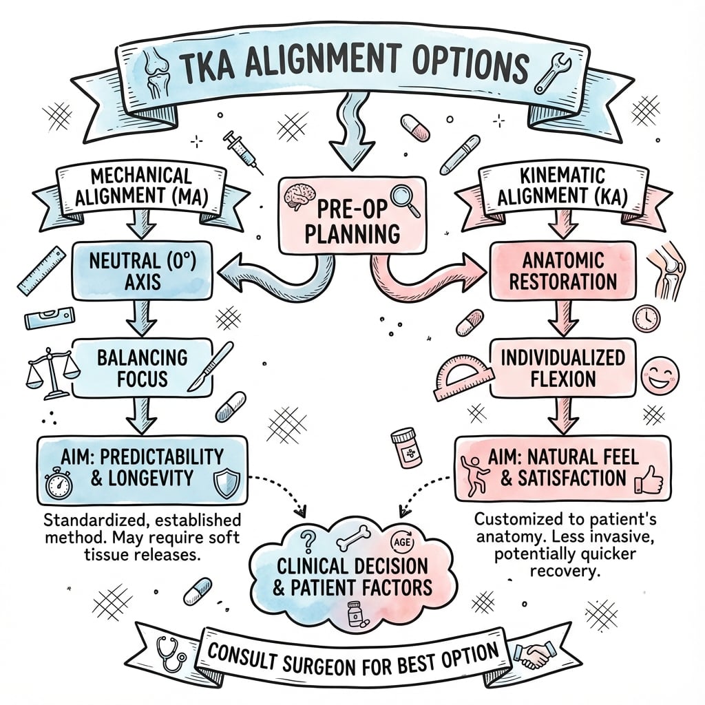
Mechanical vs Kinematic vs Restricted Kinematic | Patient-Specific Alignment | Evolution of TKA Philosophy
- Mechanical alignment targets neutral 0° ± 3° hip-knee-ankle axis with perpendicular cuts - gold standard for 40+ years
- Kinematic alignment restores native joint line obliquity and ligament isometry - gaining popularity since 2014
- Restricted kinematic alignment combines KA principles with safety boundaries (HKA 0° ± 3°) to avoid extreme outliers
- 20% of patients report dissatisfaction with MA-TKA despite correct alignment - driver for alternative techniques
- No Level 1 evidence shows long-term superiority of any alignment philosophy - decision based on surgeon experience and patient anatomy
- “Mechanical axis: line from femoral head center to ankle center - MA-TKA aims for 0° deviation
- “Kinematic axis: reproduces native joint line orientation - typically 3° varus in constitutional alignment
- “Safe zone debate: Restricted KA limits alignment to HKA 0° ± 3° to avoid catastrophic outliers
- “Forgotten joint score higher with KA in short-term studies - less clear at 5+ years
Neutral mechanical axis 0° ± 3°. Perpendicular cuts to mechanical axis. Femur 5-6° valgus cut (relative to anatomic axis), tibia 0° perpendicular. Ignores constitutional alignment. Traditional gold standard since 1970s.
Restore native joint line obliquity. Co-level resection matching worn cartilage + implant thickness. Maintains constitutional alignment (typically 3° varus). Preserves ligament isometry. Higher patient satisfaction in short-term.
Hip-Knee-Ankle (HKA) angle on standing long-leg radiograph. Mechanical axis: femoral head center to ankle center. Anatomic axis: femoral shaft midline. Difference approximately 5-6° valgus (anatomic axis is more varus).
Restricted KA: HKA 0° ± 3°. Avoids extreme varus (greater than 3°) or valgus (greater than 3°). Component position limits: femur 5° varus to 10° valgus, tibia 5° varus to 5° valgus. Prevents edge loading and catastrophic failure.
- Constitutional Alignment
- HKA 0° ± 2° on contralateral knee
- Recommended Approach
- Mechanical Alignment (MA)
- Key Consideration
- Traditional approach, proven long-term outcomes
- Constitutional Alignment
- HKA 3-5° varus bilaterally
- Recommended Approach
- Restricted Kinematic Alignment
- Key Consideration
- Restore native alignment within safe HKA 0° ± 3°
- Constitutional Alignment
- Severe bone loss, ligament incompetence
- Recommended Approach
- Adjusted Mechanical Alignment
- Key Consideration
- May need constrained implants, cannot safely restore KA
- Constitutional Alignment
- Well-preserved ligaments, minimal deformity
- Recommended Approach
- True Kinematic Alignment (if experienced)
- Key Consideration
- Requires advanced technique, no long-term data beyond 10 years
PERPENDICULARMechanical Alignment (MA-TKA) Principles
Hook:MA-TKA is PERPENDICULAR - every cut is 90° to mechanical axis, creating uniform neutral alignment regardless of patient's native anatomy!
RESTOREKinematic Alignment (KA-TKA) Principles
Hook:KA-TKA aims to RESTORE the knee's native anatomy - the joint line obliquity and ligament tension that existed before arthritis!
SAFERestricted Kinematic Alignment Boundaries
Hook:Restricted KA keeps alignment SAFE - within 3° of neutral to avoid catastrophic edge loading and early polyethylene wear!
Overview and Evolution of TKA Alignment
Historical Context
The debate over alignment in total knee arthroplasty represents one of the most significant philosophical shifts in orthopedic surgery over the past decade. For over 40 years, mechanical alignment was the undisputed gold standard, based on the principle that a neutral mechanical axis (0° ± 3°) would distribute loads evenly across medial and lateral compartments, maximizing implant longevity.
Despite excellent survivorship with mechanical alignment (greater than 95% at 15 years), 20% of patients report dissatisfaction with their TKA. Common complaints include feeling the knee is "not natural," inability to forget the joint, and persistent anterior knee pain. These outcomes drove the search for alternative alignment philosophies.
Constitutional Alignment Concept
Average HKA angle: 1.3° varus (range 3° varus to 3° valgus), Joint line obliquity: Distal femur 3° valgus, proximal tibia 3° varus, Natural ligament tension: Isometric throughout range of motion, Individual variation: Wide spectrum of "normal" alignment
Changes joint line: Creates horizontal joint line in most patients, Alters ligament lengths: Requires releases to balance gaps, Modifies kinematics: Changes patellofemoral and tibiofemoral tracking, One-size-fits-all: Ignores individual constitutional variation
The Three Main Philosophies
Alignment Philosophy Evolution
Goal: Neutral 0° ± 3° hip-knee-ankle axis with perpendicular cuts to mechanical axis. Technique: Distal femur cut 5-7° valgus (relative to anatomic axis), proximal tibia cut perpendicular to mechanical axis (0°). Soft tissue balancing via ligament releases. Outcomes: Excellent survivorship (greater than 95% at 15 years), but 20% patient dissatisfaction.
Goal: Restore pre-arthritic joint line obliquity and ligament isometry. Technique: Co-level resections matching worn cartilage thickness + implant thickness. Preserve constitutional alignment (typically 3° varus). Minimal ligament releases. Outcomes: Higher forgotten joint scores and patient satisfaction short-term, limited data beyond 10 years.
Goal: Kinematic principles within safe boundaries (HKA 0° ± 3°). Technique: Start with kinematic approach, but cap alignment at 3° varus or 3° valgus. Adjust component position if exceeding safe zone. Outcomes: Emerging evidence suggests similar satisfaction to KA with potentially safer boundaries. Compromise approach.
Anatomy and Biomechanics
Anatomical Axes and Measurements
- Definition
- Line from femoral head center to ankle center
- Normal Value
- 0° ± 2° (neutral to slight varus)
- Clinical Significance
- MA-TKA target - used for load distribution assessment
- Definition
- Midline of femoral or tibial shaft
- Normal Value
- Femur: 5-7° valgus to mechanical axis
- Clinical Significance
- Intramedullary alignment guides reference this axis
- Definition
- Angle between femoral and tibial mechanical axes
- Normal Value
- 1.3° varus average (range 3° varus to 3° valgus)
- Clinical Significance
- Primary alignment measurement - defines constitutional alignment
- Definition
- Angle of distal femur and proximal tibia surfaces
- Normal Value
- Distal femur 3° valgus, proximal tibia 3° varus
- Clinical Significance
- KA-TKA aims to restore this native obliquity
The CPAK Classification and Arithmetic HKA
Modern alignment selection and reporting use the Coronal Plane Alignment of the Knee (CPAK) classification (developed in the MacDessi 2021 study cited below), which the topic relies on in its guidelines and evidence sections but never actually lays out. CPAK describes a knee by two measures of its native coronal anatomy, both derived from the bony joint-line angles (lateral distal femoral angle, LDFA, and medial proximal tibial angle, MPTA):
- Arithmetic HKA (aHKA) estimates the constitutional limb alignment and is calculated as MPTA minus LDFA: a negative value indicates constitutional varus, a positive value valgus, and a value near zero a neutral limb. It is used because the true pre-arthritic HKA cannot be measured once the knee is worn.
- Joint line obliquity (JLO) describes the orientation of the joint line and is the sum MPTA plus LDFA: a low sum is an apex-distal (more horizontal) joint line, a high sum an apex-proximal one.
Combining aHKA (varus / neutral / valgus) with JLO (low / neutral / high) yields nine phenotypes (Types I to IX). In both healthy and osteoarthritic populations the distribution is similar and Types I, II and V predominate (varus-to-neutral limbs with a neutral-to-apex-distal joint line), while Types VII to IX are rare.
CPAK matters because it gives a single reproducible language for the native knee phenotype and helps predict which knees gain most from a kinematic or restricted strategy (in the MacDessi data, kinematic alignment balanced best in Types I, II and IV) and which phenotypes already sit inside the restricted safe zone.
CPAK classifies the native knee by arithmetic HKA (constitutional alignment = MPTA minus LDFA) and joint line obliquity (= MPTA plus LDFA) into nine phenotypes; varus-with-neutral-JLO types (I, II, V) dominate worldwide. It is the modern shared language for choosing and reporting an alignment strategy - varus phenotypes that fall inside the safe zone are the natural candidates for restricted kinematic alignment.
Biomechanical Load Distribution
Equal 50/50 distribution across medial and lateral compartments.
- Theoretical advantage: Uniform polyethylene wear
- Perpendicular cuts create horizontal joint line
- Requires ligament releases to achieve balanced gaps
- May alter native kinematics and ligament tension
Reproduces native load distribution (typically 60/40 medial/lateral).
- Matches pre-arthritic loading pattern
- Oblique joint line maintains constitutional alignment
- Preserves native ligament lengths and tension
- Concern: Potential for asymmetric polyethylene wear
Extreme varus or valgus alignment (greater than 5°) concentrates loads on polyethylene rim, leading to accelerated wear, deformation, and potential catastrophic failure. This is the primary concern with unrestricted kinematic alignment and drives the "safe zone" concept in restricted KA.
Ligament Considerations
Ligament Behavior Across Alignment Philosophies
Changes ligament lengths due to altered joint line orientation. Medial collateral ligament (MCL) typically lengthened in varus knees corrected to neutral. Lateral collateral ligament (LCL) lengthened in valgus knees. Requires systematic ligament releases to achieve rectangular extension gap and parallel flexion gap.
Maintains native ligament lengths by restoring joint line obliquity. MCL and LCL retain isometry throughout range of motion. Minimal to no ligament releases required. Theoretically improves proprioception and patient satisfaction.
Preserves ligaments when possible, but releases if needed to stay within HKA 0° ± 3° safe zone. Hybrid approach accepting some ligament modification to avoid extreme alignment.
Classification of Alignment Philosophies
Traditional Mechanical Alignment (MA-TKA)
Core Principles
Neutral mechanical axis: Create 0° hip-knee-ankle alignment ± 3° tolerance. This distributes loads equally (50/50) across medial and lateral compartments.
Perpendicular cuts: Distal femur and proximal tibia cuts made 90° to their respective mechanical axes, creating horizontal joint line.
Gap balancing: Rectangular extension gap and parallel flexion gap achieved through systematic ligament releases.
Surgical Technique
- Reference
- Intramedullary rod (anatomic axis)
- Target
- 5-7° valgus from anatomic axis
- Technical Detail
- 9mm typical resection (8-10mm range)
- Reference
- Extramedullary alignment rod
- Target
- 0° perpendicular to mechanical axis
- Technical Detail
- 8-10mm resection from least worn side
- Reference
- Anteroposterior (AP) axis
- Target
- 3° external rotation from posterior condylar axis
- Technical Detail
- Whiteside's line or epicondylar axis alternative
- Reference
- Transepicondylar axis preferred
- Target
- Parallel to flexion gap in balanced knee
- Technical Detail
- Avoid internal rotation - causes patella maltracking
Soft Tissue Balancing in MA-TKA
Goal: Rectangular gap, equal medial and lateral.
- Tight medial: Release superficial MCL off proximal tibia
- Tight lateral: Release iliotibial band (ITB), popliteus
- Severe varus: May need MCL "pie-crusting" or posteromedial release
- Severe valgus: LCL, popliteus, lateral head gastrocnemius release
Goal: Parallel to extension gap, equal medial and lateral.
- Tight flexion gap: Downsize femur or reduce distal femoral resection
- Loose flexion gap: Thicker polyethylene insert
- Asymmetric flexion gap: Adjust femoral component rotation
- Severe imbalance: Consider posterior stabilized or constrained implant
Advantages and Disadvantages
- Advantages
- Excellent long-term data (greater than 95% survivorship at 15 years)
- Disadvantages
- 20% patient dissatisfaction despite correct alignment
- Advantages
- Well-established, reproducible, taught universally
- Disadvantages
- Requires systematic ligament releases, alters native anatomy
- Advantages
- Predictable mechanical outcomes
- Disadvantages
- Lower forgotten joint scores, less natural feeling
- Advantages
- Proven longevity, wide acceptable alignment range
- Disadvantages
- No clear advantage over KA in randomized trials to 5 years
Understanding mechanical alignment remains essential for all TKA surgeons - it is the foundation from which alternative techniques have evolved.
Functional Alignment — The Fifth Philosophy
The four philosophies above are increasingly joined by a fifth - functional alignment (FA) - which the topic names in passing (the Winnock de Grave / Vendittoli approach above and the European-guidance and practice-variation tables) but never develops.
- Concept: rather than fixed bony targets (mechanical alignment's neutral) or pure native anatomy (kinematic alignment), FA uses intra-operative assessment of the soft-tissue envelope (gap and laxity data from navigation or a robot) and then co-adjusts bone resections, implant position and selective releases together to balance the knee in flexion and extension, while keeping limb and component alignment within a defined safe boundary.
- It is the "balance-first" philosophy: the plan is refined live from measured gaps and laxity, which is why FA is, in practice, dependent on computer navigation or robotics.
- Where it sits: like restricted KA it caps coronal outliers; unlike pure KA it will adjust bony cuts (not just reproduce anatomy) to achieve balance; unlike MA it does not force everyone to neutral. Its proponents regard it as the most individualised of the strategies.
- Caveats: it requires enabling technology, its boundaries are not standardised between authors, and - like KA and restricted KA - it lacks long-term comparative survivorship data.
Functional alignment is the balance-first fifth philosophy: it uses navigation/robotic gap-and-laxity data to co-adjust bone cuts, component position and selective releases so the knee balances within a defined safe boundary. It sits between kinematic alignment (pure anatomy) and mechanical alignment (fixed neutral), depends on enabling technology, and shares the KA/restricted-KA limitation of immature long-term survivorship evidence.
Clinical Assessment and Pre-operative Planning
Patient Evaluation for Alignment Choice
Constitutional alignment: Review old radiographs if available, Contralateral knee: Is other knee arthritic? What alignment?, Activity level: High-demand patient may benefit from KA "natural feel", Expectations: Discuss forgotten joint concept vs implant longevity, Prior surgery: Previous osteotomy, fracture - may limit options
Deformity magnitude: Mild (less than 5°), moderate (5-10°), severe (greater than 10°), Deformity correctability: Varus/valgus stress - fixed vs correctable?, Ligament competence: Medial and lateral collateral integrity, Patellofemoral tracking: Maltracking may influence rotational alignment, Range of motion: Severe flexion contracture may limit KA options
Radiographic Planning
Imaging Protocol for Alignment Planning
Both legs if possible to assess constitutional alignment of contralateral knee. Measure hip-knee-ankle (HKA) angle. Assess for extra-articular deformity (femoral or tibial shaft bowing). Determine mechanical axis deviation (MAD) - distance from knee center to mechanical axis line.
Weight-bearing full-extension view. Measure joint line convergence angle (JLCA). Assess medial and lateral joint space - estimate cartilage wear. Identify osteophytes and bone loss. Measure tibial varus/valgus angle.
Assess posterior slope of native tibia. Identify patella height (Insall-Salvati ratio). Check for posterior femoral condylar wear. Assess flexion contracture on lateral view.
Assess patellofemoral tracking and tilt. Identify patella subluxation or dysplasia. Influences decision on femoral component rotation and alignment.
Decision Algorithm for Alignment Philosophy
- Constitutional Alignment
- Neutral HKA 0-2°, mild arthritis
- Recommended Philosophy
- Mechanical Alignment
- Rationale
- Traditional approach works well - proven longevity
- Constitutional Alignment
- Bilateral 3° varus, no previous trauma
- Recommended Philosophy
- Restricted Kinematic Alignment
- Rationale
- Restore native alignment within HKA 0° ± 3° safe zone
- Constitutional Alignment
- Any alignment, high expectations for natural feel
- Recommended Philosophy
- Kinematic Alignment (if experienced surgeon)
- Rationale
- Maximize patient satisfaction and forgotten joint
- Constitutional Alignment
- HKA greater than 10° varus or valgus, bone loss
- Recommended Philosophy
- Adjusted Mechanical Alignment
- Rationale
- Safety concern with KA - may need constrained implant
- Constitutional Alignment
- MCL or LCL deficiency, varus/valgus instability
- Recommended Philosophy
- Mechanical Alignment with constrained implant
- Rationale
- Cannot achieve stability with KA - need implant constraint
- Constitutional Alignment
- Prior fracture, malunion, retained hardware
- Recommended Philosophy
- Adjusted Mechanical Alignment
- Rationale
- Anatomy already altered - difficult to define native alignment
The choice of alignment philosophy must be individualized based on patient anatomy, surgeon experience, and patient expectations. No single approach is universally superior. Beware the surgeon who uses only one technique for all patients - this ignores the spectrum of knee pathology.
Investigations
Radiographic Assessment for Alignment Planning
Imaging Protocol for Alignment Planning
Both legs if possible to assess constitutional alignment of contralateral knee. Measure hip-knee-ankle (HKA) angle. Assess for extra-articular deformity (femoral or tibial shaft bowing). Determine mechanical axis deviation (MAD) - distance from knee center to mechanical axis line.
Weight-bearing full-extension view. Measure joint line convergence angle (JLCA). Assess medial and lateral joint space - estimate cartilage wear. Identify osteophytes and bone loss. Measure tibial varus/valgus angle.
Assess posterior slope of native tibia. Identify patella height (Insall-Salvati ratio). Check for posterior femoral condylar wear. Assess flexion contracture on lateral view.
Assess patellofemoral tracking and tilt. Identify patella subluxation or dysplasia. Influences decision on femoral component rotation and alignment.
Advanced Imaging for Kinematic Alignment
Three-dimensional reconstruction: CT scan of entire lower limb from hip to ankle. Software reconstructs anatomy and identifies pre-arthritic joint line. Custom patient-specific cutting guides manufactured. Allows reproducible kinematic alignment without intraoperative caliper technique.
Cartilage mapping: MRI sequences can visualize remaining cartilage thickness on medial and lateral compartments. Helps predict bone resection amounts for kinematic co-level technique. May identify subchondral bone edema suggesting overload pattern.
Understanding pre-operative imaging is critical for alignment planning - the choice between MA, KA, or restricted KA often depends on constitutional alignment measured on long-leg radiographs.
Management Algorithm
Decision Algorithm for Alignment Philosophy Selection
- Constitutional Alignment
- Neutral HKA 0-2°, mild arthritis
- Recommended Philosophy
- Mechanical Alignment
- Rationale
- Traditional approach works well - proven longevity
- Constitutional Alignment
- Bilateral 3° varus, no previous trauma
- Recommended Philosophy
- Restricted Kinematic Alignment
- Rationale
- Restore native alignment within HKA 0° ± 3° safe zone
- Constitutional Alignment
- Any alignment, high expectations for natural feel
- Recommended Philosophy
- Kinematic Alignment (if experienced surgeon)
- Rationale
- Maximize patient satisfaction and forgotten joint
- Constitutional Alignment
- HKA greater than 10° varus or valgus, bone loss
- Recommended Philosophy
- Adjusted Mechanical Alignment
- Rationale
- Safety concern with KA - may need constrained implant
- Constitutional Alignment
- MCL or LCL deficiency, varus/valgus instability
- Recommended Philosophy
- Mechanical Alignment with constrained implant
- Rationale
- Cannot achieve stability with KA - need implant constraint
- Constitutional Alignment
- Prior fracture, malunion, retained hardware
- Recommended Philosophy
- Adjusted Mechanical Alignment
- Rationale
- Anatomy already altered - difficult to define native alignment
The choice of alignment philosophy must be individualized based on patient anatomy, surgeon experience, and patient expectations. No single approach is universally superior. Beware the surgeon who uses only one technique for all patients - this ignores the spectrum of knee pathology.
Understanding patient-specific factors is critical for alignment philosophy selection.
Surgical Technique by Alignment Type
Mechanical Alignment Surgical Steps
MA-TKA Step-by-Step
Intramedullary alignment: Enter femoral canal at intercondylar notch anterior to PCL insertion. Choose rod diameter 1mm smaller than canal width. Set distal femoral cutting block at 5-7° valgus from anatomic axis (rod is along anatomic axis). Typical resection 9mm from least worn condyle (measure with cutting block caliper). Verify cut is perpendicular to mechanical axis if using navigation.
Extramedullary alignment: Place alignment rod along tibial crest, aiming at center of ankle (talus dome). Adjust medial-lateral to center of tibial spines. Set cutting block at 0° perpendicular to mechanical axis (confirm with ankle alignment). Typical resection 8-10mm from least worn plateau. Verify posterior slope matches native (typically 3-5°).
Rectangular gap goal: Place spacer blocks in extension gap. Measure medial and lateral gap heights with tensiometer or calipers. Goal is equal medial and lateral tension. If asymmetric, perform ligament releases: Tight medial in varus knee - release superficial MCL from proximal tibia incrementally. Tight lateral in valgus knee - release ITB, popliteus, lateral head gastrocnemius.
3° external rotation from posterior condylar axis (Whiteside's line parallel). Alternatives: Transepicondylar axis (most accurate), or parallel to cut tibial surface with balanced flexion gap. Avoid internal rotation - causes patella maltracking and anterior knee pain. Set anterior-posterior (AP) dimension based on bone cuts and implant size.
Parallel to extension gap goal: Place femoral trial and assess flexion gap at 90° flexion. Should be equal to extension gap height and equal medial-lateral. If flexion gap tight: Downsize femoral component or reduce distal femoral resection. If flexion gap loose: Thicker polyethylene insert. If asymmetric flexion gap: Adjust femoral rotation.
Verify stability through full range of motion with trial components. Check patella tracking - should centralize without lateral tilt or subluxation. Confirm mechanical axis passes through center of knee (navigation or alignment rod). Insert definitive components with cement technique per implant design.
Common Ligament Releases in MA-TKA
- Tight Side
- Medial compartment tight
- Release Sequence
- 1. Osteophytes. 2. Deep MCL (posterior capsule). 3. Superficial MCL (pie-crust). 4. Semimembranosus
- Endpoint
- Equal medial-lateral tension in extension
- Tight Side
- Lateral compartment tight
- Release Sequence
- 1. Osteophytes. 2. ITB. 3. Popliteus. 4. Lateral head gastrocnemius. 5. LCL (rare)
- Endpoint
- Equal medial-lateral tension in extension
- Tight Side
- Posterior capsule tight
- Release Sequence
- 1. Osteophytes. 2. Posterior capsule release. 3. Increase distal femoral resection (last resort)
- Endpoint
- Full extension achieved with balanced gaps
Understanding mechanical alignment technique remains the foundation - even surgeons using alternative techniques must know MA-TKA principles.
Complications and Concerns by Alignment Type
- MA-TKA Risk
- Symmetric 50/50 loading - predictable wear pattern
- KA-TKA Risk
- Asymmetric loading in varus knees - long-term wear unknown
- Prevention Strategy
- Restricted KA limits extreme alignment - stay within HKA 0° ± 3°
- MA-TKA Risk
- Requires releases to achieve rectangular gaps - risk of over-release
- KA-TKA Risk
- Minimal releases - preserves native tension, but may leave asymmetry
- Prevention Strategy
- Careful gap assessment and incremental releases in MA
- MA-TKA Risk
- 20% report knee does not feel natural despite correct alignment
- KA-TKA Risk
- Higher forgotten joint scores - more natural feeling
- Prevention Strategy
- Set realistic expectations pre-operatively regardless of technique
- MA-TKA Risk
- Ligament over-release common - mid-flexion laxity
- KA-TKA Risk
- Preserved ligament lengths - theoretically less mid-flexion instability
- Prevention Strategy
- Avoid aggressive releases - consider thicker insert if unstable
- MA-TKA Risk
- Altered Q-angle and trochlear orientation with neutral alignment
- KA-TKA Risk
- Preserved native trochlear groove orientation - less maltracking risk
- Prevention Strategy
- Femoral rotation critical in both techniques - avoid internal rotation
- MA-TKA Risk
- MA safe zone 0° ± 3° prevents edge loading in vast majority
- KA-TKA Risk
- Unrestricted KA may create HKA greater than 5° - edge loading risk
- Prevention Strategy
- Restricted KA with HKA 0° ± 3° boundary mitigates this concern
- MA-TKA Risk
- Traditional concern with MA malalignment greater than 3°
- KA-TKA Risk
- Unknown long-term risk with constitutional varus alignment
- Prevention Strategy
- Long-term registry data needed - currently limited to 10 years KA
Mechanical alignment has 40+ years of registry data showing excellent survivorship (greater than 95% at 15 years). Kinematic alignment has only 10 years maximum follow-up, with most studies under 5 years. The true test of KA will be 15-20 year survivorship - whether asymmetric loading and constitutional varus alignment lead to accelerated polyethylene wear or aseptic loosening. This uncertainty must be disclosed to patients when choosing KA.
Failure Patterns
Malalignment greater than 3°: Increased aseptic loosening risk, Ligament over-release: Instability, mid-flexion laxity, Patella maltracking: Altered Q-angle, anterior knee pain, Patient dissatisfaction: "Unnatural" feel despite correct alignment, Stiffness: Aggressive gap balancing, scar tissue
Edge loading: Extreme varus (greater than 5°) concentrates loads on medial rim, Accelerated wear: Asymmetric loading pattern - long-term unknown, Aseptic loosening: Constitutional varus may overload medial bone-implant interface, Technique error: Incorrect caliper measurement - fails to restore joint line, Severe deformity: Cannot achieve stability without releases
Differential Diagnosis of the Painful / Dissatisfied TKA
Before attributing a poor result to the alignment philosophy, the dissatisfied or painful TKA must be worked up systematically - malalignment is only one of several causes and infection must always be excluded first.
- Key Distinguishing Features
- Rest/night pain, warmth, effusion, early or persistent symptoms
- Decisive Investigation
- ESR/CRP, then aspiration (cell count, culture, alpha-defensin)
- Relation to Alignment
- Must exclude first - unrelated to alignment philosophy
- Key Distinguishing Features
- Activity-related pain, varus/valgus thrust, asymmetric wear
- Decisive Investigation
- Standing long-leg radiograph (HKA), CT for component position
- Relation to Alignment
- Directly related - HKA outside safe zone, tibial component outlier
- Key Distinguishing Features
- Anterior knee pain, patellar maltracking, stiffness
- Decisive Investigation
- CT rotational profile (Berger protocol)
- Relation to Alignment
- Rotational, not coronal - distinct from KA/MA debate
- Key Distinguishing Features
- Giving way, recurrent effusion, sense of insecurity
- Decisive Investigation
- Stress radiographs, examination under anaesthesia
- Relation to Alignment
- Over-release in MA or unbalanced gaps in any technique
- Key Distinguishing Features
- Start-up pain, progressive symptoms, radiolucent lines
- Decisive Investigation
- Serial radiographs, bone scan if equivocal
- Relation to Alignment
- May follow extreme alignment but also fixation/implant factors
- Key Distinguishing Features
- Hip or spine pathology, neuropathic features, normal knee imaging
- Decisive Investigation
- Hip and spine assessment, diagnostic injection
- Relation to Alignment
- Unrelated - a common cause of 'unexplained' TKA pain
Postoperative Care and Follow-Up
Rehabilitation Protocol (Common to All Alignment Types)
TKA Rehabilitation Timeline
Mobilization: Same-day or day 1 mobilization with physiotherapy. Weight-bearing as tolerated with walking aid. DVT prophylaxis: Aspirin 100mg daily (Australian standard) or LMWH if high risk. Pain management: Multimodal analgesia (paracetamol, NSAIDs, opioids as needed). Range of motion: Passive and active-assisted exercises commenced.
Discharge: Typically day 2-4 depending on mobility and social support. Goals: Walk 50+ meters, negotiate stairs safely, ROM 0-90°. Exercises: Quadriceps strengthening, knee flexion exercises, gait re-education. Follow-up: 2-week wound check (suture/staple removal).
ROM goal: 0-110° by 6 weeks. Strengthening: Progressive resistance exercises, stationary bike. Walking: Wean off walking aid by 4-6 weeks. Return to driving: Typically 4-6 weeks (right knee TKA).
ROM goal: 0-120° by 12 weeks (may plateau at 115-120° - acceptable). Activity: Return to low-impact activities (golf, swimming, cycling). Work: Return to sedentary work by 6-12 weeks depending on demands.
Functional improvement: Continues up to 12 months post-op (pain, stiffness, function). Activity: Return to higher-impact activities (tennis, skiing) by 6-12 months if desired and cleared. Follow-up: 6-week, 3-month, 12-month clinical and radiographic review.
Alignment-Specific Considerations
Standard protocol: Same as above - no specific modifications, Ligament releases: May have more stiffness if extensive medial/lateral releases performed, ROM focus: Emphasize early flexion exercises if posterior capsule released, Stability: Test for mid-flexion laxity if over-released - may need brace
Faster ROM: Often achieve ROM goals earlier due to preserved ligament lengths, Less stiffness: Typically less post-op stiffness compared to MA (fewer releases), Proprioception: May feel more "natural" earlier - emphasize balance exercises, Monitoring: Same radiographic follow-up - watch for alignment drift
Long-Term Surveillance
- Clinical Assessment
- Wound healing, ROM, gait, pain level
- Radiographic Assessment
- Standing AP and lateral knee - component position
- Red Flags
- Wound dehiscence, excessive pain, ROM under 70°
- Clinical Assessment
- ROM, function, return to activities
- Radiographic Assessment
- Standing long-leg radiograph - confirm alignment
- Red Flags
- Persistent instability, ROM plateau under 90°
- Clinical Assessment
- Oxford Knee Score, Forgotten Joint Score, satisfaction
- Radiographic Assessment
- Standing AP/lateral - radiolucent lines, wear
- Red Flags
- Radiolucent lines greater than 2mm, component migration
- Clinical Assessment
- Symptoms, function, any changes
- Radiographic Assessment
- Every 2-5 years or if symptomatic
- Red Flags
- New pain, swelling, instability, loss of function
Kinematic alignment patients require specific attention to alignment on follow-up long-leg radiographs. Confirm HKA angle is maintained (should match immediate post-op). Any drift toward further varus (in constitutional varus patients) may indicate medial component subsidence or polyethylene wear - requires close monitoring and may warrant earlier revision.
Patient Education and Expectations
Regardless of alignment philosophy, counsel patients that TKA outcomes mature over 12 months. Peak pain relief at 3-6 months, peak function at 6-12 months. Approximately 80-90% patient satisfaction overall (varies by alignment type - KA may be higher). Realistic ROM expectations: 0-115° is acceptable, 0-120° is excellent. Activities to avoid: High-impact running, jumping sports, contact sports. Implant longevity: Greater than 95% survivorship at 15 years with mechanical alignment - kinematic alignment long-term data still emerging.
Postoperative care for TKA is similar across alignment philosophies - the key difference is patient-reported satisfaction and "naturalness" which may be higher with kinematic alignment.
Guidelines, Registries & Global Practice
Global Epidemiology and the Case for Individualised Alignment
In asymptomatic young adults, 32% of men and 17% of women have constitutional varus (native mechanical alignment 3° varus or more) - Bellemans 2011. The CPAK study of 1,000 knees confirmed only a minority of healthy or arthritic knees sit at true neutral, and the same nine phenotype distribution is seen worldwide in healthy and osteoarthritic populations.
Knee osteoarthritis is among the leading global causes of years lived with disability, and TKA volumes are rising steeply across high-income countries and increasingly in middle-income settings. The major joint registries (AOANJRR, NJR, AJRR and others) together capture well over a million primary TKAs, making them the dominant source of survivorship evidence globally.
Guidance and Position Statements Side by Side
- Position on Alignment
- No single alignment target mandated; emphasises shared decision-making and registry-tracked outcomes
- Evidence Level
- Consensus / observational
- Practical Message
- Neutral MA remains the benchmark; alternative strategies acceptable with informed consent
- Position on Alignment
- Focus on use of proven, registry-monitored implants (ODEP-rated) rather than prescribing an alignment philosophy
- Evidence Level
- Registry / HTA-based
- Practical Message
- Implant survivorship and unit audit prioritised over alignment dogma
- Position on Alignment
- Recognise the shift from systematic MA toward individualised (kinematic / restricted-kinematic / functional) alignment
- Evidence Level
- Level 1-2 RCTs, short-mid term
- Practical Message
- Restricted boundaries advocated to avoid extreme outliers
- Position on Alignment
- Promote phenotype-based individualised alignment within a defined safe zone
- Evidence Level
- Level 1 (balance) + classification
- Practical Message
- Use CPAK to describe native anatomy and select alignment strategy
No major body endorses a single universal alignment target. Across guidelines the common thread is: use a registry-monitored implant, avoid extreme coronal outliers, counsel on the immature long-term data for KA, and share the decision with the patient. This is the globally defensible position to take in any viva.
Registry Evidence at a Glance
- Region
- Australia
- Relevant Signal
- Marked coronal malalignment historically linked to higher revision in MA-TKA; high overall survivorship
- Limitation for Alignment Research
- Alignment philosophy (KA vs MA) not separately coded
- Region
- England, Wales, NI, IoM
- Relevant Signal
- Drives ODEP implant benchmarking; both well-aligned MA implants show excellent 10-15 year survival
- Limitation for Alignment Research
- Records implant and fixation, not surgeon alignment target
- Region
- USA
- Relevant Signal
- Largest TKA volume captured; supports the neutral safe-zone concept for MA
- Limitation for Alignment Research
- Heterogeneous reporting, alignment intent not captured
Global Practice Variation
- Predominant Practice
- Mechanical alignment taught as the foundation
- Driver
- Reproducibility, exam syllabi, established survivorship
- Direction of Travel
- Gradual uptake of restricted-KA / functional alignment
- Predominant Practice
- Rising kinematic, restricted-kinematic and functional alignment
- Driver
- Access to navigation, robotics and PSI; patient demand for 'natural feel'
- Direction of Travel
- Phenotype-based individualised alignment expanding
- Predominant Practice
- Manual mechanical alignment predominates
- Driver
- Cost, lack of navigation/robotics, surgeon familiarity
- Direction of Travel
- MA likely to remain standard; conventional instrumentation reliable
Wherever they practise, surgeons should master mechanical alignment as the foundation and understand kinematic and restricted-kinematic philosophies - the global trend is toward individualised alignment guided by native phenotype and patient expectation, within evidence-based safe boundaries.
MCQ Practice Points
Q: What is the mechanical axis of the lower limb and how does it differ from the anatomical axis? A: The mechanical axis is a line drawn from the center of the femoral head to the center of the ankle (talus dome) on a standing long-leg radiograph. It represents the load-bearing axis of the lower limb. The anatomical axis is the midline of the femoral or tibial shaft. The femoral mechanical axis typically differs from the femoral anatomical axis by approximately 5-7° in the coronal plane (anatomical axis is more varus). This 5-7° difference is why the distal femoral cut in mechanical alignment TKA is made 5-7° valgus relative to the anatomical axis (referenced by intramedullary alignment rod) to achieve a cut perpendicular to the mechanical axis.
Q: What is the normal HKA angle and what does deviation from neutral signify? A: The HKA angle is measured on a standing long-leg radiograph as the angle between the femoral mechanical axis and the tibial mechanical axis. Normal average is 1.3° varus (range 3° varus to 3° valgus) - representing constitutional alignment. Mechanical alignment TKA targets 0° ± 3° (neutral alignment). Deviations beyond 3° from neutral (i.e., greater than 3° varus or greater than 3° valgus) are associated with increased revision rates in mechanical alignment TKA based on registry data. Kinematic alignment accepts constitutional alignment (typically 3° varus) as the target, while restricted kinematic alignment limits to HKA 0° ± 3° to avoid extreme outliers.
Q: What is the fundamental principle of kinematic alignment and how does it differ from mechanical alignment? A: The fundamental principle of kinematic alignment is to restore the native joint line obliquity and ligament isometry by performing co-level bone resections that account for cartilage wear. This reproduces the patient's pre-arthritic (constitutional) alignment, typically 3° varus HKA. The technique uses caliper measurement of remaining cartilage on medial and lateral sides, then resects bone equal to implant thickness PLUS cartilage wear on each side independently. This contrasts with mechanical alignment which creates perpendicular cuts to the mechanical axis (horizontal joint line) and targets neutral 0° HKA alignment regardless of constitutional alignment. KA preserves native ligament lengths (minimal releases), while MA requires systematic ligament balancing to achieve rectangular gaps.
Q: What are the safe zone boundaries for restricted kinematic alignment and what is the rationale? A: Restricted kinematic alignment (rKA) combines kinematic principles with defined safety boundaries to avoid extreme outliers. The HKA safe zone is 0° ± 3° (same as traditional mechanical alignment acceptable range). Component position limits are: femoral component 5° varus to 10° valgus from mechanical axis, tibial component 5° varus to 5° valgus from mechanical axis, and tibial slope 0-7° posterior. The rationale is based on (1) historical mechanical alignment registry data showing increased failure rates with HKA deviation beyond 3°, (2) concern for edge loading with extreme varus or valgus causing rim contact on polyethylene, and (3) theoretical risk of accelerated wear or aseptic loosening with asymmetric loading patterns. rKA allows patient-specific alignment restoration when constitutional anatomy is within safe boundaries, but adjusts alignment (via bone cuts or ligament releases) if exceeding thresholds.
Q: What is the Forgotten Joint Score and what does the evidence show comparing kinematic vs mechanical alignment? A: The Forgotten Joint Score (FJS) is a patient-reported outcome measure (scale 0-100, higher is better) that assesses how often a patient is aware of their artificial joint during activities of daily living. Questions focus on joint awareness during walking, stairs, sitting, and recreation. Several randomised trials and cohorts report a trend toward higher patient-reported and "natural feel" scores with KA at 2-5 years (Dossett 2014 showed better Oxford, WOMAC and Knee Society scores; Blakeney 2018 showed higher KOOS). However, the pooled individual-data meta-analysis (Woon and Young 2018) found no significant WOMAC or KSS function difference and only a small KSS pain advantage. The patient-reported edge for KA is therefore real but modest and inconsistent, whether it is sustained beyond 5-10 years is unknown, and FJS is a surrogate that does not measure implant survivorship.
Q: What is the longest follow-up data available for kinematic alignment TKA and how does it compare to mechanical alignment? A: Mechanical alignment has decades of registry and cohort data, with well-designed implants commonly reporting greater than 95% survivorship at 15 years - the benchmark for long-term implant survivorship. Kinematic alignment evidence is dominated by randomised trials and cohorts with 2-5 year follow-up, with relatively few series reaching 10 years and none reaching mature 15-year comparative survivorship. The highest-level synthesis, the Woon and Young (2018) individual-data meta-analysis of four RCTs, found broadly similar patient-reported outcomes between KA and MA at short to mid-term. The critical knowledge gap is whether KA's restored constitutional (often varus) alignment and more asymmetric loading leads to accelerated polyethylene wear or aseptic loosening at 15-20 years - this remains unproven either way. National registries do not yet separately code alignment philosophy, so registry-level long-term comparison is immature. Patients must be counselled about this uncertainty when choosing KA.
Exam Viva Scenarios
Practise clinical reasoning and management decisions out loud
“A 65-year-old active male presents with end-stage medial compartment osteoarthritis. Standing long-leg radiograph shows constitutional varus alignment of 4° (HKA 4° varus) bilaterally. He has read about kinematic alignment online and asks if it is appropriate for him. Discuss your approach to TKA alignment in this patient.”
“You have decided to perform kinematic alignment TKA using the caliper technique (Howell method). Walk me through your surgical technique for distal femoral and proximal tibial bone cuts in a patient with medial compartment osteoarthritis.”
“You performed a kinematic alignment TKA on a patient with constitutional varus of 5°. You restored the full 5° varus alignment. At 18 months post-operatively, the patient presents with medial knee pain and difficulty with activities. Radiographs show maintained 5° varus alignment and subtle medial tibial component subsidence. Discuss your assessment and management.”
Alignment Definitions
- Mechanical axis = Hip center to ankle center (load-bearing axis)
- HKA angle = Angle between femoral and tibial mechanical axes (normal 1.3° varus)
- Anatomical axis = Femoral/tibial shaft midline (differs from mechanical by 5-7°)
- Joint line obliquity = Native distal femur 3° valgus, proximal tibia 3° varus
Three Main Philosophies
- Mechanical Alignment (MA): Neutral 0° ± 3° HKA, perpendicular cuts, 40+ years data
- Kinematic Alignment (KA): Restore native joint line, co-level resection, 10 years max data
- Restricted KA: Kinematic within HKA 0° ± 3° safe zone - compromise approach
- MA = 95%+ survivorship 15 years | KA = Higher forgotten joint scores 2-5 years
MA-TKA Technique
- Distal femur: 5-7° valgus from anatomic axis (9mm resection), perpendicular to mechanical
- Proximal tibia: 0° perpendicular to mechanical axis (8-10mm resection)
- Femoral rotation: 3° external rotation from posterior condylar axis
- Gap balancing: Rectangular extension gap, parallel flexion gap via ligament releases
KA-TKA Technique
- Measure cartilage wear with calipers on medial and lateral sides
- Co-level resection: Bone cut = Implant thickness + Cartilage wear (each side independent)
- Femoral rotation: 0° from posterior condylar axis (not 3° external rotation)
- No ligament balancing - preserve native tension, minimal releases
Restricted KA Boundaries
- HKA safe zone: 0° ± 3° (cap varus/valgus at 3° boundary)
- Femoral component: 5° varus to 10° valgus from mechanical axis
- Tibial component: 5° varus to 5° valgus from mechanical axis
- Edge loading risk if HKA greater than 5° - restrict to prevent catastrophic outlier
Outcomes and Evidence Base
Patient-Reported Outcomes
- Mechanical Alignment
- 78-82 at 2 years
- Kinematic Alignment
- 85-88 at 2 years
- Clinical Significance
- Higher is better - KA patients more likely to forget artificial joint
- Mechanical Alignment
- 38-42 at 2 years
- Kinematic Alignment
- 40-43 at 2 years
- Clinical Significance
- Minimal clinically important difference 5 points - no clear advantage
- Mechanical Alignment
- 85-90 at 2 years
- Kinematic Alignment
- 88-92 at 2 years
- Clinical Significance
- Small improvement with KA - unclear if sustained long-term
- Mechanical Alignment
- 80% satisfied (20% dissatisfied)
- Kinematic Alignment
- 85-90% satisfied (10-15% dissatisfied)
- Clinical Significance
- Consistent trend favoring KA in multiple studies
The Forgotten Joint Score asks patients how often they are aware of their artificial joint during activities of daily living. Higher scores (range 0-100) indicate better ability to "forget" the joint. KA consistently shows 5-10 point advantage over MA at 2-5 years - this is the strongest patient-reported outcome difference between alignment philosophies.
Radiographic Outcomes and Survivorship
Evidence Timeline by Follow-Up Duration
Multiple RCTs (Dossett 2014; Calliess 2016) show at least non-inferior, and in some series superior, patient-reported outcomes with KA versus MA, with no excess of complications, revision or radiographic loosening at 1-2 years. Dossett reported better Oxford, WOMAC and Knee Society scores with KA at 2 years.
Woon and Young 2018 individual-data meta-analysis of four RCTs (229 PSI-KA vs 229 MA) found no significant difference in WOMAC, KSS function or KSS combined, with only a small KSS pain advantage for KA. No patient subgroup benefited preferentially - tempering claims of clear KA superiority.
Long-term comparative data remain sparse: most KA series have follow-up under 10 years, and no large randomised cohort has reached 15-year survivorship. This is the principal evidence gap when counselling patients on KA.
Major national registries (AOANJRR, NJR, AJRR and others) collectively capture over a million TKAs and historically link marked coronal malalignment to higher revision in mechanically aligned TKA. KA and restricted-KA are not yet separately coded, so registry-level long-term comparison is immature.
Current evidence is Level 1-2 for short-term (2-5 years) showing non-inferior or superior patient satisfaction with KA. No Level 1 evidence beyond 5 years comparing KA to MA. No Level 1 evidence beyond 10 years for any alignment philosophy - we rely on registry data and case series for long-term survivorship.
Evidence Base and Key Studies
Randomised Controlled Trial of Kinematically and Mechanically Aligned TKR: Two-Year Clinical Results (Dossett et al.)
- Blinded RCT: 88 patients randomised to KA (patient-specific guides) vs MA (conventional instruments)
- At 2 years all outcomes favoured KA: mean Oxford Knee Score 40 vs 33 (p equals 0.005)
- Mean WOMAC 15 vs 26 (p equals 0.005) and combined Knee Society Score 160 vs 137 (p equals 0.005)
- Greater mean flexion with KA (121° vs 113°, p equals 0.002); odds ratio for a pain-free knee 3.2-4.9
PSI Kinematic versus Non-PSI Mechanical Alignment in TKA: A Prospective Randomised Study (Calliess et al.)
- RCT: 200 patients randomised (100 KA with custom guides vs 100 MA, manual)
- WOMAC and combined Knee Society Score improved in both, significantly favouring KA at 12 months
- KA restored the pre-morbid flexion-extension axis without altering the joint line
- More poor-outcome outliers occurred in the KA group; deviation from the plan correlated with worse outcomes
Kinematic Alignment in TKA Better Reproduces Normal Gait Than Mechanical Alignment (Blakeney et al.)
- Matched case-control: 18 KA vs 18 MA TKAs vs healthy controls, 3D gait analysis
- KA knee kinematics did not differ significantly from healthy controls in sagittal ROM, max flexion or rotation
- MA knees showed reduced ROM, lower max flexion and increased external tibial rotation versus healthy
- Post-operative KOOS was higher after KA than MA (74.2 vs 60.7, p equals 0.034)
Restoring Constitutional Alignment with a Restrictive Kinematic Protocol Improves Soft-Tissue Balance: An RCT (MacDessi et al.)
- Superiority RCT: 63 patients (70 knees) restricted KA vs 62 patients (68 knees) MA, sensor-quantified balance
- Mean intercompartmental pressure difference at 10° flexion 11.7 psi (KA) vs 32.0 psi (MA), difference 20.3 psi (p less than 0.001)
- Optimal balance (ICPD 15 psi or less) achieved in 80% of KA vs 35% of MA knees (p less than 0.001)
- Bone recuts needed in 9% (KA) vs 49% (MA); tibiofemoral lift-off in 13% vs 43% (both p less than 0.001)
Coronal Plane Alignment of the Knee (CPAK) Classification (MacDessi et al.)
- Radiological analysis of 500 healthy and 500 osteoarthritic knees defining nine CPAK phenotypes
- Phenotypes based on arithmetic HKA (constitutional alignment) and joint line obliquity
- Types I, II and V predominate; similar distribution in healthy and arthritic knees
- In a 138-knee randomised cohort, KA achieved optimal balance more often than MA, especially CPAK Types I, II and IV
Constitutional Varus: Neutral Mechanical Alignment Is Not Normal for Many Patients (Bellemans et al.)
- Analysis of 250 asymptomatic adults aged 20-27 for native lower-limb alignment
- 32% of men and 17% of women had constitutional varus (natural mechanical alignment 3° varus or more)
- Constitutional varus emerged at the growth spurt and was associated with high sporting activity
- Forcing such knees to neutral at TKA may be unnatural for that individual
Kinematic versus Mechanical Alignment in TKA: Meta-Analysis of Randomised Trials (Woon, Young et al.)
- Collaborative individual-data meta-analysis of four RCTs: 229 PSI-KA vs 229 MA patients
- No significant difference in WOMAC, KSS function or KSS combined change scores
- Small advantage for KA in KSS pain (mean difference 3.6, 95% CI 0.2-7.1)
- No subgroup (varus, valgus or neutral) benefited preferentially from KA
National Joint Registry Evidence on TKA Coronal Alignment and Revision Risk
- Registries collectively capture well over a million primary TKAs, providing the largest survivorship datasets worldwide
- Marked coronal malalignment outside roughly neutral ± 3° has historically been linked to higher revision in mechanically aligned TKA
- Modern designs and patient-specific/computer-assisted techniques show high overall 10-15 year survivorship across registries
- Kinematic and restricted-kinematic alignment are not yet separately coded, so registry-level long-term comparison remains immature