Flexion Gap | Extension Gap | Constraint | Revision
- Gap imbalance most common cause
- Flexion instability: loose in flexion
- Extension instability: recurvatum tendency
- Component malposition key factor
- Revision with increased constraint if failed
- “Flexion gap: posterior femoral condyle resection
- “Extension gap: distal femur resection
- “PCL incompetent = flexion instability in CR
- “Increase constraint level for revision
Overview and Epidemiology
Why the knee is unstable. Most TKA instability results from gap imbalance: a mismatch between the flexion and extension gaps, or global ligamentous laxity. Component malpositioning is the underlying cause.
How it is named. Instability is described by the position in which the knee is loose: in flexion, in extension, in mid-flexion (30-60 degrees) or globally, in all positions. Each pattern has its own causes, set out under Classification.
Pathophysiology and Mechanisms
Two gaps. The stability of a TKA depends on equal and balanced flexion and extension gaps. Knowing which structures contribute to each gap is the basis of both preventing instability and treating it.
The extension gap is assessed with the knee fully extended, at 0 degrees. The MCL and LCL are its primary stabilisers, with the posterior capsule, and a tight posterior capsule limits the extension gap.
The flexion gap is assessed at 90 degrees of flexion. The collaterals contribute to stability in flexion as well, and in a cruciate-retaining (CR) knee the retained PCL is a major flexion stabiliser.
What changes each gap. The biomechanical principles:
- Distal femoral resection primarily affects the extension gap
- Posterior condylar resection primarily affects the flexion gap
- Femoral sizing affects the flexion gap: a larger femur gives a smaller flexion gap
- Posterior tibial slope affects the flexion gap: more slope gives a larger flexion gap
- The collateral ligaments contribute to both gaps
Classification and Mechanism
A flexion gap that is too large. The knee feels loose in flexion and gives way on stairs and when sitting, and the tibia may subluxate posteriorly. Causes:
- Excessive posterior condylar resection
- Femoral component too small (anterior referencing)
- PCL rupture or incompetence in a CR TKA
- Excessive posterior tibial slope
- Component malrotation
Treatment reduces the flexion gap or increases constraint.
Bone Defect Classification (AORI)
Revision for instability frequently uncovers metaphyseal bone loss that dictates the reconstruction. The Anderson Orthopaedic Research Institute (AORI) classification grades the defect separately for the femur (F) and tibia (T) and maps directly onto the fixation strategy.
- Defect
- Intact metaphyseal bone; minor contained defect; joint line near normal
- Typical reconstruction
- Cement, morselised graft, small modular augments
- Defect
- Damaged metaphyseal bone of ONE condyle or plateau
- Typical reconstruction
- Modular augments, with or without cement; stem
- Defect
- Damaged metaphyseal bone of BOTH condyles or plateaus
- Typical reconstruction
- Augments plus metaphyseal cones/sleeves; stem
- Defect
- Deficient metaphyseal segment, often with collateral or extensor-mechanism compromise
- Typical reconstruction
- Metaphyseal cones/sleeves, structural allograft, or a hinge
Grade F1-F3 and T1-T3 and let the grade drive fixation. Modern revision aims for zonal fixation: secure purchase in at least two of the three zones (epiphysis, metaphysis, diaphysis), the metaphyseal zone being the one that cones and sleeves restore.
Clinical Assessment
History. The timing of symptoms helps classify the instability. Establish:
- Giving-way episodes, and whether they come on stairs, walking or rising
- Pain location and character
- Time from the primary operation
- Previous surgery
Examination. Document both the degree of laxity and the position in which it occurs, comparing with the contralateral knee.
- Varus and valgus stress at 0 and 30 degrees
- Anterior and posterior drawer
- Recurvatum
- Range of motion
- Gait
The differential. The patient who reports "giving way" or recurrent effusion does not always have mechanical instability. Distinguishing true ligamentous or gap instability from its mimics changes management entirely.
- Discriminating Features
- Reproducible laxity on stress; effusion; stairs or recurvatum symptoms
- Key Test
- Stress exam at 0 and 90 deg, stress radiographs
- Pitfall if Missed
- Revising the wrong way (e.g. liner alone) fails
- Discriminating Features
- Rest pain, warmth, persistent effusion, raised CRP/ESR
- Key Test
- Aspiration: synovial WCC and differential, cultures
- Pitfall if Missed
- Missed infection turns a 1-stage into a disaster
- Discriminating Features
- Anterior knee pain, maltracking, instability 30-60 deg
- Key Test
- CT rotational profile (epicondylar axis, tibial tubercle)
- Pitfall if Missed
- Constraint added without correcting rotation re-fails
- Discriminating Features
- Start-up pain, progressive radiolucency/subsidence
- Key Test
- Serial weight-bearing radiographs, bone scan
- Pitfall if Missed
- Mislabelled as instability; loose component missed
- Discriminating Features
- Extension lag, quadriceps weakness, buckling
- Key Test
- Active extension lag, patellar tracking assessment
- Pitfall if Missed
- Quads-avoidance buckling mistaken for laxity
- Discriminating Features
- Late onset (usually over 10 years), effusion, lysis on film
- Key Test
- Radiographs for lysis, insert thickness assessment
- Pitfall if Missed
- Late wear can coexist with secondary laxity
Every painful or effusing TKA must have periprosthetic joint infection (PJI) excluded before it is attributed to mechanical instability. Screen with serum CRP and ESR; if either is raised, or clinical suspicion is high, aspirate the joint. Widely used 2018 International Consensus Meeting (ICM) thresholds for chronic knee PJI are a synovial-fluid white-cell count above ~3,000 cells/microlitre and polymorphonuclear cells above ~70%, supported by a positive alpha-defensin, raised synovial CRP, and culture.
Investigations
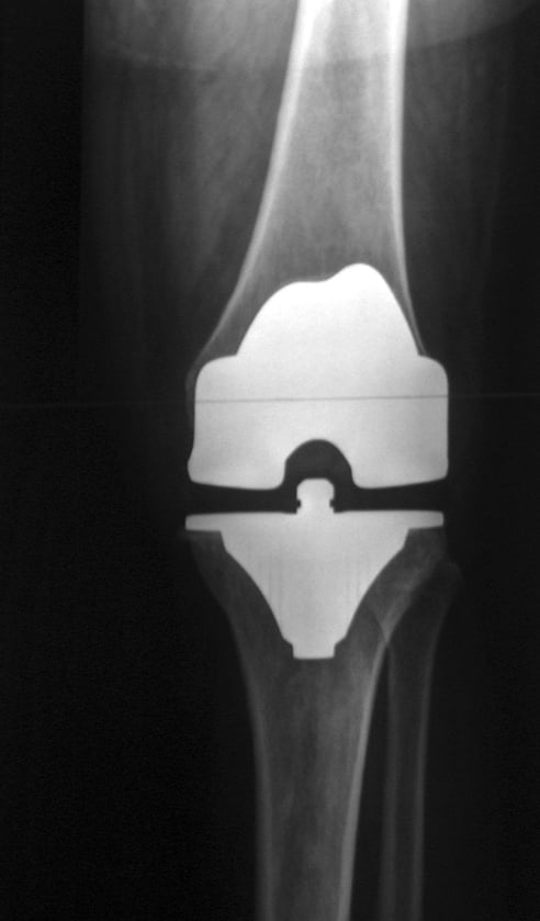
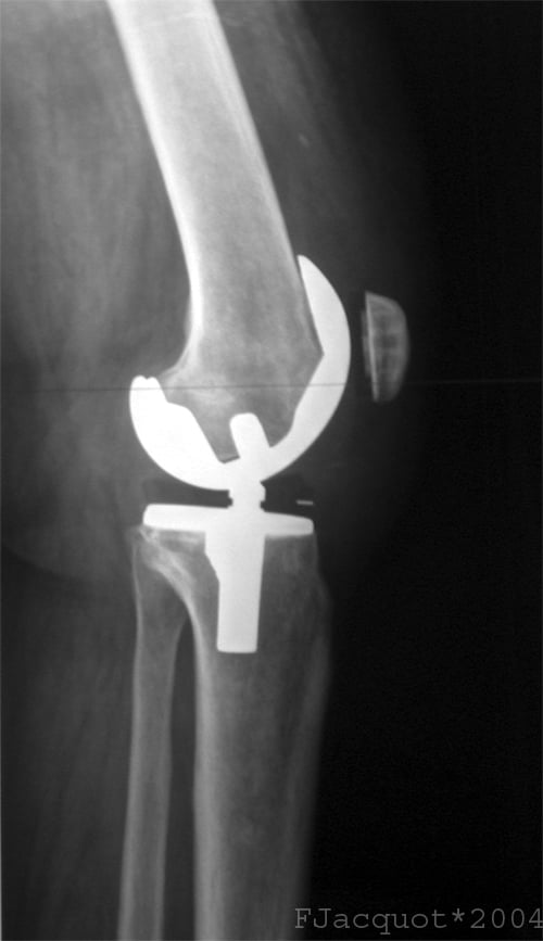
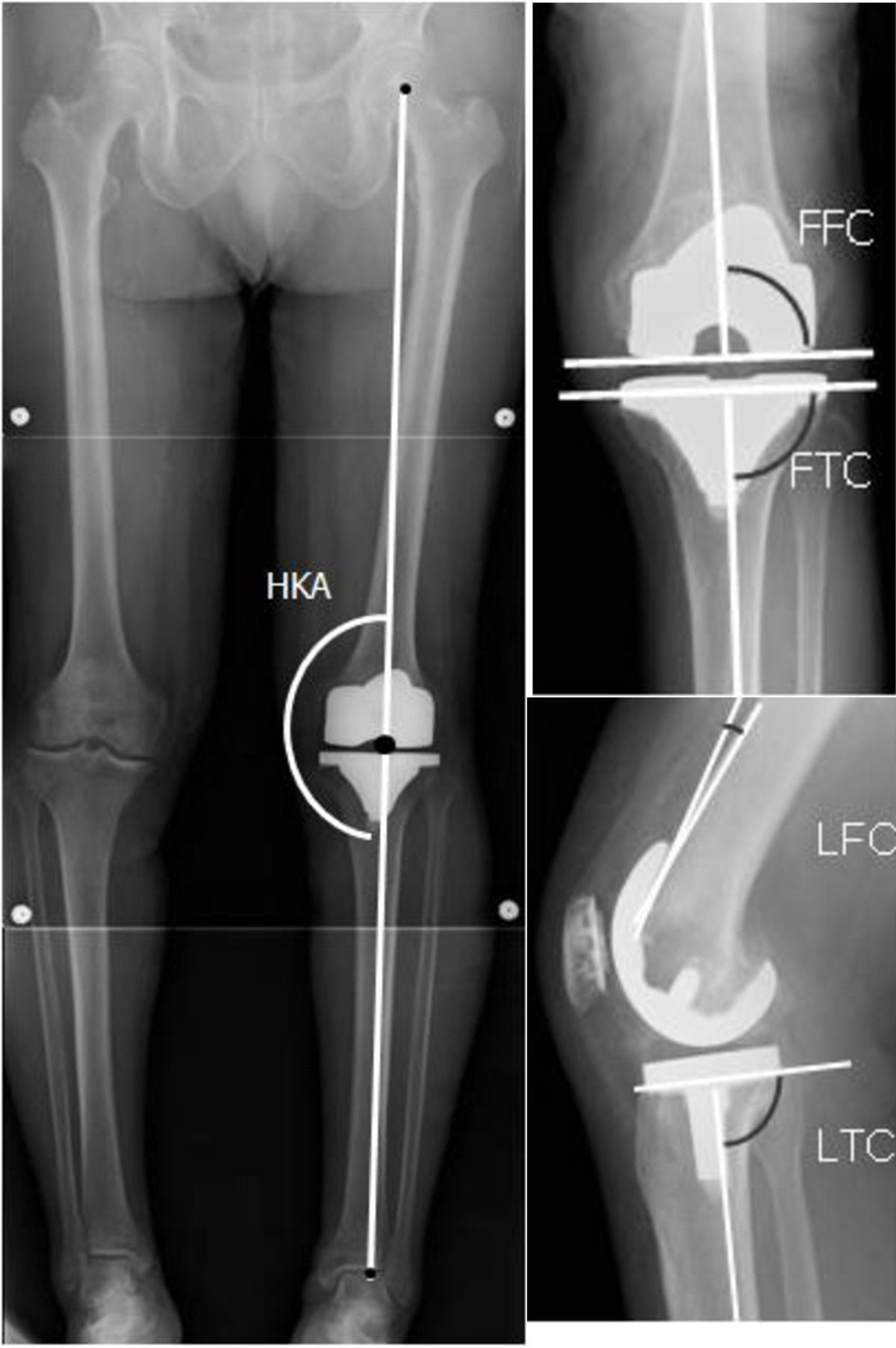
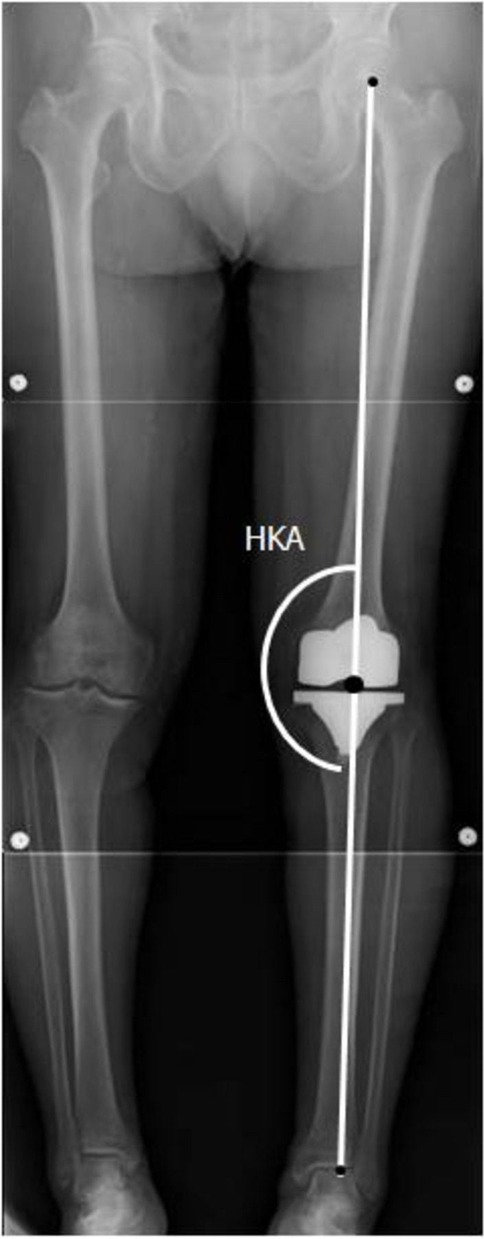
Radiographs. Weight-bearing AP, lateral and skyline views, with full-length standing films for alignment. Component position and limb alignment are central to diagnosing instability, malalignment and loosening. On the films, assess:
- Component position
- Joint-line position
- Polyethylene wear
- Alignment
Stress views may demonstrate the laxity itself.
CT. Indicated when component malrotation is suspected and for preoperative planning, and critical for rotational assessment. It measures:
- Femoral rotation, referenced to the surgical transepicondylar axis (medial sulcus to lateral epicondyle) and cross-checked against the posterior condylar axis and Whiteside's (anteroposterior) line
- Tibial rotation, referenced to the tibial tubercle (the Akagi line, from the PCL centre to the medial third of the tubercle)
- Component position and combined rotational mismatch
Berger's thresholds. Combined femoral and tibial internal rotation correlates with the severity of patellofemoral complication, and well-functioning knees sit in combined external rotation:
- Roughly 1-4° of combined internal rotation: patellar tilt and maltracking
- 3-8°: subluxation
- 7-17°: frank dislocation or late patellar failure
Why rotation matters for stability. Internal femoral rotation also opens the medial flexion space, linking malrotation to mid-flexion instability. That is why malrotation must be corrected at revision rather than simply adding constraint.
Examination under anaesthesia. Performed at revision, EUA quantifies laxity in all positions, compares the flexion gap with the extension gap and identifies the unstable arc. It guides the intraoperative decisions.
Management Algorithm
The choice. Treatment escalates from a conservative trial through polyethylene exchange to component revision, and the level of constraint is matched to the degree of instability.
- Key Finding
- Loose in flexion, stairs difficulty
- First-line Treatment
- Thicker polyethylene
- Revision Approach
- PS conversion or upsize femur
- Key Finding
- Recurvatum, loose standing
- First-line Treatment
- Bracing trial
- Revision Approach
- Distal femoral augment
- Key Finding
- 30-60 degree laxity
- First-line Treatment
- PT, bracing
- Revision Approach
- Correct rotation, VVC
- Key Finding
- All positions unstable
- First-line Treatment
- Hinged brace
- Revision Approach
- VVC or rotating hinge
Conservative trial. For mild instability, the poor surgical candidate, and the recent operation that should be given time to stabilise. A hinged knee brace, quadriceps strengthening physiotherapy and activity modification have limited success for true mechanical instability.
Polyethylene exchange. Exchange to a thicker insert, considering a more constrained option, is indicated for well-fixed, well-positioned components with mild to moderate laxity, or where polyethylene wear is identified. On its own it addresses only minimal laxity.
Component revision. For moderate to severe instability, component malposition, or a failed polyethylene exchange. The principles are to correct component position, balance the gaps and increase constraint as needed.
Surgical Technique
The constraint ladder. Implants provide varying degrees of intrinsic constraint:
- Indication
- Normal ligaments
- Mechanics
- Relies on an intact PCL
- Indication
- PCL deficiency
- Mechanics
- Cam-post replaces the PCL
- Indication
- Moderate collateral laxity
- Mechanics
- Taller post, deeper box
- Indication
- Severe or global instability
- Mechanics
- Linked axis
Use the minimum constraint necessary. Higher constraint transfers more stress to the fixation interface, potentially increasing loosening risk. Balance it against the need for stability.
Balancing the gaps. Identify the imbalance, correct it, and balance the gaps before closing:
- Cause
- Excessive posterior condyle resection
- Solution
- Upsize femur, reduce posterior tibial slope, PS if CR (PCL issue), thicker polyethylene
- Cause
- Excessive distal resection
- Solution
- Distal femoral augments; accept slight flexion contracture
- Cause
- Global ligament laxity
- Solution
- Increase constraint level
- Cause
- Under-resection
- Solution
- Additional bone cuts
Stems and augments. Stems are used in most revision cases, to bypass stress risers and improve fixation. Metal augments replace deficient condyles where bone has been lost.
Joint Line Restoration and Pseudo-Patella Baja
Why the joint line matters. Restoring the native joint line is one of the most important and most often missed goals of revision. Raising it, for example by over-resecting the distal femur or over-stuffing the tibia to fill a gap, shifts the flexion-extension kinematics and is a recognised cause of mid-flexion instability.
Pseudo-patella baja. Joint-line elevation also leaves the patella low. The patellar tendon length is unchanged, but because the joint line has moved proximally the patella now sits low relative to the new joint line, causing anterior knee pain, reduced flexion and extensor dysfunction. This is distinct from true patella baja, a genuinely shortened or scarred patellar tendon.
Finding the native joint line. The landmarks:
- About 1 cm proximal to the fibular head
- About 1.5 cm distal to the medial epicondyle and about 2.5 cm distal to the lateral epicondyle
- About 3 cm distal to the adductor tubercle
- The meniscal scar and the inferior pole of the patella also help
Aim to restore the joint line to within about 5 to 8 mm of native. Suspect pseudo-patella baja when the patella looks low after a revision: the Insall-Salvati ratio is normal (the tendon is unchanged) but a joint-line-referenced index (Blackburne-Peel or the modified Insall-Salvati) is abnormal, the give-away that the joint line, not the tendon, has shifted. Joint-line elevation and patella baja both correlate with worse function, so re-establish the joint line rather than just chasing the gaps.
Complications
- Incidence
- 5-10%
- Prevention/Management
- Appropriate constraint, good balance
- Incidence
- Variable
- Prevention/Management
- Aggressive early ROM
- Incidence
- Increased with constraint
- Prevention/Management
- Adequate fixation, stems
- Incidence
- 2-3% revision
- Prevention/Management
- Prophylaxis, staged if indicated
Prevention at revision. Confirm gap balance before final cementation, and assess stability through the full range of motion under anaesthesia. Use adequate stem fixation for constrained implants, document why the constraint level was chosen, and consider a staged approach if there is any concern about infection.
Follow-up. Regular clinical and radiographic review identifies recurrent symptoms early, and a recurrent effusion should lower the threshold for aspiration. Long-term outcome depends on the initial balance.
Postoperative Care
Revision TKA Rehabilitation
Weight-bearing as tolerated. Brace if needed. ROM exercises.
Progressive strengthening. ROM focus. Stairs training.
Full activities. Quad and hamstring focus. Balance training.
Return to normal activities. Long-term follow-up.
Outcomes and Prognosis
Prognostic factors. A correct diagnosis, appropriate constraint and good bone stock predict a better outcome. Global instability, multiple prior surgeries and poor soft tissues predict a worse one.
Guidelines, Registries & Global Practice
Global epidemiology of TKA instability:
Instability is consistently reported as the second most common indication for TKA revision after aseptic loosening, accounting for roughly 15-20% of all revision TKAs across multi-centre and registry datasets. It is disproportionately an early failure: more than a third of all revisions occur within 2 years of the index procedure, and around half of these early revisions are attributable to instability, malposition or fixation failure - all surgeon-dependent technical factors.
National joint registry signals (cross-registry consensus):
- Region
- UK
- Instability-related observation
- Instability among leading non-infective revision indications; constraint escalation common at revision
- Region
- USA
- Instability-related observation
- Instability a top-three revision cause; documents rising use of constrained and hinge designs at revision
- Region
- Australia/New Zealand
- Instability-related observation
- Instability a major revision indication; younger age at revision associated with higher re-revision risk
- Region
- Sweden
- Instability-related observation
- Long-term survivorship data; emphasises balanced gaps and alignment to reduce revision
- Region
- New Zealand
- Instability-related observation
- Captures constraint level and re-revision; corroborates higher re-revision in younger patients
Registries agree on three points: instability is largely preventable at the index operation, adequate constraint at revision reduces re-revision for instability, and younger patients carry a higher re-revision risk.
Side-by-side guidance from major societies:
- Region
- USA
- Position relevant to TKA instability
- Evidence-based work-up of the painful TKA; rule out infection before attributing symptoms to instability; CT for suspected malrotation
- Region
- UK
- Position relevant to TKA instability
- Revision arthroplasty should be undertaken in networks with appropriate expertise and implant availability (constraint ladder, stems, augments)
- Region
- Global
- Position relevant to TKA instability
- Stepwise constraint principle - use the minimum constraint that restores stability; protect fixation with stems
- Region
- Europe
- Position relevant to TKA instability
- Systematic diagnostic algorithm for the unstable TKA; gap balancing and component re-positioning before defaulting to high constraint
Every society places exclusion of periprosthetic joint infection (serum CRP/ESR, aspiration with synovial WBC and differential) ahead of attributing a painful, effusing TKA to mechanical instability.
- Well-resourced settings: ready access to CT rotational profiling, the full constraint ladder (PS, CCK/VVC, rotating hinge), metaphyseal cones/sleeves and modular augments, and revision arthroplasty networks.
- Limited-resource settings: CT and the higher-constraint/hinge inventory may be scarce; greater reliance on clinical examination and stress radiographs, prolonged bracing trials, and primary-implant-based solutions. The biomechanical principles (balance the gaps, correct rotation, use the least constraint that works) are universal even where the implant menu is narrower.
- Instability as the second leading cause of revision and its early, technique-related nature
- The constraint ladder (CR to PS to CCK/VVC to rotating hinge) and the "minimum constraint" principle
- Why a CR knee with an incompetent PCL becomes flexion-unstable and is converted to PS
- Interpretation of CT rotational studies (epicondylar axis, tibial tubercle)
- Mandatory exclusion of infection before revising for presumed instability
Controversies and Areas of Uncertainty
There is no agreed quantitative threshold for diagnosing flexion instability. Testing positions and laxity grades vary between surgeons, and revision for flexion instability shows the least improvement of any failure etiology - partly because the diagnosis is imprecise. Mechanical symptoms, recurrent effusion and posterior sag remain the practical triad.
Isolated polyethylene liner exchange is attractive (shorter stay, fewer complications) but carries a higher re-revision rate for instability than full component revision. The debate centres on patient selection: well-fixed, well-positioned, correctly rotated components are the only reasonable candidates.
Whether gap-balancing or measured-resection technique better prevents instability remains unsettled; large series report acceptable results with both. Surgeon familiarity and accurate rotational referencing likely matter more than the philosophy chosen.
Higher constraint restores stability but transfers load to the fixation interface and the stems. The "minimum necessary constraint" principle is widely endorsed, yet the exact threshold for stepping from CCK/VVC to a rotating hinge in borderline collateral incompetence is judgement-based, not evidence-defined.
MCQ Practice Points
Q: What does resecting more posterior femoral condyle do? A: Increases the flexion gap. Posterior condyle resection primarily affects flexion gap.
Q: What does resecting more distal femur do? A: Increases the extension gap. Distal femur resection primarily affects extension gap.
Q: What happens if PCL is incompetent in CR TKA? A: Flexion instability. PCL is primary flexion restraint. Need to revise to PS.
Q: What constraint level for moderate collateral laxity? A: VVC (varus-valgus constrained). Taller post provides more coronal stability.
Q: What does internal rotation of femoral component cause? A: Mid-flexion instability. The tibia externally rotates relative to internally rotated femur.
Q: Why use stems with constrained TKA revision? A: To bypass stress transfer to metaphysis. Higher constraint increases fixation stress.
Exam Viva Scenarios
Practise clinical reasoning and management decisions out loud
“A patient 6 months after primary CR TKA reports instability going downstairs and difficulty rising from a chair. Examination shows increased anterior-posterior translation in flexion but stable in extension. X-rays show well-fixed components. What is your diagnosis and management?”
“A 70-year-old woman with severe RA has had 2 previous TKA revisions. She now has gross instability in all positions and cannot walk without aids. What are your options?”
“A 62-year-old man, 18 months after a primary PS TKA, complains of anterior knee pain, a feeling of the knee 'shifting', and recurrent effusions. He is stable on varus/valgus stress at full extension and reasonably stable at 90 degrees, but feels unstable in the mid-arc. Radiographs show well-fixed components and a slightly laterally tracking patella. How do you work this up and manage him?”
Types
- Flexion: loose in flexion, stairs/sitting
- Extension: recurvatum, loose standing
- Mid-flexion: 30-60 degree laxity
- Global: all positions unstable
Gap Balancing
- Posterior condyle = flexion gap
- Distal femur = extension gap
- Equal gaps for stability
- Flexion gap tight = limited bend
- Extension gap tight = flex contracture
Causes
- Gap imbalance most common
- Component malposition
- PCL incompetence (CR)
- Ligament laxity
Constraint Levels
- CR: relies on PCL
- PS: cam-post for PCL deficiency
- VVC: collateral laxity
- Hinge: global instability
Treatment Principles
- Minimum constraint needed
- Balance gaps at revision
- Address malposition
- Use stems in revision
Outcomes
- 80-85% good after revision
- Re-revision 5-10%
- Match constraint to laxity
- Early instability: technical error likely
- Late instability: polyethylene wear/laxity
Evidence Base and Key Studies
Instability as a Leading Cause of TKA Failure (Insall Award)
- 212 revision TKAs reviewed; instability was a leading failure mode after polyethylene wear and aseptic loosening
- More than half of revisions occurred under 2 years from the index operation; the series overall spanned 9 days to 28 years, averaging 3.7 years
- 50% of early revisions were due to instability, malalignment/malposition or failure of fixation
- Early failure mechanisms are predominantly technique-related
Etiology of Modern TKA Revision (Multicentre)
- Six-centre review: aseptic loosening 31.2% and instability 18.7% were the two leading revision causes
- Instability was the second most common indication overall, ahead of infection at 16.2%, polyethylene wear at 10.0%, arthrofibrosis 6.9% and malalignment 6.6%
- Mean time to failure was 5.9 years (range 10 days to 31 years)
- 35.3% of revisions occurred under 2 years and 60.2% within 5 years of index surgery
- Polyethylene wear was uncommon before 15 years; early failure is surgeon-dependent
Across the failure-etiology literature instability is consistently a leading indication for TKA revision - the second commonest after aseptic loosening in contemporary multicentre data (Schroer 2013, 18.7%) and a top-three cause historically (Sharkey 2002, after polyethylene wear and aseptic loosening) - and most early instability is technical. Revision strategy follows a constraint ladder: re-balance and convert CR to PS for flexion instability (Pagnano 1998), constrained condylar for collateral laxity (Mancino 2020), and rotating hinge as salvage for global instability (Kouk 2018).