Midfoot Pain | Progressive Deformity | Surgical Reconstruction
- TMT arthritis most commonly follows Lisfranc injury (missed or inadequate treatment)
- Second TMT joint is recessed - keystone preventing dorsal subluxation
- Fusion gold standard - motion at TMT 1-3 minimal (2-3 degrees), unlike TMT 4-5 (10-15 degrees)
- Selective fusion - fuse stiff medial column (TMT 1-3), preserve mobile lateral column (TMT 4-5)
- Non-union rate 5-15% with TMT arthrodesis - requires rigid fixation
- “Distinguish primary vs post-traumatic (most common after Lisfranc injury)
- “Piano key sign on exam - dorsal subluxation of metatarsal bases
- “Weight-bearing radiographs essential - reveal instability not seen non-WB
- “Fusion extends medially if involves naviculocuneiform or intercuneiform joints
TMT 1-3 form rigid medial column. TMT 2 is keystone - recessed between cuneiforms preventing dorsal/plantar translation. Loss of this architecture causes collapse deformity.
80% of TMT arthritis is post-traumatic. Most follows inadequately treated Lisfranc injuries. Missed subtle injuries progress to arthritis within 2-5 years.
Selective fusion principle. Fuse medial column (TMT 1-3) for stability, preserve lateral column (TMT 4-5) for forefoot adaptation on uneven ground.
Rigid fixation mandatory. Non-union rate 5-15%. Use compression screws or dorsal plates. Avoid crossing unfused TMT 4-5 with hardware.
- Clinical Features
- Activity pain, minimal deformity, joint space preserved
- Treatment
- Conservative: orthoses, activity modification
- Key Pearl
- 80% respond to orthotics in first 2 years
- Clinical Features
- Daily pain, walking limitation, visible osteophytes
- Treatment
- Trial conservative first, then arthrodesis
- Key Pearl
- Steroid injections diagnostic - predicts fusion success
- Clinical Features
- Constant pain, deformity, complete joint loss
- Treatment
- Arthrodesis TMT 1-3 +/- NC/IC joints
- Key Pearl
- Extend fusion if adjacent joint involvement on imaging
MIDASPost-Traumatic Arthritis Risk Factors
Hook:MIDAS touch turns Lisfranc injuries to gold - but missed injuries turn to arthritis!
FUSEArthrodesis Principles
Hook:FUSE the medial column, but don't fuse everything - selective fusion preserves function!
Overview and Epidemiology
Tarsometatarsal (TMT) arthritis affects the joints between the metatarsal bases and the three cuneiforms (medial, intermediate, lateral) and cuboid. This complex of five joints forms the anatomic and functional transition between the midfoot and forefoot.
TMT arthritis is predominantly post-traumatic, with 80% of cases following Lisfranc injuries. The majority result from inadequately treated or missed subtle injuries that progress to arthritis within 2-5 years. Primary osteoarthritis is less common and typically affects the first TMT joint in isolation.
- Post-traumatic: Equal gender distribution, age 30-50 years
- Primary OA: Female predominance (2:1), age 50-70 years
- TMT 1 most commonly affected in primary OA
- TMT 1-3 involved in post-traumatic arthritis
- Chronic disability: 60% unable to return to previous activity level
- Work limitation: 40% change occupation or reduce hours
- Progressive deformity: Forefoot abduction, arch collapse if untreated
- Adjacent joint arthritis: 30% develop NC or IC joint involvement by 5 years
Anatomy and Biomechanics
The second TMT joint is recessed 2-3mm dorsally between the intermediate cuneiform and bases of metatarsals 1 and 3. This "keystone" configuration provides inherent stability, preventing dorsal or plantar translation. Loss of this architecture (in Lisfranc injuries or arthritis) causes progressive midfoot collapse.
- Native Motion
- 2-3° sagittal
- Role
- Push-off stability
- Fusion Impact
- Minimal - stiff joint
- Native Motion
- 1-2° (least mobile)
- Role
- Keystone stability
- Fusion Impact
- No functional loss
- Native Motion
- 2-3° sagittal
- Role
- Completes medial arch
- Fusion Impact
- Minimal functional loss
- Native Motion
- 10-12° sagittal
- Role
- Forefoot flexibility
- Fusion Impact
- Significant loss if fused
- Native Motion
- 12-15° sagittal
- Role
- Ground adaptation
- Fusion Impact
- Significant loss if fused
Anatomic Subdivisions
- Rigid, minimal motion (2-3 degrees total)
- Forms longitudinal arch base
- Primary weight-bearing during stance and push-off
- Dense plantar ligamentous support (Lisfranc ligament from medial cuneiform to MT2 base)
- Mobile, 10-15 degrees sagittal motion
- Allows forefoot accommodation on uneven terrain
- Less ligamentous constraint
- Fusion significantly impairs function
- Lisfranc ligament: Strongest ligament - medial cuneiform to MT2 base (plantar)
- Intermetatarsal ligaments: Strong dorsal and plantar connections between MT bases 2-5
- No intermetatarsal ligament between MT1-2: Explains common Lisfranc injury pattern
- Keystone loss: TMT 2 dorsal subluxation causes forefoot abduction deformity
- Medial column collapse: Loss of longitudinal arch with planovalgus foot
- Lateral column overload: Fusion TMT 1-3 increases stress on TMT 4-5
KRISTMT Joint Stability Anatomy
Hook:KRIS keeps the midfoot stable - Keystone anatomy and Rigid medial column contrasts with mobile lateral column!
Pathophysiology
Post-Traumatic Arthritis (80% of cases)
The most common pathway to TMT arthritis follows inadequately treated Lisfranc injuries:
Post-Traumatic Arthritis Progression
Lisfranc injury with ligamentous disruption or fracture-dislocation. Subtle injuries often missed on initial radiographs if not weight-bearing views obtained.
Inadequate reduction (greater than 2mm displacement) or hardware failure allows persistent malalignment. Abnormal joint loading begins.
Cartilage breakdown at areas of abnormal contact stress. Joint space narrowing visible on radiographs. Intermittent activity-related pain.
Progressive joint space loss, subchondral sclerosis, osteophyte formation. Daily pain, walking limitation. Deformity may be visible.
Complete joint space loss, subchondral cysts, collapse deformity. Adjacent naviculocuneiform and intercuneiform joints often involved. Constant pain.
Primary Osteoarthritis (20% of cases)
Primary TMT arthritis typically affects the first TMT joint in isolation, likely due to mechanical factors:
- Hypermobility: First ray instability from ligamentous laxity
- Hallux valgus association: Lateral thrust force from great toe deformity
- Inflammatory arthritis: Rheumatoid, psoriatic arthritis can involve TMT joints
Classification
Arthritis Severity (Descriptive)
No universally accepted classification system exists for TMT arthritis severity. Typically described by radiographic changes:
Mild Arthritis
- Joint space narrowing (under 50% loss)
- Minimal osteophytes
- No subchondral cysts
- Preserved alignment
- Activity-related pain
- Minimal functional limitation
- Conservative management often successful
Conservative with orthoses, activity modification, NSAIDs. Surgery only if conservative fails after 6 months.
Clinical Presentation
- Pain location: Dorsal midfoot, worse with weight-bearing
- Onset: Gradual worsening over months to years
- Trauma history: Previous Lisfranc injury or high-energy foot trauma
- Functional limitation: Difficulty walking on uneven ground, stairs
- Footwear issues: Shoes feel tight dorsally, pressure over prominences
- Inspection: Dorsal prominence, forefoot abduction, arch collapse
- Palpation: Tenderness over TMT joints, palpable osteophytes
- Piano key test: Dorsal-plantar translation of metatarsal bases (instability)
- Single limb stance: Unable to perform or significant pain
- Range of motion: Limited or painful dorsiflexion at TMT joints
Key Clinical Signs
Grasp metatarsal shaft and attempt dorsal-plantar translation at TMT joint. Positive test shows increased motion and pain compared to contralateral foot. Indicates instability or advanced arthritis.
Observe patient standing on tiptoes from behind. Normal foot shows smooth arch contour. Arthritic foot shows "break" or collapse at TMT level.
TMT arthritis pain must be differentiated from naviculocuneiform arthritis, posterior tibial tendon dysfunction, and plantar fasciitis. Specific tenderness localized to TMT joints with pain on metatarsal manipulation suggests TMT arthritis.
Differential Diagnosis
- Distinguishing Features
- Dorsal midfoot pain over TMT joints, often post-traumatic, positive piano-key/instability
- Best Discriminating Test
- Weight-bearing radiographs; fluoroscopic intra-articular anaesthetic relieves pain
- Distinguishing Features
- Pain more proximal/medial, sag at NC joint on lateral view
- Best Discriminating Test
- Lateral weight-bearing radiograph; selective NC injection
- Distinguishing Features
- Medial pain, progressive flatfoot, weak single-heel-rise, too-many-toes sign
- Best Discriminating Test
- Single-heel-rise test; MRI/ultrasound of PTT
- Distinguishing Features
- Focal bony tenderness, activity-related, athletes/military
- Best Discriminating Test
- MRI or CT; bone scan if early
- Distinguishing Features
- Acute red hot swollen joint or polyarticular pattern, raised inflammatory markers
- Best Discriminating Test
- Joint aspirate (crystals), serology
- Distinguishing Features
- Plantar heel pain worst on first steps, not dorsal midfoot
- Best Discriminating Test
- Clinical - tenderness at calcaneal origin
Investigations
Imaging Protocol
Essential views: AP, lateral, and oblique of foot, weight-bearing mandatory. Assess joint space narrowing, osteophytes, subluxation, alignment. Compare to contralateral foot.
Gold standard for pre-operative planning. Evaluates extent of arthritis, involvement of adjacent NC/IC joints, assesses reducibility of deformity. 3D reconstruction helpful.
Useful if diagnosis uncertain or suspecting soft tissue pathology (tendon, ligament). Shows bone marrow edema indicating active arthritis. Less useful than CT for bony architecture.
Local anesthetic + steroid injection into TMT joints under fluoroscopy guidance. Complete pain relief confirms TMT arthritis as pain source. Predicts fusion success.
Radiographic Features
- Significance
- Early arthritis
- Treatment Implication
- Consider conservative trial first
- Significance
- Established arthritis
- Treatment Implication
- Fusion likely needed if symptomatic
- Significance
- Chronic arthritis
- Treatment Implication
- May require dorsal cheilectomy at fusion
- Significance
- Post-traumatic
- Treatment Implication
- Fusion with deformity correction
- Significance
- Advanced disease
- Treatment Implication
- Extend fusion proximally
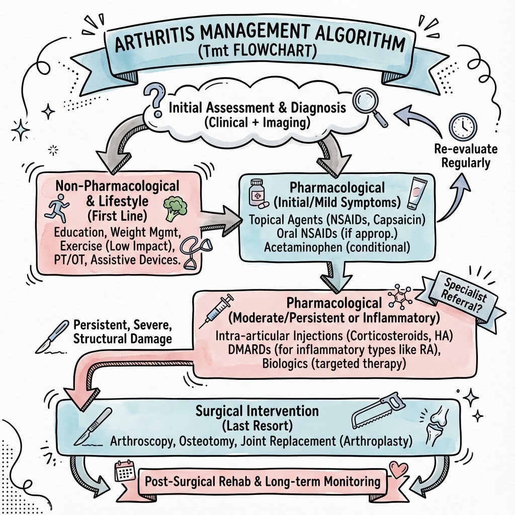
Management
Conservative Management
Conservative management is first-line for mild to moderate TMT arthritis without significant deformity.
- Rigid custom orthoses: Control midfoot motion, redistribute pressure
- Full-length carbon fiber inserts: Maximum rigidity for severe symptoms
- Rocker-bottom shoe modification: Reduces force through TMT joints
- Accommodative padding: Offload prominent osteophytes
- NSAIDs: Regular use for 2-3 months trial (if no contraindications)
- Activity modification: Avoid impact activities, prolonged walking
- Weight loss: Reduces midfoot loading (5-10kg loss significantly helps)
- Physical therapy: Strengthening intrinsic foot muscles, gait training
Injection Therapy
- Useful for both diagnostic and therapeutic purposes
- Local anesthetic + steroid (triamcinolone 40mg or equivalent)
- Fluoroscopic guidance ensures accurate placement
- Complete pain relief confirms TMT arthritis as pain source
- Therapeutic effect typically 3-6 months
- Can be repeated up to 3 times
If complete pain relief achieved, arthrodesis likely to be successful. If no improvement, consider alternative diagnosis or adjacent joint pathology.
Surgical Management
Indications for Surgery
- Failed conservative management (6 months adequate trial)
- Significant functional limitation affecting daily life
- Progressive deformity
- Severe pain limiting walking
- Failed multiple injection attempts
- High functional demand requiring return to activity
- Patient preference after informed consent
- Adjacent joint involvement developing
Surgical Options
TMT Arthrodesis (Gold Standard)
Fusion of affected TMT joints to eliminate painful motion while preserving adjacent joint function.
- Fuse TMT 1-3 (medial column): Minimal functional loss due to low native motion
- Preserve TMT 4-5 (lateral column): Maintains forefoot flexibility for ground adaptation
- Extend proximally if needed: Include NC or IC joints if arthritic on pre-op imaging
Operative Steps
Dual dorsal longitudinal incisions over TMT 1-2 and TMT 2-3 intervals. Protect superficial peroneal nerve branches. Expose joints, perform joint debridement to bleeding bone.
Remove all cartilage with osteotomes and curettes. Fashion flat apposing surfaces. Avoid excessive bone resection (causes shortening, transfer metatarsalgia). Preserve metatarsal length.
Correct deformity. Restore longitudinal arch height. Ensure metatarsal alignment in coronal plane (no forefoot abduction). Check with intraoperative fluoroscopy in multiple planes.
Compression screws: 4.0mm cannulated screws across each TMT joint. Direct compression essential. Or dorsal plates: Low-profile 2.7-3.5mm plates if poor bone quality. Avoid crossing TMT 4-5.
Layered closure. Consider drain if significant oozing. Bulky compressive dressing. Below-knee non-weight-bearing cast applied.
- Compression screws: Preferred for good bone quality, maximal compression
- Dorsal plates: Better for osteoporotic bone, deformity correction
- Combination: Plates + screws for severe deformity or revision
- Fusion rate: 85-95%
- Good-excellent results: 80-90%
- Return to walking: 3-4 months
- Full recovery: 6-12 months
Selective fusion TMT 1-3 preserves lateral column motion and provides good functional outcomes.
Technical Pearls
- Adequate debridement: Remove all cartilage to bleeding subchondral bone
- Preserve length: Avoid excessive bone resection - causes transfer metatarsalgia
- Compression fixation: Use lag screws for maximal compression across fusion site
- Restore alignment: Check arch height and forefoot alignment with fluoroscopy
- Bone graft: Consider if gaps remain after reduction - promotes fusion
- Don't fuse TMT 4-5: Causes significant functional impairment - preserve lateral column
- Don't cross unfused joints: Hardware crossing TMT 4-5 causes pain, breakage
- Don't under-resect: Leaving cartilage islands causes non-union
- Don't rush weight-bearing: 6-8 weeks NWB mandatory for fusion success
- Don't ignore NC/IC arthritis: Check pre-op CT and extend fusion if involved
Complications
- Incidence
- 5-15%
- Risk Factors
- Smoking, diabetes, inadequate fixation
- Management
- Revision fusion with bone graft, rigid fixation
- Incidence
- 5-10%
- Risk Factors
- Inadequate reduction, hardware failure
- Management
- May require revision if symptomatic deformity
- Incidence
- 10-15%
- Risk Factors
- Excessive bone resection, malposition
- Management
- Orthotic management, rarely revision surgery
- Incidence
- 15-20% at 5y
- Risk Factors
- Altered biomechanics
- Management
- Extend fusion if symptomatic and severe
- Incidence
- 2-5%
- Risk Factors
- Diabetes, poor wound healing
- Management
- Antibiotics, debridement, may need hardware removal
- Incidence
- 10-20%
- Risk Factors
- Prominent dorsal hardware
- Management
- Remove after fusion (1 year post-op)
Non-union is the most common major complication of TMT arthrodesis. Prevention strategies include: adequate cartilage debridement to bleeding bone, rigid fixation with compression, bone grafting for gaps, minimum 6-8 weeks non-weight-bearing, smoking cessation pre-operatively. If non-union occurs, revision with iliac crest bone graft and revised fixation typically successful.
Postoperative Care and Rehabilitation
Rehabilitation Timeline
Immobilization: Below-knee cast, strict non-weight-bearing. Elevate limb above heart level. DVT prophylaxis (aspirin or LMWH). Remove drain if placed (48 hours). First dressing change 2 weeks.
Continue NWB: New cast or CAM boot. Sutures removed 2-3 weeks. Repeat radiographs at 6 weeks. Check for early signs of healing (callus formation). Maintain NWB until radiographic healing seen.
Weight-bearing begins: If radiographs show healing callus, start progressive WB in CAM boot. Gradual increase from toe-touch to full WB over 4-6 weeks. Physical therapy for gait training, edema management. CT scan if healing uncertain at 12 weeks.
Transition to shoes: When full WB comfortable and radiographs confirm solid fusion (3-4 months). Custom orthoses for support. Gradual return to activities. Avoid high-impact until 6 months. May remove hardware if prominent after fusion solid (1 year).
Full recovery: 6-12 months for maximal improvement. Annual follow-up to monitor adjacent joints. Orthotic management indefinitely. Modify activities as needed. Monitor for transfer metatarsalgia.
The prolonged non-weight-bearing period is critical for fusion success.
Outcomes and Prognosis
TMT arthrodesis provides reliable pain relief and functional improvement for appropriately selected patients.
- Key Outcomes
- 60-80% initial success, 40% eventually require surgery
- Notes
- Best for mild arthritis, patient must accept activity limitations
- Key Outcomes
- 85-95% fusion rate, 80-90% good-excellent results
- Notes
- Gold standard - selective fusion preserves lateral column
- Key Outcomes
- Similar outcomes but longer recovery
- Notes
- Indicated if adjacent joints involved - check pre-op CT
Prognostic Factors
Favorable factors include: non-smoker, normal BMI, good bone quality, isolated TMT 1-3 arthritis (no adjacent joint involvement), compliant patient able to remain NWB for 6-8 weeks, adequate surgical technique with rigid fixation and compression.
Unfavorable factors include: smoking (doubles non-union risk), diabetes, obesity, poor bone quality (osteoporosis), extensive adjacent joint arthritis, inadequate fixation or poor surgical technique, patient non-compliance with weight-bearing restrictions.
Isolated First-TMT Arthritis, First-Ray Hypermobility and the Lapidus Connection
The topic states that primary (atraumatic) OA "typically affects the first TMT joint in isolation", names "first-ray instability from ligamentous laxity" and the "hallux valgus association", but never develops this distinct first-ray entity - which behaves and is treated differently from the post-traumatic TMT 1-3 pattern.
- A different disease from post-traumatic TMT arthritis. Isolated first-tarsometatarsal (medial-cuneiform-MT1) OA is the commonest primary midfoot arthritis, with a female predominance in the fifth-to-seventh decades. It presents as dorso-medial pain over the first TMT joint and a dorsal osteophyte/bump (which can cause dorsal-cutaneous-nerve irritation and shoe conflict), rather than the diffuse dorsal-midfoot pain of the TMT 1-3 post-traumatic pattern.
- First-ray hypermobility is the mechanical driver - and the link to hallux valgus. Excess sagittal (and rotational) motion at the first TMT joint (first-ray hypermobility) overloads the joint and is strongly associated with hallux valgus: the unstable, dorsiflexed, everted first ray both fails to bear its share of load (transfer to the lesser rays / metatarsalgia) and lets the first metatarsal drift into varus, driving the bunion. So first-TMT arthritis, first-ray hypermobility and hallux valgus form an interlinked triad.
- The Lapidus procedure treats both. A first-TMT arthrodesis (the Lapidus procedure) simultaneously (a) fuses the painful arthritic first TMT joint and (b) corrects the deforming force by stabilising the hypermobile first ray and correcting first-metatarsal varus - which is why it is a workhorse for hallux valgus with demonstrable first-ray hypermobility or coexisting first-TMT arthritis, and the definitive treatment for symptomatic isolated first-TMT OA. Technical caveats mirror the medial-column fusion above: preserve/set correct first-metatarsal length and plantarflexion (avoid dorsal malunion → transfer metatarsalgia), rigid compression fixation, and the usual non-union caution.
Q: How does isolated first-TMT arthritis differ from post-traumatic TMT arthritis, and what links it to hallux valgus? A: Isolated first-TMT (primary) OA is the commonest atraumatic midfoot arthritis - dorso-medial first-TMT pain with a dorsal osteophyte, female-predominant - driven by first-ray hypermobility, which also promotes hallux valgus (unstable dorsiflexed first ray + metatarsus primus varus). The Lapidus procedure (first-TMT arthrodesis) treats both by fusing the arthritic joint and stabilising/realigning the hypermobile first ray; set correct length and plantarflexion to avoid transfer metatarsalgia.
Is it Arthritis or Charcot? The Lisfranc Joint as the Classic Neuroarthropathy Site
The topic's own account of "collapse deformity", the "midfoot break sign", forefoot abduction and progressive midfoot deformity describes exactly the picture that a Charcot neuroarthropathy produces at this joint - yet Charcot is never mentioned, and it is the single most important thing to exclude before calling a collapsing midfoot "arthritis", because the management is completely different.
- Why it matters at the TMT joint specifically. The tarsometatarsal (Lisfranc) joint is the classic site of midfoot Charcot (Eichenholtz/Brodsky pattern I), typically in a diabetic with peripheral neuropathy. Untreated, it collapses into the pathognomonic rocker-bottom (plantar-convex) foot with a plantar-medial bony prominence that ulcerates - a limb-threatening problem, not just a painful joint.
- How to tell them apart. Charcot classically presents as a warm, swollen, erythematous, often relatively PAINLESS foot in a neuropathic patient (pain out of proportion-ly low for the destruction), whereas degenerative TMT arthritis is painful in a sensate foot without the florid inflammatory signs. The acute Charcot foot mimics infection/cellulitis; the dependent-rundown test (elevation settles the erythema of Charcot but not of infection) and the neuropathy help. Radiographs in Charcot show fragmentation, debris, dislocation and disorganisation far exceeding the patient's pain.
- Why you must not treat them the same. In active (acute) Charcot the cornerstone is offloading and immobilisation in a total-contact cast until the process consolidates - operating on, or simply fusing, an actively inflamed Charcot joint as if it were degenerative arthritis courts hardware failure, non-union and catastrophic wound problems. Surgical reconstruction (with far more robust "superconstruct" fixation than a standard TMT fusion) is reserved for unstable deformity, recurrent ulceration or instability once the Charcot has consolidated - and the general Charcot work-up/management belongs to the dedicated neuroarthropathy topic.
Q: A diabetic presents with a warm, swollen, deformed, relatively painless collapsing midfoot - is this TMT arthritis? A: Suspect Charcot neuroarthropathy of the Lisfranc joint (the classic midfoot Charcot site), not degenerative TMT arthritis. Charcot is a warm, swollen, erythematous, disproportionately PAINLESS neuropathic foot collapsing to a rocker-bottom with radiographic fragmentation/dislocation exceeding the pain; degenerative TMT arthritis is painful in a sensate foot. Management differs completely - active Charcot needs offloading/total-contact casting until consolidation, NOT the standard TMT fusion, which would fail. Reconstruct only once consolidated, with robust fixation.
Guidelines, Registries & Global Practice
Global Epidemiology
- Midfoot (Lisfranc) injuries account for roughly 0.2% of all fractures, with an incidence around 1 per 55,000 person-years; up to 20-40% of low-energy injuries are missed at first presentation.
- Post-traumatic arthritis is the dominant pathway to symptomatic TMT arthritis worldwide; the strongest modifiable predictor is the quality of initial reduction.
- Primary (atraumatic) TMT osteoarthritis is less common, has a female predominance, and most often isolates to the first TMT joint, frequently in association with hallux valgus or first-ray hypermobility.
Side-by-Side Society Guidance
- Position on midfoot injury / TMT arthritis
- Anatomic reduction and stable fixation of Lisfranc injuries; primary arthrodesis favoured for comminuted or purely ligamentous patterns
- Practical message
- Restore the medial-column keystone to prevent later arthritis
- Position on midfoot injury / TMT arthritis
- Weight-bearing and stress imaging to detect subtle instability; arthrodesis is the standard salvage for established arthritis
- Practical message
- Do not rely on non-weight-bearing films
- Position on midfoot injury / TMT arthritis
- Early senior review of suspected midfoot injuries, weight-bearing radiographs, prompt definitive management
- Practical message
- Minimise diagnostic delay
- Position on midfoot injury / TMT arthritis
- Stability-based (not classification-based) treatment; primary arthrodesis for ligamentous instability
- Practical message
- Stability drives the decision
The unresolved controversy across all societies is bony unstable Lisfranc injuries: ORIF vs primary arthrodesis. For purely ligamentous instability the evidence (Ly and Coetzee RCT) favours primary arthrodesis; for bony injuries there is no Level 1 consensus and practice varies by surgeon and region.
Registry and Outcome Notes
- TMT arthritis and Lisfranc injuries are not implant-registry tracked the way hip/knee arthroplasty is (no NJR/AJRR/AOANJRR equivalent), so the evidence base rests on RCTs and case series rather than national registries.
- Across published series, medial-column arthrodesis achieves union in roughly 85-95% with good-to-excellent results in 80-90%; non-union (5-15%) is driven mainly by smoking, diabetes, and inadequate fixation.
High- vs Limited-Resource Practice
- Well-resourced settings: weight-bearing CT for planning, low-profile locking plates and lag screws, fluoroscopy, and protected non-weight-bearing with formal physiotherapy.
- Limited-resource settings: weight-bearing plain radiographs and clinical examination guide treatment; transarticular screws or K-wires substitute for plates; emphasis on accurate primary reduction is even greater because salvage surgery and imaging are less available.
- Universal principle regardless of resources: anatomic reduction of the medial-column keystone and early definitive management are the single most important determinants of avoiding post-traumatic arthritis.
Controversies and Areas of Uncertainty
Level 1 evidence favours primary arthrodesis for purely ligamentous Lisfranc injuries. For bony fracture-dislocations the question is unresolved - no RCT shows clear superiority, and many surgeons still favour anatomic ORIF to preserve native joints.
How far medially/proximally to extend fusion (TMT 1-3 alone vs including NC/IC joints) is judgement-based. Over-fusion sacrifices motion; under-fusion risks residual painful arthritis. Komenda found extent of fusion did not significantly affect outcome.
Transarticular lag screws vs dorsal locking plates (which spare articular cartilage) is debated. Plates may suit osteoporotic bone and deformity correction; screws give direct compression. No construct has proven superior union rates in high-quality trials.
Whether the mobile lateral column (TMT 4-5) should ever be fused remains contentious because of functional loss. Interposition arthroplasty and resection are described alternatives, but evidence is limited to small series.
The classic viva trap is to over-generalise the Ly and Coetzee RCT to all Lisfranc injuries. State clearly that its conclusion (primary arthrodesis superior) is established only for isolated, primarily ligamentous injuries.
MCQ Practice Points
Q: Which TMT joint is the keystone of the midfoot, recessed dorsally between adjacent metatarsals to provide stability? A: TMT 2 (second tarsometatarsal joint). The second metatarsal base is recessed 2-3mm dorsally between the intermediate cuneiform and the bases of MT1 and MT3. This keystone architecture provides inherent stability preventing dorsal or plantar translation.
Q: What is the rationale for selective fusion of TMT 1-3 while preserving TMT 4-5 in TMT arthrodesis? A: TMT 1-3 have minimal native motion (2-3 degrees each) forming the rigid medial column, so fusion causes minimal functional loss. TMT 4-5 have significantly more motion (10-15 degrees) forming the mobile lateral column which allows forefoot adaptation to uneven terrain. Fusing the lateral column causes significant functional impairment.
Q: What percentage of TMT arthritis is post-traumatic, and what is the most common preceding injury? A: 80% of TMT arthritis is post-traumatic, most commonly following inadequately treated Lisfranc injuries. Missed subtle injuries or residual subluxation greater than 2mm progresses to arthritis within 2-5 years. Anatomic reduction of Lisfranc injuries is critical to prevent arthritis.
Q: What is the fusion rate for TMT 1-3 arthrodesis and what is the most common major complication? A: Fusion rate is 85-95% with good-excellent results in 80-90% of patients. The most common major complication is non-union (5-15% incidence). Risk factors include smoking, diabetes, inadequate fixation, and insufficient post-operative non-weight-bearing period.
Q: What is the critical technical principle to prevent non-union in TMT arthrodesis? A: Complete cartilage removal to bleeding subchondral bone is the most critical factor. All cartilage must be debrided with osteotomes and curettes, leaving no cartilage islands. Other important factors include rigid compression fixation, bone grafting for gaps, and minimum 6-8 weeks non-weight-bearing post-operatively.
Q: What complication occurs in 10-15% of TMT arthrodesis patients due to excessive bone resection at the fusion site? A: Transfer metatarsalgia. Excessive bone resection during joint preparation causes metatarsal shortening, which alters weight distribution and causes overload of adjacent metatarsals. Prevention requires preserving metatarsal length by avoiding over-resection of bone.
Exam Viva Scenarios
Practise clinical reasoning and management decisions out loud
“A 45-year-old manual laborer presents with 2 years of progressive midfoot pain following a crush injury. He has tried orthotics and NSAIDs without benefit. Examination shows dorsal tenderness over TMT joints and positive piano key test at TMT 2-3. Weight-bearing radiographs show joint space loss at TMT 1-3 with subchondral sclerosis. How would you manage this patient?”
“You are performing TMT 1-3 arthrodesis for post-traumatic arthritis. Walk me through your surgical approach, joint preparation, and fixation technique. What are the key technical points to ensure fusion success?”
“A 52-year-old patient had TMT 1-3 arthrodesis 9 months ago. She followed weight-bearing restrictions but now complains of persistent midfoot pain with walking. Radiographs show lucency at TMT 2 fusion site with no bridging bone. CT confirms non-union at TMT 2. How would you manage this complication?”
Key Anatomy
- TMT 2 = Keystone - recessed 2-3mm dorsally between cuneiforms prevents translation
- Medial column (TMT 1-3) = Rigid, minimal motion (2-3° each) - safe to fuse
- Lateral column (TMT 4-5) = Mobile (10-15° motion) - preserve function
- Lisfranc ligament = Strongest ligament from medial cuneiform to MT2 base plantar
Classification
- Mild = Joint space narrowing under 50%, minimal osteophytes → Conservative trial
- Moderate = 50-75% joint space loss, sclerosis → Trial conservative, then fuse if fails
- Severe = Complete joint space loss, cysts, deformity → Arthrodesis indicated
- Post-traumatic = 80% of cases - follows inadequately treated Lisfranc injury (residual displacement greater than 2mm)
Treatment Algorithm
- First-line: Custom rigid orthoses + NSAIDs + activity modification (6 months trial)
- Diagnostic injection: Confirms diagnosis, predicts fusion success if complete relief
- Surgery: TMT 1-3 arthrodesis with compression screws or dorsal plates
- Extend fusion: Include NC/IC joints if arthritic on pre-op CT
Surgical Pearls
- Dual dorsal incisions protecting superficial peroneal nerve branches
- Complete cartilage removal to bleeding bone - leave no islands
- Preserve metatarsal length - avoid over-resection (causes transfer metatarsalgia)
- Rigid compression fixation - 4.0mm screws preferred, plates if poor bone
- NEVER cross unfused TMT 4-5 with hardware - causes pain and breakage
Complications
- Non-union 5-15% - smoking, diabetes, inadequate fixation - revise with bone graft
- Transfer metatarsalgia 10-15% - from excessive bone resection/shortening
- Adjacent joint arthritis 15-20% at 5y - altered biomechanics - extend fusion if severe
- Hardware irritation 10-20% - remove after fusion solid (1 year post-op)
Evidence Base and Key Studies
Primary Arthrodesis vs ORIF for Ligamentous Lisfranc Injury (Landmark RCT)
- Prospective randomised trial of 41 isolated primarily ligamentous Lisfranc injuries
- Primary arthrodesis of the medial two-to-three rays vs open reduction and screw fixation
- At 2 years, mean AOFAS Midfoot score 88 (arthrodesis) vs 68.6 (ORIF), p less than 0.005
- 5 of 20 ORIF patients developed deformity or arthrosis and were salvaged with fusion
Primary Arthrodesis vs ORIF - Reoperation Burden (RCT)
- 40 acute tarsometatarsal fracture/fracture-dislocations randomised to ORIF or primary arthrodesis
- Secondary surgery (including routine hardware removal) 78.6% after ORIF vs 16.7% after arthrodesis
- No significant difference in SF-36 or SMFA functional scores between groups
- Satisfaction comparable at mean 53 months
Arthrodesis for Post-Traumatic Midfoot Arthritis
- Retrospective series of 32 patients fused for intractable midfoot pain after trauma (mean 35 months post-injury)
- Mean AOFAS Midfoot score improved from 44 to 78 of 100 (p = 0.02)
- Only one asymptomatic non-union; complications included neuritis (3), metatarsalgia (2), malunion (2)
- Extent of fusion and injury mechanism did not significantly affect functional outcome
Salvage Arthrodesis of the Lisfranc Joint
- 16 patients (49 joints) salvaged by arthrodesis after failed initial Lisfranc treatment
- Good-to-excellent result in 11 of 16 (69%); symptomatic non-union at 4 sites in 3 patients
- Accurate reduction and early treatment correlated with better outcome
- Work-related injury and long delay to treatment correlated with worse outcome
Midfoot Joint Motion - Biomechanical Basis for Selective Fusion
- In vitro study of 10 cadaveric below-knee specimens with reference-pin tracking
- Medial column near-rigid: middle cuneiform-MT2 0.6 degrees and medial cuneiform-MT1 3.5 degrees of sagittal motion
- Lateral column mobile: cuboid-MT4 9.6 degrees and cuboid-MT5 10.2 degrees of sagittal motion
- Confirms TMT2 as the least mobile (keystone) articulation
Lisfranc Injury - Diagnosis and Treatment Algorithm (Review)
- Narrative review proposing a classification-free, stability-based treatment algorithm
- Delay in diagnosis worsens outcome; weight-bearing radiographs best assess stability
- Stable injuries treated non-operatively; unstable ligamentous injuries favour primary arthrodesis
- For bony unstable injuries, ORIF vs primary arthrodesis remains unresolved - more RCTs needed