Diagnosis | Imaging | Medical Management | Surgical Indications
- Thoracolumbar junction (T10-L2) is most common site
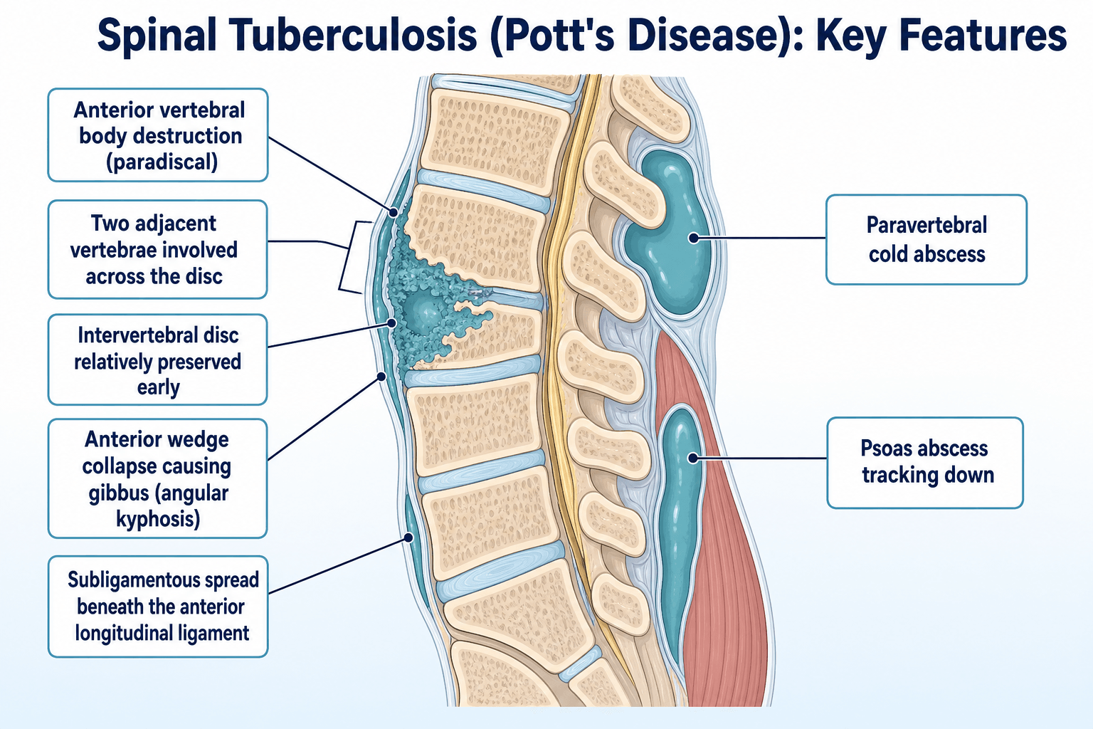
- Disc preservation early distinguishes from pyogenic infection
- Cold abscess does not contain pus - caseous material
- MRI is the imaging modality of choice - high sensitivity, detects marrow, abscess and cord involvement
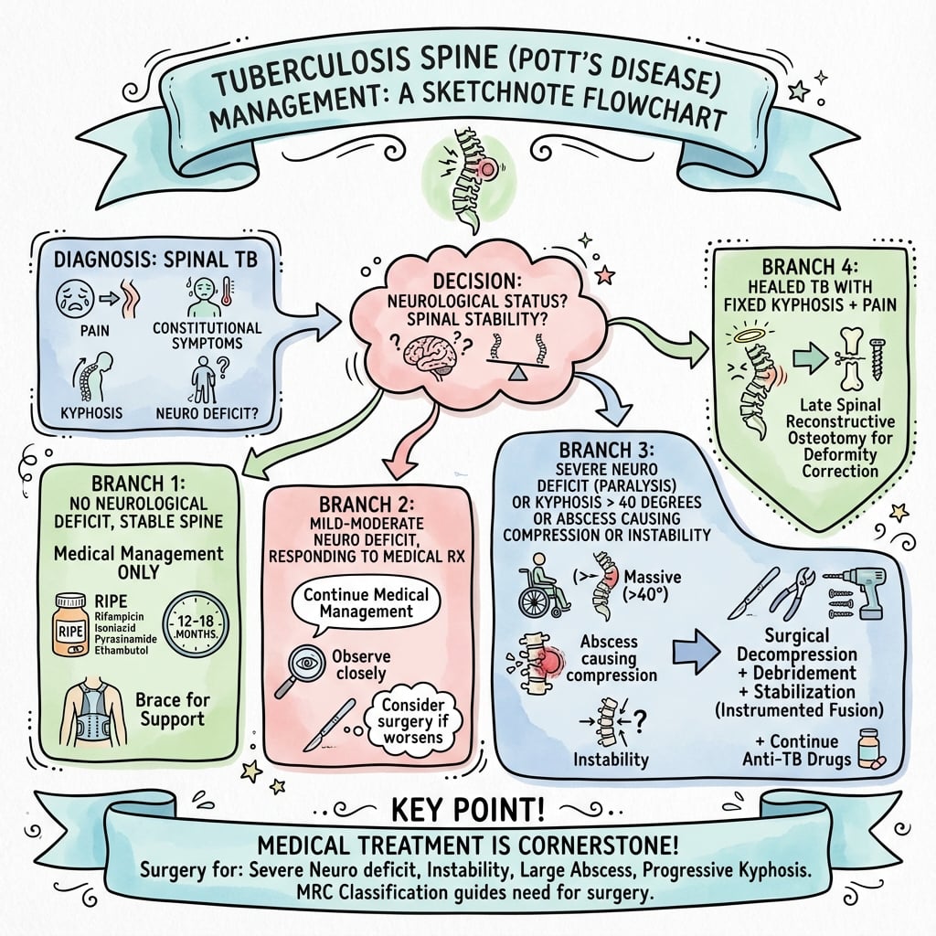
- Medical treatment first - surgery for specific indications
- “Paradiscal type is most common - adjacent vertebrae + disc involvement
- “Skip lesions in 10-15% - always image whole spine
- “Psoas abscess is pathognomonic when combined with spine findings
- “Neurological deficit from granulation tissue, abscess, or kyphosis
Early TB preserves the disc while pyogenic infection destroys it early. This is a key differentiating feature. However, late-stage TB can involve the disc. Always compare with clinical tempo - TB is insidious, pyogenic is acute.
Cold abscess is characteristic of TB - it lacks acute inflammatory features and contains caseous material, not pus. It can track along fascial planes (psoas abscess). The absence of local warmth and systemic toxicity distinguishes it from pyogenic abscess.
MRI is essential - high sensitivity for marrow oedema, paravertebral and epidural abscess, and cord compression, and earlier than plain films. X-rays become positive only once substantial trabecular bone is destroyed (months of delay). Always image the whole spine for skip lesions.
Medical treatment is primary - 85-90% respond to antitubercular therapy (ATT) alone. Surgery reserved for: progressive neurology, instability, failure of medical treatment, severe kyphosis. Even with neurology, medical treatment often effective.
- Tuberculosis
- Insidious (weeks-months)
- Pyogenic
- Acute (days-weeks)
- Tuberculosis
- Common (93%)
- Pyogenic
- Uncommon (24%)
- Tuberculosis
- 94%
- Pyogenic
- 18%
- Tuberculosis
- 77%
- Pyogenic
- 25%
- Tuberculosis
- 68%
- Pyogenic
- 24%
- Tuberculosis
- 22%
- Pyogenic
- 59%
- Tuberculosis
- 82%
- Pyogenic
- 95%
COLDCOLD ABSCESS - TB Features
Hook:COLD ABSCESSES lack heat, redness, and acute toxicity - unlike pyogenic
SPINESPINE TB - Imaging Findings
Hook:MRI is imaging modality of choice - high sensitivity for marrow, abscess and cord
SURGERYSURGERY - Indications for Surgery
Hook:Most patients (85-90%) respond to medical treatment alone
Overview and Epidemiology
Spinal tuberculosis (Pott's disease) is the most common form of skeletal tuberculosis, first described by Percivall Pott in 1779. It represents a significant cause of morbidity, particularly in endemic regions, and remains important in low-incidence high-income settings due to immigration patterns.
Epidemiology:
- Spinal TB accounts for 50% of all skeletal TB cases
- Second most common form of extrapulmonary TB
- Thoracolumbar junction (T10-L2) is most commonly affected
- Approximately 10-15% have skip lesions (non-contiguous involvement)
- Male to female ratio approximately 1.5:1
- Can occur at any age but peaks in second and third decades
Global Burden:
- Estimated Incidence
- More than 100/100,000
- Risk Factors
- Crowding, poverty, HIV
- Estimated Incidence
- 10-100/100,000
- Risk Factors
- Immigration, immunosuppression
- Estimated Incidence
- Less than 10/100,000
- Risk Factors
- Imported cases, reactivation
Pathophysiology and Anatomy
Route of Infection
- Haematogenous spread from pulmonary focus (most common)
- Arterial dissemination to vertebral bodies
- Paradiscal arteries supply adjacent vertebrae (paradiscal type)
- Batson's venous plexus may facilitate spread
- Subligamentous extension under ALL
- Epidural spread causing cord compression
- Paravertebral spread forming cold abscess
- Psoas tracking along muscle sheath
Anatomical Patterns of Involvement
- Most common pattern
- Involves adjacent vertebral bodies and intervening disc
- Arterial spread via paradiscal arteries
- Late disc destruction (unlike pyogenic)
- May extend to multiple levels
- Isolated vertebral body involvement
- Concertina collapse
- May produce ivory vertebra
- Spares disc initially
- Spreads under anterior longitudinal ligament
- Multi-level involvement with anterior scalloping
- Can produce skip lesions
- Extensive yet may spare vertebral integrity initially
- Involves neural arch (pedicle, lamina, spinous process)
- More common in lower lumbar spine
- May cause early neurological deficit
Pathological Changes
- Granulomatous inflammation
- Caseous necrosis
- Bone destruction and sequestration
- Minimal new bone formation (unlike pyogenic)
- Cold abscess formation (paravertebral, epidural, psoas)
- Granulation tissue proliferation
- Fibrosis with healing
- Early: Epidural granulation tissue, abscess
- Late: Bony compression from kyphotic deformity
Classification Systems
Kumar Classification (Neurological Status)
- Description
- No deficit
- Neurological Findings
- Normal
- Description
- Sensory
- Neurological Findings
- Sensory loss only
- Description
- Motor - ambulatory
- Neurological Findings
- Motor weakness, can walk
- Description
- Motor - non-ambulatory
- Neurological Findings
- Cannot walk
- Description
- Complete paraplegia
- Neurological Findings
- No function below lesion
Tuli Classification (Paraplegia Type)
Type A: Active Disease with Paraplegia
- A1: Minimal bone loss, severe deficit
- A2: Moderate bone loss, severe deficit
- A3: Extensive bone loss with deficit
Type B: Healed Disease with Paraplegia
- B1: Cord compression from healed kyphosis
- B2: Reactivation in previously healed lesion
This classification guides surgical approach and prognosis for neurological recovery.
Clinical Assessment
History
Presenting Symptoms:
- Frequency
- 90-95%
- Characteristics
- Insidious, localized, constant
- Frequency
- 50-70%
- Characteristics
- Weight loss, night sweats, fever
- Frequency
- 20-30%
- Characteristics
- Weakness, sensory changes
- Frequency
- 30-40%
- Characteristics
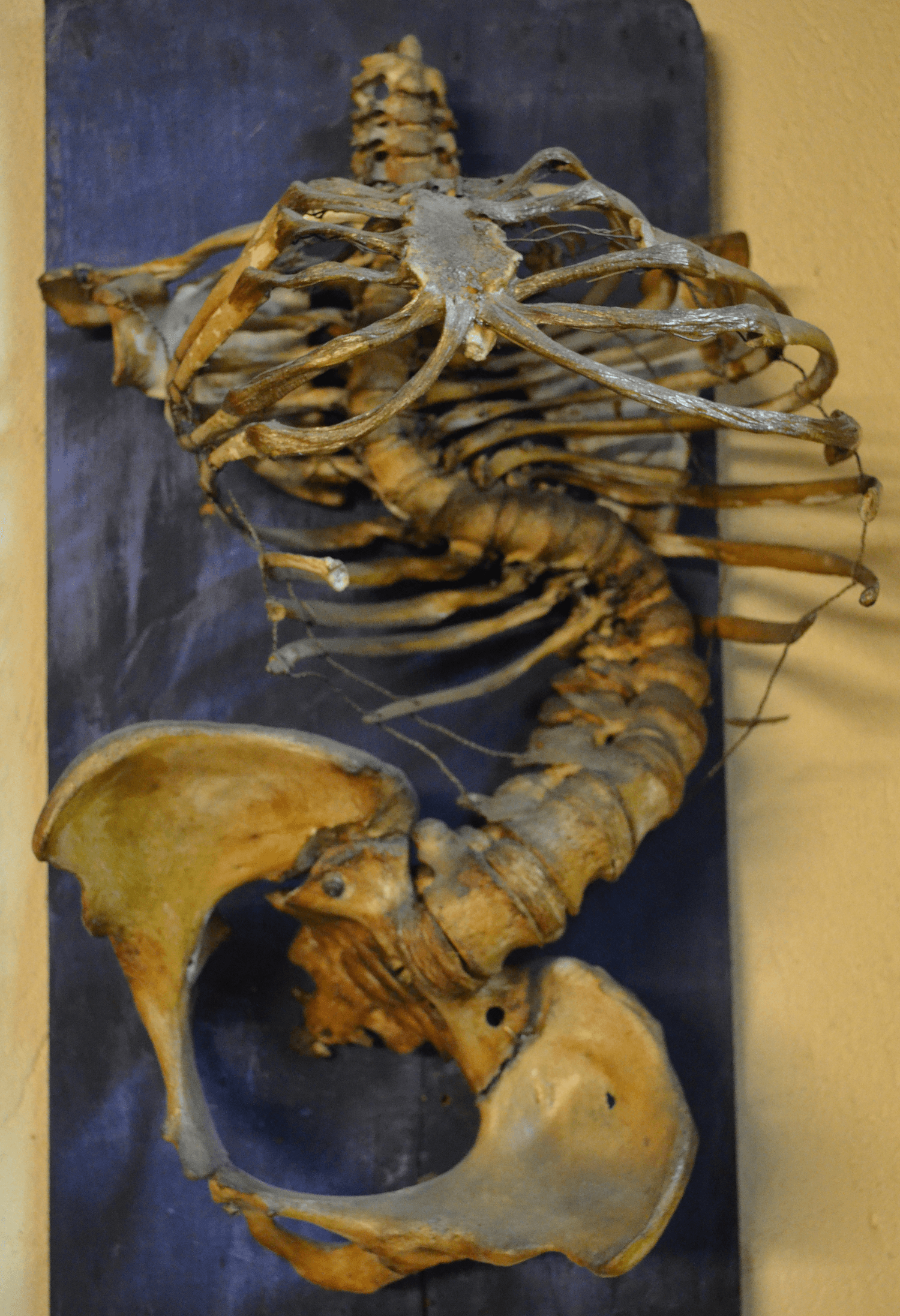
- Visible kyphosis (gibbus)
- Frequency
- 20-30%
- Characteristics
- Swelling (groin, flank)
Key History Elements:
- Duration of symptoms (typically weeks to months)
- Constitutional symptoms (weight loss, night sweats, low-grade fever)
- Neurological symptoms (weakness, numbness, bowel/bladder)
- Contact history (TB exposure)
- Country of origin and travel
- Immunocompromise (HIV, diabetes, immunosuppressants)
- Previous TB treatment
Physical Examination
Spinal Assessment:
- Gibbus deformity (angular kyphosis)
- Localized tenderness
- Paraspinal muscle spasm
- Restricted range of motion
- Cold abscess (paravertebral, groin, flank)
- Motor power (myotomes)
- Sensory level
- Reflexes
- Long tract signs (spasticity, clonus, Babinski)
- Bladder/bowel function
- Gait assessment
- Lymphadenopathy
- Pulmonary findings (primary TB)
- Peripheral cold abscess
- Signs of other organ involvement
A cold abscess lacks the cardinal signs of inflammation (calor, rubor, dolor, tumor). It presents as a non-tender, fluctuant swelling that may track to distant sites (groin in psoas abscess). The absence of acute inflammatory features is characteristic.
Red Flags
- Rapidly progressive neurological deficit
- Complete paraplegia
- Bladder/bowel dysfunction
- Respiratory compromise (cervical lesions)
- Signs of MDR-TB or treatment failure
Investigations
Laboratory Investigations
Essential Tests:
- Expected Finding
- Elevated (often more than 50)
- Notes
- Useful for monitoring
- Expected Finding
- Elevated
- Notes
- Less specific than ESR
- Expected Finding
- Usually positive
- Notes
- Does not confirm active disease
- Expected Finding
- May be positive
- Notes
- If pulmonary involvement
- Expected Finding
- Rule out coinfection
- Notes
- Important for management
Imaging Protocol
- First-line imaging
- Positive only when 50% trabecular bone destroyed
- May take 4-6 months to show changes
- Shows: vertebral destruction, collapse, kyphosis
- High sensitivity; detects disease earlier than plain radiographs
- Essential for soft tissue assessment
- Detects epidural extension and cord compression
- Identifies skip lesions
- T1: Hypointense marrow signal
- T2/STIR: Hyperintense marrow edema
- Contrast: Rim enhancement of abscess
- Epidural extension
- Skip lesions
- Psoas/paravertebral abscess
- Superior bone detail
- Calcification within soft tissue (pathognomonic)
- Bony destruction pattern
- CT-guided biopsy assistance
Tissue Diagnosis
- Atypical presentation
- Negative Mantoux/IGRA
- MDR-TB suspected
- Exclude malignancy
- Failed empirical treatment
- CT-guided percutaneous biopsy
- Open surgical biopsy
- Abscess aspiration
- Caseous necrosis
- Epithelioid granulomas
- Langhans giant cells
- AFB staining
- PCR (GeneXpert)
Differential Diagnosis
- Discriminating features
- Insidious, subligamentous spread, disc relatively spared early, thin-walled cold abscess, calcification
- Key test
- Biopsy: caseating granuloma, Xpert/culture
- Discriminating features
- Acute, high fever, early disc destruction, thick irregular abscess wall
- Key test
- Blood cultures, biopsy culture
- Discriminating features
- Endemic / livestock contact, lower lumbar, gas in disc, little deformity
- Key test
- Serology, blood culture
- Discriminating features
- Older, disc characteristically spared, posterior element / pedicle destruction, no abscess
- Key test
- MRI, biopsy, serum electrophoresis
- Discriminating features
- Immunocompromised, indolent, can mimic TB
- Key test
- Biopsy fungal stain/culture
A key trap is that metastatic disease and myeloma also spare the disc, so disc preservation alone does not confirm infection; the presence of a paravertebral cold abscess and subligamentous spread points to TB, whereas pedicle destruction without abscess favours malignancy.

Management
Antitubercular Therapy (ATT)
Standard Regimen (WHO):
- Duration
- 2 months
- Drugs
- HRZE (4 drugs)
- Duration
- 4-10 months
- Drugs
- HR (2 drugs)
- H (Isoniazid): 5 mg/kg (max 300 mg)
- R (Rifampicin): 10 mg/kg (max 600 mg)
- Z (Pyrazinamide): 25 mg/kg (max 2 g)
- E (Ethambutol): 15 mg/kg (max 1.2 g)
- WHO endorses a 6-month regimen for drug-susceptible disease, supported by the MRC ambulant-chemotherapy RCT, in which 6 months of isoniazid plus rifampicin gave a 94% favourable outcome at 10 years
- CNS/spinal-focused guidance (e.g. British Infection Society) recommends 12 months: 2 months of four drugs then at least 10 months of isoniazid and rifampicin
- Many spinal units extend to 9-12 months in extensive or slow-responding disease
- Drug-resistant disease requires substantially longer, specialist-directed regimens
- Baseline: LFTs, uric acid, visual acuity
- Monthly: LFTs during intensive phase
- Clinical response assessment
- Radiological healing (MRI at 6 months)
- ESR trending
- MDR-TB requires specialist management
- Second-line drugs (fluoroquinolones, injectables)
- Extended duration (18-24 months)
- Specialist input essential
Monitor LFTs closely during ATT. Risk factors for hepatotoxicity include age more than 35, pre-existing liver disease, alcohol use, and concurrent hepatotoxic medications. Stop ATT if ALT rises more than 5x ULN or symptoms develop.
Medical therapy alone achieves good outcomes in 85-90% of cases when neurological deficit is not severe.
Complications
Disease-Related Complications
- Incidence
- 30-50%
- Management
- Surgical correction if severe
- Incidence
- 20-30%
- Management
- Decompression ± steroids
- Incidence
- 20-30%
- Management
- Drainage if large
- Incidence
- 5-10%
- Management
- Debridement, prolonged ATT
- Incidence
- Variable
- Management
- Instrumented fusion
Neurological Complications
Pott's Paraplegia:
- Early onset (active disease): Better prognosis
- Late onset (healed disease): Worse prognosis
- Causes: Abscess, granulation tissue, bony compression
Early-onset (active-disease) paraplegia results from reversible anterior compression by an epidural cold abscess, caseous granulation tissue, or sequestrum/debris, and usually recovers well with decompression and ATT.
Late-onset (healed-disease) paraplegia appears years after the disease has healed and carries a worse prognosis. The cord is compressed by the internal salient (internal gibbus) — the bony ridge of the healed kyphus projecting into the canal at the apex — and by dural/peridural fibrosis, with the cord stretched and draped over the bony ridge; rarely it is due to reactivation or, late, cord atrophy/syringomyelia. Treatment is anterior decompression that removes the internal salient (an anterior problem needs an anterior solution).
Because the compression is almost always anterior, laminectomy does NOT relieve it and removes the only remaining intact posterior tension band, accelerating kyphosis and worsening neurology. Laminectomy is reserved for the uncommon case of posterior-element (neural-arch) disease causing posterior compression.
Prognosis Factors:
- Duration of deficit (shorter = better)
- Completeness (incomplete = better)
- Age (younger = better)
- Vertebral destruction extent
Treatment-Related Complications
- Hepatotoxicity (2-10%)
- Drug reactions
- Drug interactions
- MDR development
- Infection
- Neurological injury
- Hardware failure
- Pseudarthrosis
- Recurrence
Long-Term Sequelae
- Residual kyphosis
- Chronic pain
- Residual neurological deficit
- Adjacent segment disease
- Growth disturbance (paediatric)
Early recognition and appropriate treatment can prevent severe kyphosis. Children are at higher risk of progressive deformity. Consider early surgery in children with significant vertebral destruction to prevent late kyphotic deformity.
Outcomes and Prognosis
Neurological Recovery
Prognosis by Presentation:
- Recovery Rate
- 70-80%
- Timeframe
- 3-6 months
- Recovery Rate
- 50-70%
- Timeframe
- 6-12 months
- Recovery Rate
- 20-30%
- Timeframe
- Variable
Favourable Factors:
- Short duration of deficit
- Incomplete paraplegia
- Younger age
- Compression by soft tissue (not bone)
- Early treatment initiation
Treatment Outcomes
- Success rate: 85-90%
- Healing time: 6-12 months
- Residual kyphosis: 30-50%
- Neurological improvement: 70-90%
- Fusion rate: 90-95%
- Deformity correction maintained
Long-Term Prognosis
- Extent of initial disease
- Timing of treatment
- Adequacy of ATT
- Surgical technique
- Patient compliance
- Light activities: 3-6 months
- Full activities: 6-12 months
- Depends on neurological status
With appropriate treatment, most patients with spinal TB achieve good outcomes. Medical treatment alone is sufficient in 85-90%. Neurological recovery is expected in 70-80% of those with incomplete deficits. Surgery improves outcomes in selected cases with specific indications.
Guidelines, Registries & Global Practice
Global Epidemiology
Tuberculosis remains a leading infectious cause of death worldwide, with around 1.3 million deaths annually. Spinal TB is the most common musculoskeletal manifestation, affecting roughly 1 to 2% of all cases of TB, and skeletal involvement occurs in approximately 10% of those with active disease. Burden is concentrated in South and South-East Asia, Sub-Saharan Africa and the Western Pacific. In low-incidence high-income settings, the overwhelming majority of cases are in people born in high-burden countries, frequently presenting as reactivation of latent infection years after migration. HIV co-infection, which is endemic in parts of Sub-Saharan Africa, adds substantially to both the burden and complexity of management.
Major Guidance Side-by-Side
- Drug regimen
- 2 months HRZE then 4 months HR (6 months total)
- Surgical trigger
- Not a surgical body; medical cure expected in uncomplicated disease
- Drug regimen
- 4 drugs for 2 months then isoniazid + rifampicin for at least 10 months (12 months total)
- Surgical trigger
- Decompression for cord compression with deficit
- Drug regimen
- Ambulant 6-9 months HR adequate without paraplegia
- Surgical trigger
- Routine radical surgery confers no healing benefit in uncomplicated disease
- Drug regimen
- Chemotherapy continued through and after surgery
- Surgical trigger
- Deficit, instability, severe / progressive kyphosis, failed medical therapy, diagnostic doubt
The most important guideline divergence to recognise is duration: WHO endorses a 6-month regimen for drug-susceptible disease (supported by the MRC ambulant-chemotherapy RCT), whereas neurological/CNS-focused guidance such as the British Infection Society recommends a longer 12-month course (2 months of four drugs then at least 10 months of isoniazid and rifampicin) for spinal and CNS disease. Many spinal units extend treatment to 9-12 months in extensive or slow-responding disease.
Diagnostic Standards
A tissue diagnosis (culture, histology and nucleic-acid amplification) is the worldwide gold standard before committing a patient to prolonged therapy, and is especially important in low-prevalence settings and to detect drug resistance. Xpert MTB/RIF on bone or abscess material gives rapid results and simultaneously reports rifampicin resistance, but its sensitivity on bone is only moderate when used alone, so it should be paired with histopathology and culture rather than replacing them.
Practice Variation by Resource Setting
- Diagnosis
- MRI plus image-guided biopsy with Xpert and culture; drug-susceptibility testing routine
- Surgery
- Instrumented single- or two-stage reconstruction; titanium implants
- Diagnosis
- Often clinical/radiographic diagnosis; empirical ATT where confirmation is impractical
- Surgery
- Middle-path regimen; surgery rationed to deficit and major deformity
In resource-limited high-burden regions, empirical antitubercular therapy on clinical and radiographic grounds is common where biopsy is impractical, and a conservative "middle-path" approach (chemotherapy with selective surgery) predominates. In low-incidence settings, mandatory public-health notification, contact tracing, directly observed therapy for adherence, and multidisciplinary input from infectious diseases, spinal surgery, radiology and rehabilitation are standard.
MCQ Practice Points
Q: What is the most common location for spinal tuberculosis?
A: The thoracolumbar junction (T10-L2) is the most common site for spinal TB. The thoracic spine is involved in approximately 50% of cases, lumbar in 35%, and cervical in 15%. The paradiscal region is the most common pattern of involvement.
Q: Which feature distinguishes spinal TB from pyogenic spondylitis?
A: Early disc preservation distinguishes TB from pyogenic infection. In pyogenic spondylitis, the disc is destroyed early as bacteria can survive in disc tissue. In TB, the avascular disc is relatively resistant to mycobacterial infection, though late TB can involve the disc.
Q: Which MRI features best distinguish tuberculous from pyogenic spondylitis?
A: A pooled meta-analysis found subligamentous spread (93% vs 24%), a thin, smooth-walled abscess (94% vs 18%) and epidural extension (77% vs 25%) favour TB, whereas disc signal change and disc height loss are more common in pyogenic infection. MRI is the modality of choice because it detects disease far earlier than plain films, which become positive only once substantial trabecular bone is destroyed.
Q: What percentage of spinal TB patients have skip lesions?
A: 10-15% of patients have skip lesions (non-contiguous vertebral involvement). This is why whole spine imaging with MRI is recommended in all suspected cases of spinal TB to detect all levels of involvement.
Q: What percentage of patients with uncomplicated spinal TB respond to medical treatment alone?
A: 85-90% of patients with uncomplicated spinal TB respond to antitubercular therapy alone without surgery. Surgery is reserved for specific indications including progressive neurology, instability, and failure of medical treatment.
Clinical Decision Scenarios
Practise clinical reasoning and management decisions out loud
“A 35-year-old man from India presents with 4 months of progressive back pain, night sweats, and weight loss. He has low-grade fever and difficulty walking due to leg weakness. MRI shows T11-12 vertebral destruction with anterior soft tissue collection.”
“A 50-year-old man presents with back pain and low-grade fever. MRI shows L3-4 vertebral body involvement with disc changes and paravertebral collection. Both TB and pyogenic infection are being considered.”
“A 28-year-old woman presents with complete paraplegia of 2 weeks duration. MRI shows T7-8 vertebral destruction with large epidural collection causing severe cord compression. She has confirmed pulmonary TB on sputum.”
Key Facts
- 50% of skeletal TB is spinal
- T10-L2 most common location
- Paradiscal type most common (50-75%)
- Skip lesions in 10-15% - image whole spine
TB vs Pyogenic
- TB: Insidious onset, low-grade fever, disc spared early
- Pyogenic: Acute onset, high fever, early disc destruction
- TB: Thin smooth abscess wall, calcification
- Pyogenic: Thick irregular wall, no calcification
Imaging
- MRI gold standard: 96% sensitivity, 93% specificity
- Changes visible on MRI in 3-5 days
- X-ray: positive only when 50% bone destroyed (4-6 months)
- Always image whole spine for skip lesions
Management
- Medical first: 85-90% respond to ATT alone
- ATT: HRZE 2 months, then HR 4-10 months
- Surgery: progressive neurology, instability, large abscess
- Combined approach for complex cases
Surgical Indications (SURGERY)
- Severe kyphosis (more than 40-50 degrees)
- Unstable spine
- Resistant (MDR) TB
- Granulation tissue compressing cord
- Extensive abscess
- Regressing neurology despite ATT
Evidence and Guidelines
MRC Trial: Ambulant Chemotherapy vs Radical Surgery (10-Year Report)
- Randomised trial of 235 patients without paraplegia, three arms: radical anterior resection + 6 months HR (Rad6), ambulant 6 months HR (Amb6), ambulant 9 months HR (Amb9)
- At 10 years a favourable outcome was reached in 90% (Rad6), 94% (Amb6) and 99% (Amb9)
- Ambulant 6-month isoniazid plus rifampicin was effective for spinal TB without paraplegia
- Exception: children under 15 with an initial kyphosis angle over 30 degrees, in whom kyphosis increased substantially
Comprehensive Review for the Modern Spine Surgeon
- Skeletal TB affects roughly 10% of those with active disease; infection begins in the anterior vertebral body
- Tissue diagnosis (culture, histology, PCR) is the gold standard for confirmation
- Multidrug chemotherapy is the cornerstone of management and can be curative with minimal residual kyphosis
- Surgery is reserved for neurological deficit or severe kyphosis: debridement, deformity correction and stable fusion
MRI Features Differentiating Tuberculous from Pyogenic Spondylitis (Meta-analysis)
- 32 studies pooled; subligamentous spread far more common in TB (93% vs 24%)
- Thin and regular abscess wall favours TB (94% vs 18%); epidural extension also favours TB (77% vs 25%)
- Vertebral collapse (68% vs 24%) and kyphosis (39% vs 3%) favour TB
- Disc signal change (82% vs 95%) and disc height loss (22% vs 59%) favour pyogenic infection
Xpert MTB/RIF on Bone Specimens for Spinal TB
- Prospective study of 106 patients with suspected spinal TB using a composite reference standard
- Xpert MTB/RIF alone: sensitivity 63.3% against the composite standard, with high specificity
- Combining Xpert with histopathology raised pooled sensitivity to 95% and specificity to 97.8%
- Xpert simultaneously detects rifampicin resistance, aiding rapid identification of MDR disease
Prediction of Final Gibbus Deformity (Rajasekaran Formula)
- 90 patients with thoracic and thoracolumbar lesions followed for 6 years
- Initial vertebral body loss correlated strongly with final gibbus angle (correlation coefficient 0.83)
- Final kyphosis was predictable with about 90% accuracy in non-operatively treated patients
- Allows selection of patients who need radical resection and grafting to prevent severe kyphosis
Single-Stage Anterior Decompression with Posterior Instrumentation
- 38 patients with panvertebral disease, deficit or severe kyphosis treated via an anterolateral extrapleural approach
- Single-stage anterior decompression, posterior instrumentation and circumferential grafting avoided staged surgery
- Mean kyphosis corrected by 25 degrees in those operated for deformity
- All but one patient with a neurological deficit recovered complete motor and sensory function