Vancouver Classification of Periprosthetic Femoral Fractures
The single most examined point is distinguishing B1 from B2. Both present with a fracture at or near the stem tip with a stem that appears radiographically well-fixed. If you commit to ORIF and the stem is actually loose, the fixation will fail and the patient returns to theatre for revision. State clearly: you assess radiographs for cement mantle fracture, stem subsidence, and radiolucent lines at interfaces, but if in any doubt, you test the stem intra-operatively before locking in your treatment plan. If the stem moves, it is B2 or B3 — revise, do not plate.
The Vancouver Classification System
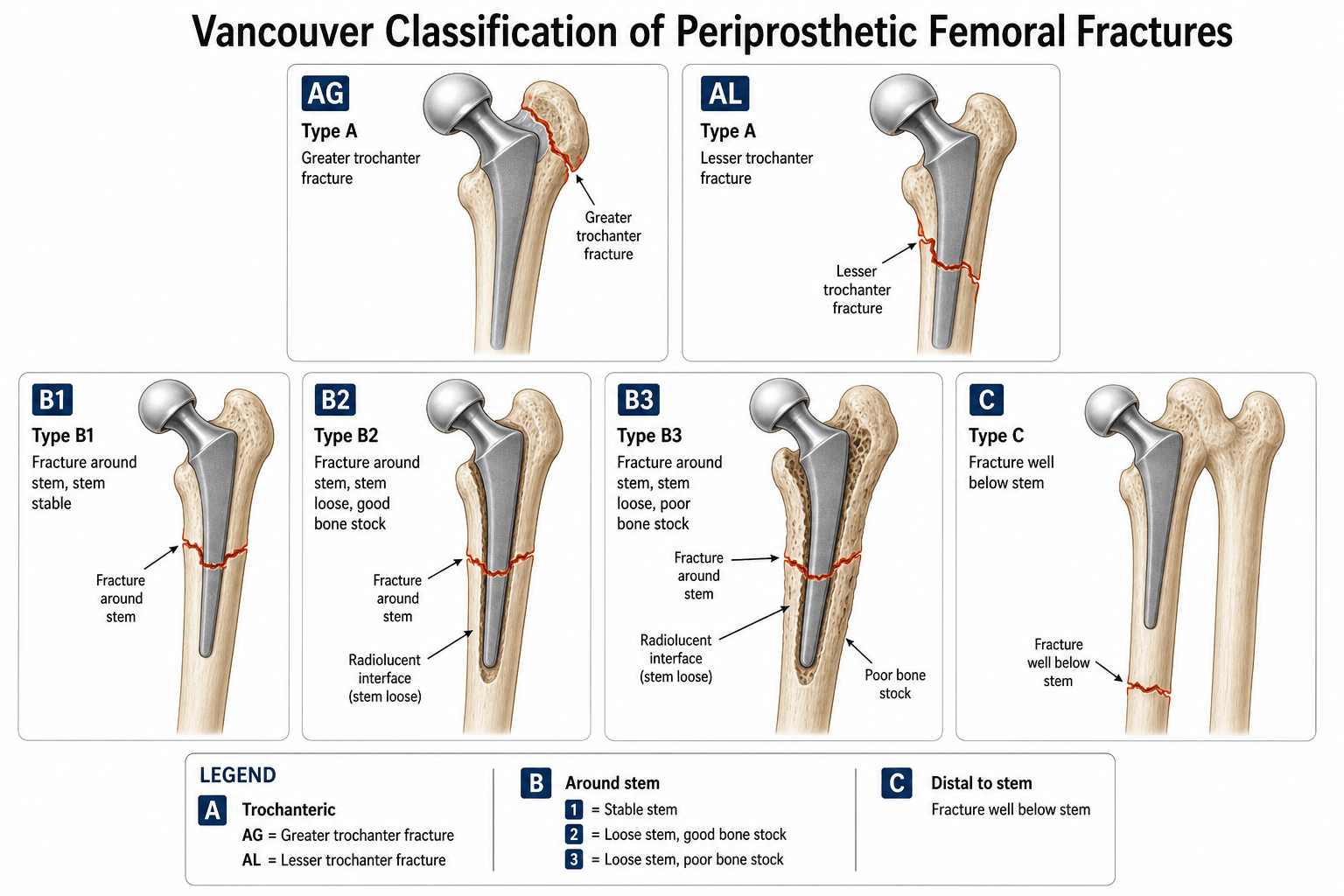
The classification uses the fracture location relative to the femoral stem and the fixation status of the implant. Every examiner expects you to recite the full scheme and then state the two key treatment questions: is the stem fixed or loose, and is the bone stock adequate.
- Location
- Greater trochanter
- Stem Status
- Stable (not relevant)
- Bone Stock
- Not relevant
- Principle
- Cable/plate fixation if displaced
- Location
- Lesser trochanter
- Stem Status
- Stable (not relevant)
- Bone Stock
- Not relevant
- Principle
- Conservative or cable fixation
- Location
- At or just below stem tip
- Stem Status
- Well-fixed
- Bone Stock
- Adequate
- Principle
- Open reduction and internal fixation (plate and cables)
- Location
- At or just below stem tip
- Stem Status
- Loose
- Bone Stock
- Adequate
- Principle
- Revision arthroplasty with long femoral stem
- Location
- At or just below stem tip
- Stem Status
- Loose
- Bone Stock
- Poor (severe bone loss)
- Principle
- Revision with augmentation (impaction grafting, allograft-prosthesis composite, or tumour prosthesis)
- Location
- Well below stem tip
- Stem Status
- Stable (stem irrelevant)
- Bone Stock
- Variable
- Principle
- Treat as native femur fracture (retrograde nail or plate)
Above · By · CaudalThe type letters
Hook:A = Above (trochanteric), B = By the stem, C = Caudal (well below) — location first, then ask about stem fixation.
The Vancouver system is preferred over the Vancouver-AAOS and the Johansson classifications because it is treatment-directed: once you classify, the operation follows. Examiners accept Vancouver as the standard answer. State it with confidence and explain the two key questions (stem fixation and bone stock) before going into the detail.
Everything above is the post-operative classification, but there is a separate intra-operative Vancouver classification (Masri, Duncan and colleagues) for fractures that happen during the arthroplasty — most often during broaching or stem insertion of a cementless press-fit stem, or during reduction/dislocation, and more likely in osteoporotic or dysplastic bone. It uses the same location letters with a numerical severity:
- Type A — proximal metaphysis; Type B — diaphysis; Type C — distal, beyond the stem tip.
- Subtype 1 — a simple cortical perforation; Subtype 2 — an undisplaced (non-displaced) linear crack; Subtype 3 — a displaced or unstable fracture.
Management follows the severity: a perforation (1) is bone-grafted and bypassed by a longer stem if needed; an undisplaced crack (2) is protected with cerclage cables; and a displaced or unstable fracture (3) needs cerclage plus a stem that bypasses the fracture, with ORIF as required. Recognising an intra-operative fracture before closing is essential — a missed crack can propagate post-operatively.
Type A — Trochanteric Fractures
Type A fractures involve the trochanteric region. They are subdivided into AG (greater trochanter) and AL (lesser trochanter).
Common post-operatively, often from abductor pull or a fall. Small avulsions with minimal displacement are managed conservatively with restricted weight-bearing and abduction exercises. Displaced fragments requiring reattachment are fixed with cable grip or claw plates to restore abductor mechanics. Loss of abductor continuity is the long-term functional concern.
Rare in isolation; usually a small avulsion from iliopsoas pull. Generally managed non-operatively. An isolated lesser trochanter fracture in a patient with a cemented stem should raise suspicion of stem loosening (the medial cement pedestal has fractured), which reclassifies the injury to B2.
- Mechanism
- Abductor avulsion or direct blow
- Displacement
- Undisplaced or minimally displaced
- Treatment
- Conservative: protected weight-bearing, abduction orthosis
- Mechanism
- Abductor avulsion or direct blow
- Displacement
- Displaced (over 1 cm)
- Treatment
- Open fixation: cable grip, trochanteric claw plate, or tension band
- Mechanism
- Iliopsoas avulsion
- Displacement
- Usually minimally displaced
- Treatment
- Conservative; exclude stem loosening (medial cement fracture)
- Mechanism
- Medial cement pedestal fracture
- Displacement
- Any
- Treatment
- Investigate: if the stem is loose, this is actually B2 — revise
B1 · B2 · B3The B subtypes
Hook:B1 = Bone good (plate); B2 = stem loose, bone ok (long-stem revision); B3 = Bone bad (revision + augmentation).
Type B — Fractures at or Near the Stem Tip
Type B fractures are the most common and the most examined subtype. The fracture line originates at or within a few centimetres of the stem tip. The B1/B2/B3 distinction determines whether the surgeon fixes the fracture around the existing stem or revises the entire femoral component.
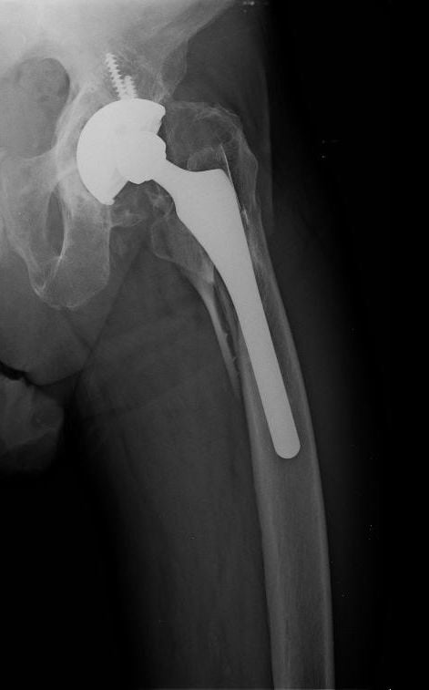
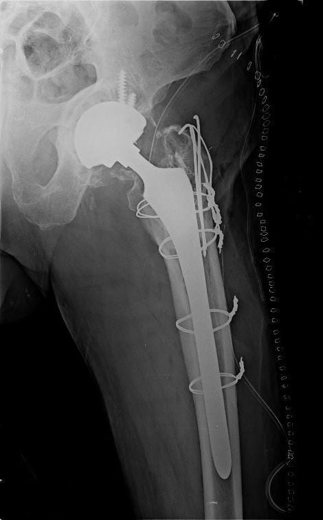
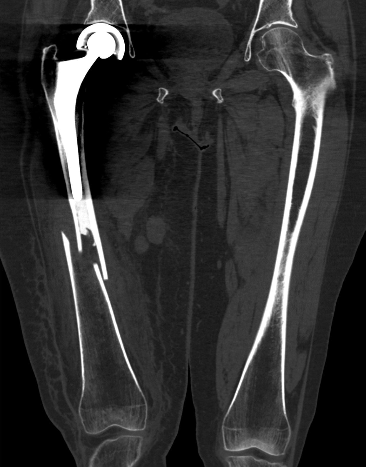
Radiographic assessment of stem stability (before deciding B1 versus B2):
For cemented stems: look for cement mantle fracture, new radiolucent lines at the cement-bone or stem-cement interface, stem subsidence or change in position, and stem-cement debonding.
For cementless stems: look for stem subsidence (compare to immediate post-operative films), change in alignment or varus migration, new radiolucent lines at the bone-prosthesis interface, broken fixation screws if present, and loss of initial osseointegration.
Always compare current radiographs to the immediate post-operative films. Many periprosthetic fractures present to units that did not perform the original arthroplasty, so request previous imaging. Change in stem position, new radiolucencies, or cement mantle fractures that were not present before are diagnostic of loosening and upgrade the classification from B1 to B2.
Type B1 — well-fixed stem, adequate bone:
Open reduction and internal fixation. The standard construct is a lateral proximal femoral locking plate combined with unicortical screws and cables around the proximal fragments to avoid perforating the stem. Cortical strut allograft augments the construct and accelerates healing. The approach requires full exposure of the fracture with the stem left in situ. Weight-bearing is protected until union is confirmed.
Type B2 — loose stem, adequate bone:
Revision arthroplasty to a long-stem (or fluted tapered) femoral component that bypasses the fracture by at least two cortical diameters. The old stem and cement are removed, the fracture is reduced, the long stem is cemented or press-fit distal to the fracture, and proximal cables or a plate may supplement fixation. The long stem acts as an internal splint stabilising the fracture while achieving durable implant fixation.
Type B3 — loose stem, poor bone stock:
Revision arthroplasty with structural bone augmentation. Options include: impaction bone grafting with a long-stem prosthesis, an allograft-prosthesis composite (APC) using a femoral allograft with a cemented stem inside it, or a megaprosthesis (tumour-type endoprosthetic replacement) when bone loss is so severe that no other option will achieve stability. The choice depends on the extent of proximal bone loss, patient age, activity level, and expected survival.
- Stem Fixation
- Well-fixed
- Bone Stock
- Adequate
- Surgical Principle
- ORIF — stem stays
- Key Fixation
- Locking plate + cables (+/- strut allograft)
- Stem Fixation
- Loose
- Bone Stock
- Adequate
- Surgical Principle
- Revision — long stem bypasses fracture
- Key Fixation
- Long fluted tapered or cemented stem + cables
- Stem Fixation
- Loose
- Bone Stock
- Poor
- Surgical Principle
- Revision + structural augmentation
- Key Fixation
- Impaction graft + long stem, APC, or megaprosthesis
If you plate a B2 fracture (loose stem) the plate will fail because the unstable stem continuously loads the fracture site. Conversely, if you revise a B1 fracture (well-fixed stem) you have performed a much larger operation than necessary with higher morbidity. Getting this wrong is the single most criticised error in the viva. When in doubt about stem fixation, test the stem intra-operatively: if it moves, revise.
TEST before you commitThe intra-operative decision
Hook:TEST the stem before you commit — feel it move and you have changed a failed plating into a successful revision.
Type C — Fractures Well Below the Stem Tip
Type C fractures occur well distal to the femoral stem tip, typically in the supracondylar or distal femoral region. The arthroplasty stem is stable and irrelevant to the fracture management.
The fracture is treated as a native femoral fracture. Options include a retrograde intramedullary nail (if the distal fragment is long enough and the canal geometry permits) or a distal femoral locking plate. The nail must avoid the stem tip — there should be sufficient intact femur between the stem tip and the fracture for stable fixation.
In a type C fracture, do not waste time classifying the stem because it is stable and far away. The clinical trap is an examiner giving you a radiograph that looks like it could be a B2 but is actually a C — look carefully at where the fracture is relative to the stem tip before committing. If the fracture is well below the stem, call it C and move on to standard distal femoral fracture management.
Pre-operative Assessment and Planning
Classification requires systematic imaging and clinical workup before the patient reaches theatre.
- Radiographs: AP pelvis, AP and lateral femur of the affected side, and the contralateral side for comparison. Include full-length films from hip to knee. Compare with all previous post-operative radiographs.
- CT scanning: Helpful for assessing fracture configuration, cortical bone stock, cement mantle integrity, and detecting subtle loosening not visible on plain film. Standard for pre-operative planning in B-type fractures.
- ** templating:** Plan for the longest stem you may need. If there is any chance the fracture is B2, have a long fluted tapered stem and revision instruments available before you start.
- Medical optimisation: These patients are typically elderly with multiple comorbidities. Coordinate with geriatric medicine, cardiology, and anaesthetics. Early surgery (within 24 to 48 hours) reduces morbidity, but the physiological priority is a safe patient.
- Bone graft and allograft availability: For B3 and selected B2 fractures, arrange proximal femoral allograft, morsellised cancellous bone for impaction grafting, or megaprosthesis availability before listing for theatre.
- Intra-operative imaging: Fluoroscopy is essential. You will need AP and lateral views of both the hip and the femur to confirm reduction, stem position, and hardware placement.
Limitations and Modern Context
- Inter-observer reliability is moderate for B-subtyping. The B1 versus B2 distinction depends on interpreting stem stability from radiographs, which is imperfect. Two surgeons reviewing the same films may disagree whether a stem is loose, which is why intra-operative testing is standard practice.
- The classification does not address fracture pattern (spiral, transverse, comminuted) or the quality of the proximal femoral bone in sufficient detail. Pattern and comminution affect fixation choice within each subtype but are not captured by the letter.
- Cementless taper designs can develop fibrous stable fixation that is not radiographically loose but is not osseointegrated. Whether this counts as well-fixed or loose for classification purposes is debated and affects B1 versus B2 treatment.
- The incidence of periprosthetic femoral fracture is rising as the ageing population expands, implant survivorship increases, and patients outlive their prostheses. Modern registries report it as one of the leading causes of revision in the second decade after arthroplasty.
- The classification was designed for cemented stems and extrapolated to cementless implants. Most modern series include a mix, but the radiographic criteria for loosening differ between the two, and the original validation studies predate widespread cementless use.
- B3 bone-loss assessment is subjective. The threshold at which adequate bone (B2) becomes poor bone (B3) is not precisely defined and varies between surgeons and centres.
The modern evolution of the Vancouver scheme is the Unified Classification System (UCS) of Duncan and Haddad (2014), which generalises the same logic to periprosthetic and peri-implant fractures around any bone and any joint (hip, knee, shoulder). It keeps the familiar grades and adds new ones:
- Type A — apophyseal (e.g. the trochanters), as in Vancouver A.
- Type B — in the bed of the implant (around or just below it), retaining the B1 (well-fixed) / B2 (loose) / B3 (loose with poor bone) subdivision.
- Type C — clear of the implant, as in Vancouver C.
- Type D — dividing the bone between two implants (an interprosthetic fracture, e.g. the femur between a hip stem and a knee component).
- Type E — each of two bones supporting one arthroplasty is fractured (e.g. femur and acetabulum, or femur and tibia).
- Type F — the surface facing/articulating with a hemiarthroplasty (e.g. an acetabular fracture against a hemiarthroplasty head).
The interprosthetic (UCS D) femoral fracture deserves special note: the bone between the two implants is a stress riser, and registry data (Chatziagorou) show these fractures fare worse, so fixation should span and overlap both implants — typically a single long plate bridging the whole femur from the hip stem to the knee component, after confirming both implants are well-fixed (revise any that is loose).
Exam Viva
Practise clinical reasoning and management decisions out loud
“A 72-year-old woman presents with a painful left thigh four years after a cemented total hip replacement. Radiographs show a spiral fracture of the proximal femur originating at the tip of the cemented stem. There is no clear cement mantle fracture and the stem position appears unchanged from the immediate post-operative film. How do you classify and manage this fracture?”
“A 78-year-old man who had a right total hip replacement for osteoarthritis nine years ago presents after a fall. Radiographs show a transverse fracture well distal to the femoral stem tip, in the distal third of the femur. The stem appears well-fixed. Outline your assessment and management.”
The classification scheme
- Type A: trochanteric (AG = greater, AL = lesser), stem stable, treat with cables or conservative
- Type B: fracture at or near stem tip — B1 (well-fixed, good bone), B2 (loose, good bone), B3 (loose, poor bone)
- Type C: fracture well below stem tip, stem is irrelevant, treat as native femur fracture
- The two critical questions: is the stem fixed or loose, and is the bone stock adequate
Treatment by type
- AG: cables or trochanteric claw plate if displaced; conservative if undisplaced
- B1: ORIF with locking plate + cables around stem (+/- cortical strut allograft)
- B2: revision to long-stem prosthesis that bypasses the fracture by at least two cortical diameters
- B3: revision with augmentation — impaction grafting, allograft-prosthesis composite, or megaprosthesis
- C: retrograde nail or distal femoral plate, treating as a native distal femoral fracture
Radiographic signs of loosening
- Cemented: cement mantle fracture, new radiolucencies at cement-bone or stem-cement interface, subsidence, stem migration
- Cementless: subsidence, change in alignment or varus migration, new radiolucent lines at bone-prosthesis interface, broken screws
- Always compare with immediate post-operative films to detect change
- If radiographs are equivocal, obtain CT and plan for intra-operative stem testing
Exam traps and high-yield points
- B1 versus B2 is the most commonly tested distinction — if in doubt, test the stem intra-operatively
- Plating a loose stem (B2 treated as B1) leads to construct failure and reoperation
- A lesser trochanter fracture with a medial cement fracture is actually B2, not AL
- Type C does not involve the stem — do not waste time classifying the prosthesis
- Have revision implants available for every B-type case before incision
Evidence Base
Every citation below has been checked against its source record in PubMed. The classic series (Beals, Springer) and the large Swedish-register studies (Lindahl, Chatziagorou) together establish that stem fixation drives treatment and that radiographic categorisation of stem stability is unreliable — hence the recommendation to TEST the stem intra-operatively. Note the nuance from Chatziagorou: in that registry it was B1 and interprosthetic fractures, not B2, that carried the higher reoperation risk.
Periprosthetic fractures of the femur. An analysis of 93 fractures
- Retrospective review of 93 periprosthetic femoral fractures (102 treatments) around hip arthroplasty
- Prosthesis type and pre-existing stress risers influenced where fractures occurred; fracture site and the pre-fracture interface guided treatment
- Fractures around a loose interface (cemented or cementless) were best treated by removing the prosthesis, reducing the fracture, and inserting a long-stemmed prosthesis — fractures around a stable stem were managed by site
Three hundred and twenty-one periprosthetic femoral fractures
- Swedish Hip Arthroplasty Register study of 321 periprosthetic femoral fractures; 88% were Vancouver type B
- A loose stem was present in 66% of fractures after primary and 51% after revision arthroplasty, but preoperative radiographic categorisation was difficult
- There was a high failure/reoperation rate (66-month survival 74.8%); the authors recommended exploring the joint to test stem stability when it is uncertain
Treatment of periprosthetic femoral fractures following total hip arthroplasty with femoral component revision
- 118 hips revised for acute Vancouver type-B periprosthetic femoral fracture at a major arthroplasty centre (mean follow-up 5.4 years)
- Implant survival (revision/removal as endpoint) was 90% at 5 years and 79.2% at 10 years; results were better with an extensively porous-coated uncemented stem
- The main long-term problems were prosthetic loosening and fracture nonunion