Late Sequela of Compartment Syndrome | Claw Hand Deformity | Irreversible Muscle Fibrosis
- Irreversible sequela of untreated or inadequately treated compartment syndrome
- Classic posture: Wrist flexion, MCP hyperextension, IP flexion, thumb adduction
- Cascade sign: Passive wrist extension causes fingers to flex further
- Prevention is key: Early fasciotomy (under 6 hours) reduces incidence to under 5%
- Reconstruction outcomes: Poor compared to prevention - 20-80% normal function
- “Volkmann's contracture is the devastating late outcome of missed compartment syndrome
- “Pathophysiology: Muscle necrosis → fibrosis → contracture (flexors stronger than extensors)
- “Tsuge classification guides treatment: Type I (mild) to Type III (severe)
- “Reconstruction requires extensive surgery but results never match prevention
Overview and Epidemiology
Volkmann's ischaemic contracture is an irreversible flexion contracture of the forearm and hand, produced by muscle fibrosis after prolonged ischaemia from compartment syndrome. It is the devastating late sequela of a compartment syndrome that was untreated or inadequately treated, and it represents a failure of early recognition.
History. Richard von Volkmann described it in 1881, in the forearm after supracondylar humerus fractures. The end result is the classic claw hand.
Who. It is most common in children, after supracondylar humerus fractures, but adults develop it too after forearm fractures and crush injuries. Once established, the contracture is permanent.
Fasciotomy within 6 hours reduces the incidence of Volkmann's contracture to under 5%; fasciotomy delayed beyond 12 hours carries an incidence of 20-40%. The contracture is largely preventable by early recognition and decompression, which is why compartment syndrome is treated as a time-critical emergency.
Anatomy and Pathophysiology
The forearm compartments. The volar compartment holds the flexors (FDP, FDS, FPL, FCR, FCU) and carries the median and ulnar nerves. The dorsal compartment holds the extensors (ECRL, ECRB, EDC and others). Compartment syndrome affects the volar compartment most severely.
From ischaemia to contracture. Prolonged ischaemia kills muscle cells. The necrotic muscle is replaced by fibrous scar, which contracts over weeks to months, and once established the contracture is permanent. Ischaemia of the nerves adds sensory loss and motor weakness.
- Process
- Compartment pressure exceeds perfusion
- Timeline
- 0-6 hours
- Reversibility
- Reversible with fasciotomy
- Process
- Muscle cells die from ischaemia
- Timeline
- 6-12 hours
- Reversibility
- Partially reversible
- Process
- Necrotic muscle replaced by scar
- Timeline
- Weeks
- Reversibility
- Irreversible
- Process
- Scar tissue contracts
- Timeline
- Months
- Reversibility
- Irreversible
Why the flexors win. The flexors are stronger and more numerous than the extensors, so the contracting scar produces a flexion contracture. The flexor side also takes the worse injury:
- The flexor compartment is more commonly affected by compartment syndrome
- Flexor muscles have a higher metabolic demand
- The flexor compartment has less collateral circulation
Beyond 6-8 hours muscle necrosis begins, and by 12 hours significant muscle death has occurred. The six-hour figure is a guide rather than a switch; the Controversies section below explains why fasciotomy should be driven by the diagnosis, not by the clock.
Classification Systems
Tsuge's classification is the most widely used, and it guides treatment selection.
- Muscle Involvement
- FDP to 2-3 fingers, FPL only
- Clinical Features
- Limited contracture, weak grip, some sensory loss
- Treatment
- Muscle slide, tendon lengthening
- Muscle Involvement
- All flexor muscles involved
- Clinical Features
- Significant deformity, intrinsic-plus posture, weak extension
- Treatment
- Muscle slide + tendon transfers
- Muscle Involvement
- Both flexors AND extensors
- Clinical Features
- Fixed contracture, claw hand, complete sensory loss, non-functional
- Treatment
- Free functioning muscle transfer
Type I preserves some function and has the best prognosis with reconstruction. Type II causes significant functional impairment and needs more extensive reconstruction. Type III leaves the hand essentially non-functional, has the worst prognosis, and may require amputation in extreme cases.
Clinical Assessment
History. The story is of a compartment syndrome that was missed or decompressed late. Ask about:
- Previous compartment syndrome, treated late or untreated
- Supracondylar humerus fracture, the most common cause in children
- Forearm fracture or crush injury
- Fasciotomy delayed beyond 6-12 hours
- Contracture progressing over weeks to months
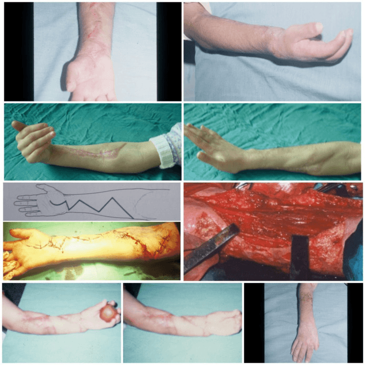
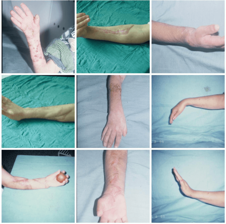
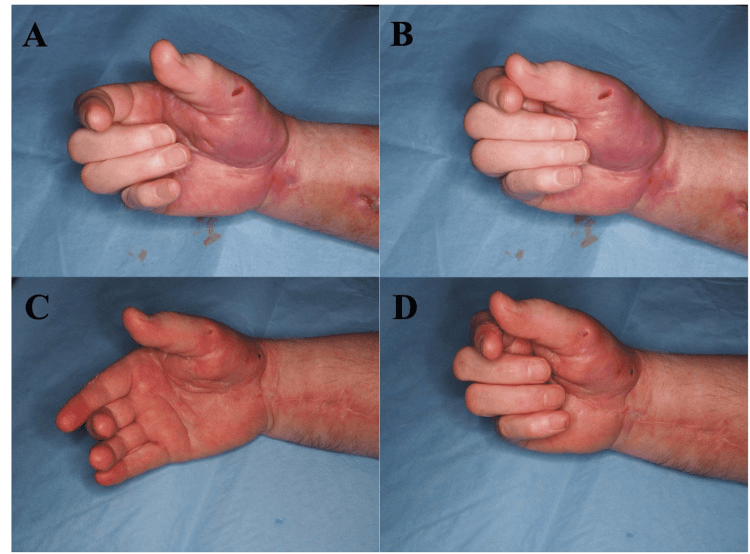
The posture. The hand is clawed. The shortened flexors also pull the elbow into flexion.
- Wrist flexion - flexor contracture
- MCP hyperextension
- IP joint flexion - FDP contracture
- Thumb adduction - FPL contracture
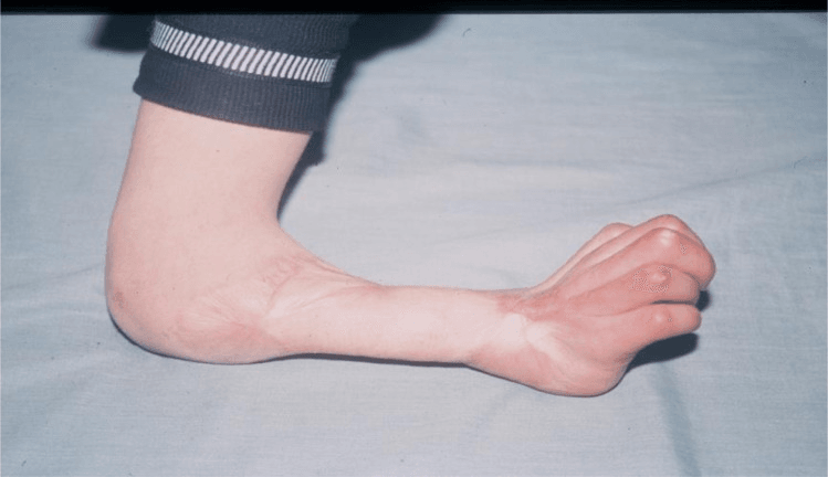
Other signs. The contracture is fixed and cannot be passively corrected. The affected muscles are wasted, sensation is lost in the median and ulnar distribution, and power grip is weak.
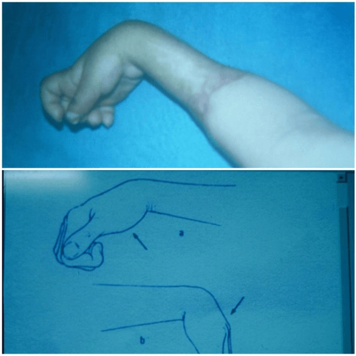
The cascade sign. Passively extending the wrist makes the fingers flex further, which indicates that the flexor muscles are shortened and fibrotic, and passive finger extension tensions the shortened flexor unit and reproduces pain. The sign is described as pathognomonic and distinguishes Volkmann's contracture from the other causes of a clawed hand below. Strictly, it shows fixed flexor muscle-tendon shortening, so interpret it with the history and the rest of the examination.
Neurological assessment. Map the deficit nerve by nerve:
- Median nerve - sensory loss in the thumb, index and middle fingers
- Ulnar nerve - sensory loss in the ring and little fingers
- Motor - loss of thumb opposition and finger abduction
- Intrinsic involvement - intrinsic-plus posture
Differential Diagnosis
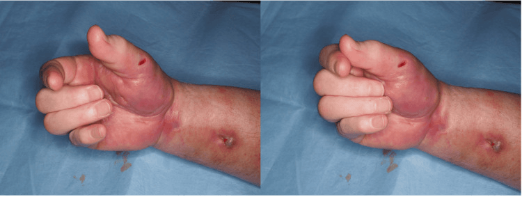
The contracted or clawed hand has several causes. What discriminates Volkmann's is the extrinsic tenodesis (cascade) effect: finger position changes with wrist position.
- Mechanism
- Extrinsic flexor fibrosis after ischaemia
- Wrist-position effect
- Fingers flex further on wrist extension (positive cascade/tenodesis)
- Distinguishing features
- History of compartment syndrome; combined motor + sensory deficit in median/ulnar territory
- Mechanism
- Tight interossei/lumbricals
- Wrist-position effect
- Deformity unchanged by wrist position; positive Bunnell-Littler test
- Distinguishing features
- MCP flexed, IP extended; PIP flexion limited more with MCP extended
- Mechanism
- Loss of ulnar intrinsics
- Wrist-position effect
- No tenodesis change; clawing of ring/little fingers
- Distinguishing features
- Sensory loss ulnar-only; Froment and Wartenberg signs; no flexor fibrosis
- Mechanism
- Palmar fascia (cord) fibrosis
- Wrist-position effect
- No tenodesis effect; skin pitting/cords
- Distinguishing features
- MCP/PIP flexion from palpable cords; no neurological deficit; no ischaemic history
- Mechanism
- Upper motor neuron spasticity
- Wrist-position effect
- Velocity-dependent resistance; partially correctable under relaxation/anaesthesia
- Distinguishing features
- Hyperreflexia, clasp-knife tone, global developmental signs
- Mechanism
- Congenital FDS/skin/joint anomaly
- Wrist-position effect
- Variable; usually little finger PIP
- Distinguishing features
- Present from childhood, no ischaemic event, often bilateral
Use joint position to localise the contracted tissue. In Volkmann's the extrinsic flexors are short, and because the shortened muscle-tendon unit crosses both the wrist and the finger joints, extending the wrist worsens finger flexion (positive cascade/tenodesis). In an intrinsic contracture the deformity is fixed regardless of wrist position and the Bunnell-Littler test is positive. Both can coexist after a severe forearm compartment syndrome.
Investigations
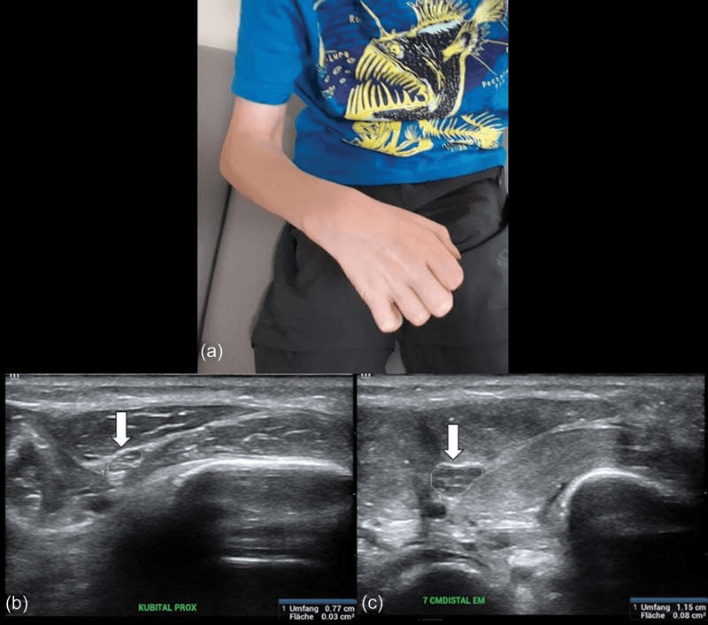
A clinical diagnosis. A history of compartment syndrome, especially one treated late, plus the classic claw deformity with a cascade sign makes the diagnosis. Imaging and tests support it and are not required for it.
Radiographs may show associated fractures and, as a late finding, muscle calcification. They are not diagnostic but may show the extent of involvement.
MRI shows muscle fibrosis and atrophy. It may help assess the extent of muscle involvement and is useful for surgical planning.
Electromyography shows denervation patterns and assesses nerve function, which helps predict recovery potential.
Functional assessment includes:
- Grip strength
- Range of motion
- Functional hand evaluation
- Activities of daily living
Management Algorithm
Prevention is the only effective treatment, and it is far superior to any reconstruction. It depends on keeping a high index of suspicion for compartment syndrome and recognising it early. Pain on passive stretch is the key early sign; pulselessness is late, and you should not wait for it.
- Pain out of proportion to the injury
- Pain on passive stretch - the earliest sign
- Paraesthesia - numbness from early nerve ischaemia
- Paresis - weakness from motor nerve ischaemia; paralysis is a late sign
- Pallor - pale or mottled skin
- Pulselessness - a late sign
- Pressure - a tense compartment; measure the compartment pressure if uncertain
Emergency fasciotomy. Decompress within 6 hours of onset. It is time-critical: do not delay.
Surgical Technique
Indication and planning. The flexor muscle slide (the Page and Scaglietti procedures) is for Tsuge Type I. Assess the extent of the contracture and identify which muscles are involved. Consent for limited improvement and possible recurrence.
- Incision, usually volar forearm, extensile if needed
- Identify the affected flexor muscles
- Release the flexor origin from the medial epicondyle
- Slide the muscles distally to lengthen them
- Lengthen tendons as needed
- Confirm that passive correction is achieved
- Splint in the corrected position
Aftercare. Splint for 4-6 weeks, then mobilise gradually; hand therapy is essential. The slide is effective in Type I, with good functional outcomes.
Sequence of release. After nerve release, adhesions are freed in turn, and only the muscle-tendon units that remain restrictive are lengthened or divided, preserving useful residual function wherever possible.
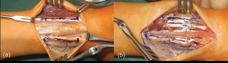
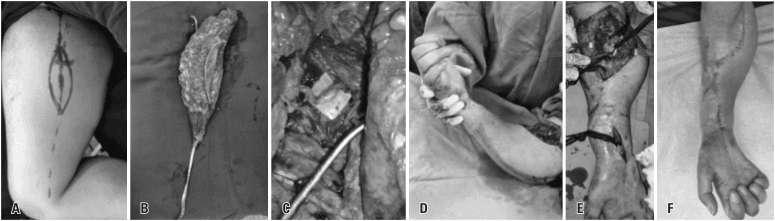
Nerve Involvement: Restoring the Sensate Hand
Why the nerves suffer. The median nerve and its anterior interosseous branch run through the deep central infarct and are injured by two mechanisms: direct ischaemic neuropathy during the acute event, and chronic compression and tethering as the dead muscle turns to dense scar. The ulnar nerve, more ulnar and superficial, is affected in more extensive disease, so the motor and sensory loss seen clinically is median-predominant.
Sensation comes first. A hand with gross grasp but no protective sensation is a hazard, prone to unfelt burns and ulcers, and functionally poor. Restoring protective sensation is therefore a primary reconstructive goal, often ranked above regaining a few degrees of motion, and that shapes both counselling and planning.
The nerve options. They are taken in order, from neurolysis onward:
- Neurolysis - releasing the median or ulnar nerve from its encasing scar is the first and often the most rewarding step, done at the time of muscle debridement or slide. A nerve in continuity within scar can recover once decompressed; its internal fascicular integrity and motor and sensory potential determine whether neurolysis alone is sufficient.
- Nerve grafting - interpositional (e.g. sural) grafting for a segment destroyed by infarct or scar.
- Nerve transfer - a healthy expendable donor fascicle (e.g. to restore key pinch or the thumb) when proximal sources are unavailable.
- Tendon transfer, arthrodesis or free-muscle neurotisation for irrecoverable motor loss, and sensory nerve transfer or a neurovascular island flap to bring sensation to the critical pinch surfaces of the thumb and index.
Do it early. Axonal regeneration is time-limited, so nerve decompression and repair are addressed at the debridement stage rather than deferred. Protecting recoverable nerve is part of why early wide excision of necrotic muscle (Stevanovic) limits distal motor and sensory loss.
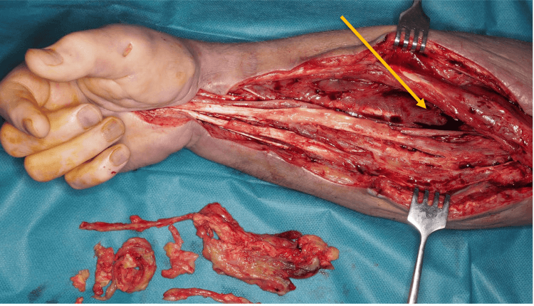
Complications
- Incidence
- Common
- Management
- May require repeat surgery
- Incidence
- 5-10%
- Management
- Nerve exploration, possible grafting
- Incidence
- 5-10%
- Management
- Antibiotics, debridement
- Incidence
- 10-15%
- Management
- Flap coverage if needed
- Incidence
- Common
- Management
- Aggressive hand therapy
- Incidence
- Common
- Management
- Realistic expectations essential
Recurrence. The contracture may recur after reconstruction, and preventing recurrence is challenging.
The nerve. Beyond injury at surgery, the nerve may be encased in scar tissue and need neurolysis or grafting.
Postoperative Care
Post-Reconstruction Protocol
- Splint in corrected position
- Elevation to reduce swelling
- Monitor neurovascular status
- Pain management
- Continue splinting
- Begin passive range of motion
- Hand therapy consultation
- Monitor for recurrence
- Active range of motion
- Strengthening exercises
- Functional training
- Serial splinting if needed
- Continue hand therapy
- Assess functional outcomes
- Plan additional procedures if needed
- Realistic goal setting
Hand therapy is essential for any functional recovery. It covers passive and active range of motion, strengthening and functional retraining, with splinting and serial casting.
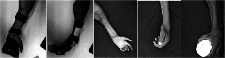
Outcomes and Prognosis
Reconstruction never restores normal function, and the expected result falls with each Tsuge type.
- Procedure
- Muscle slide, tendon lengthening
- Expected Function
- 70-80% normal
- Patient Satisfaction
- High
- Procedure
- Muscle slide + transfers
- Expected Function
- 40-60% normal
- Patient Satisfaction
- Moderate
- Procedure
- Free muscle transfer
- Expected Function
- 20-30% normal
- Patient Satisfaction
- Low to moderate
Prognostic factors. Beyond severity, where Type I does best, the result turns on:
- Timing of reconstruction, after the contracture has stabilised
- Compliance with hand therapy
- Patient age - younger patients may have better outcomes
- Associated nerve injury
Guidelines, Registries & Global Practice
Volkmann's contracture has no disease-specific registry; the relevant guidance addresses acute compartment syndrome, the preventable upstream cause.
Global epidemiology:
- Established Volkmann's contracture is now uncommon where early decompression is routine; it persists where compartment syndrome is recognised late.
- Acute compartment syndrome of the leg occurs in roughly 1-3% of tibial diaphyseal fractures in prospective series; young males are over-represented, partly reflecting tighter fascial compartments and higher-energy injury.
- In children, supracondylar humeral fractures and both-bone forearm fractures are the classic antecedents; vigilance for the "3 A's" (increasing analgesia requirement, anxiety, agitation) is emphasised in paediatric practice.
- The dominant modifiable risk factor for contracture across all settings is time from onset to fasciotomy.
Side-by-side guidance (acute compartment syndrome):
- Diagnostic emphasis
- Repeated clinical assessment; pain on passive stretch and escalating analgesia; monitoring if unreliable patient
- Decompression trigger
- Clinical diagnosis or differential pressure under 30 mmHg; do not wait for late signs
- Practical note
- Emphasises documented serial review and rapid theatre access
- Diagnostic emphasis
- Clinical signs supplemented by intracompartmental pressure when equivocal
- Decompression trigger
- Differential pressure within 30 mmHg of diastolic favoured over absolute threshold
- Practical note
- Highlights monitoring for obtunded/regional-block patients
- Diagnostic emphasis
- High index of suspicion in high-risk fractures; serial exam plus optional pressure measurement
- Decompression trigger
- Urgent four-compartment fasciotomy on diagnosis
- Practical note
- Stresses complete release and dermatofasciotomy technique
- Diagnostic emphasis
- Combine clinical findings with pressure monitoring; perfusion-pressure concept
- Decompression trigger
- Perfusion (differential) pressure threshold preferred
- Practical note
- Notes variability in equipment availability across Europe
- No implant or arthroplasty registry applies. The strongest outcome data come from prospective monitoring cohorts (Edinburgh series) showing that earlier decompression eliminates the contracture and weakness that define the late syndrome.
- Trauma databases consistently identify delay to fasciotomy and missed diagnosis (especially in sedated or anaesthetised patients) as the leading drivers of poor outcome.
- Well-resourced settings: continuous pressure monitoring available, immediate theatre access, and microsurgical free functioning muscle transfer for severe established contracture.
- Limited-resource settings: reliance on clinical diagnosis alone, longer delays to theatre, and a higher proportion of established contractures presenting late; reconstruction often limited to muscle slide, tendon transfers and tendon lengthening rather than free tissue transfer.
- Across all settings the cheapest and most effective intervention is the same: early recognition and prompt, complete fasciotomy.
Volkmann's contracture is a largely preventable end-stage of compartment syndrome. No guideline or registry offers a reconstruction that restores a normal hand. The universal message across AAOS, BOA, AO and EFORT is identical: diagnose compartment syndrome early, decompress promptly and completely, and never wait for late signs such as pulselessness.
Related pages: Compartment Syndrome and Forearm Compartment Syndrome are the preventable cause of everything on this page - Volkmann's contracture is what a missed or late-decompressed compartment syndrome becomes, which is why the differential-pressure evidence sits in this Evidence Base at all; Compartment Syndrome of the Leg carries the tibial-fracture data the McQueen cards are drawn from, and Foot Compartment Syndrome and Region-Specific Compartment Syndrome cover the other territories; Supracondylar Humerus Fracture is the classic paediatric antecedent and the injury in which the pink pulseless hand demands the most careful serial assessment, with Both-Bone Forearm Fractures and Paediatric Forearm Fractures the other common precipitants; Crush Syndrome and Rhabdomyolysis are the systemic consequences of the same ischaemic muscle injury and the reason the Mubarak card exists; Median Nerve Anatomy and Ulnar Nerve Anatomy explain the deep-compartment nerve deficits that make the hand insensate as well as contracted, and Nerve Transfers covers the reconstructive options for them; and Chronic Exertional Compartment Syndrome is the entity that shares the name and none of the urgency.
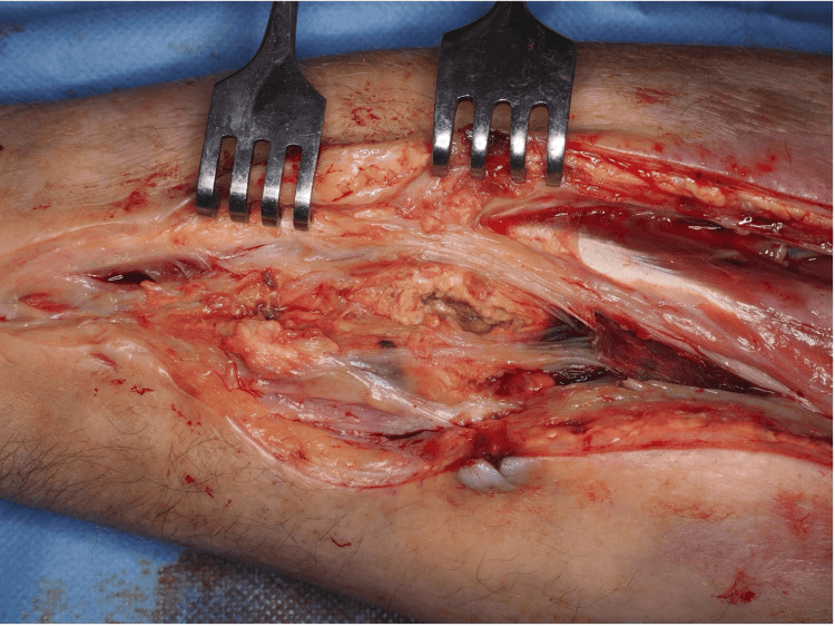
The Ellipsoid Infarct: Why the Deep Central Forearm is the Epicentre
The Tsuge staging, from mild disease confined to the deep flexors of a few fingers, through all the flexors, to the extensors, is not arbitrary. It maps the geography of the ischaemic infarct.
The shape. The classic lesion is an elliptical zone of infarction lying deep in the volar forearm, maximal at its centre and tapering at the poles. Its epicentre is the deep flexor mass, FDP and FPL, which is why the mildest disease (Tsuge I) is precisely FDP to two or three fingers plus FPL.
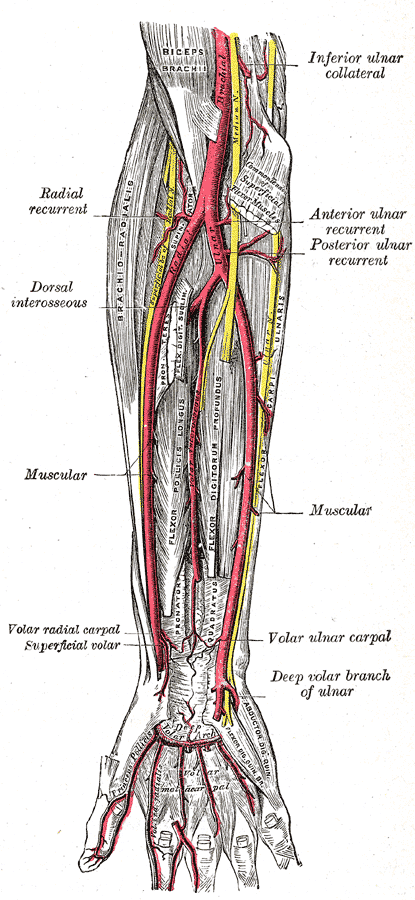
Why the deep centre. The deep flexors sit furthest from the compartment's blood supply, since the radial, ulnar and anterior interosseous vessels lie more superficially or peripherally. They are the watershed tissue, the first to infarct and the last to be spared. As ischaemia worsens, the infarct spreads outward from this core to the superficial flexors (Tsuge II) and finally the dorsal extensor mass (Tsuge III).
A gradient within the ellipse. A dense fibrotic core grades out to a rim of viable but scarred muscle. A muscle slide recruits that surviving peripheral muscle, which is why it works in mild disease; in severe disease the dead central core must be excised and replaced by a free functioning muscle transfer. Viable tissue must be distinguished from established necrosis before reconstruction is planned.
The nerve runs through it. The median nerve and its anterior interosseous branch traverse the deep central zone and are encased in the densest scar. That is the anatomical reason the median and AIN territory bears the brunt of the neurological deficit, and why the scar must be released off the nerve.
Controversies and Areas of Uncertainty
Most uncertainty in Volkmann's contracture sits upstream, in the diagnosis and decompression of acute compartment syndrome, because the contracture itself is largely a function of how that emergency was handled.
The classic absolute threshold of 30 mmHg (Mubarak, wick catheter) over-calls compartment syndrome. Continuous monitoring of the differential (perfusion) pressure with a trigger of under 30 mmHg (diastolic minus compartment pressure) missed no cases yet avoided many unnecessary fasciotomies. The optimal single number, and whether 20 or 30 mmHg is safest, remains debated.
Continuous pressure monitoring shortens time to fasciotomy and reduces sequelae, with high sensitivity and specificity in tibial fractures. Critics note false positives, cost, and that a vigilant repeated clinical exam suffices in the alert, cooperative patient. Monitoring is most defensible in the obtunded, anaesthetised, regional-block or polytrauma patient who cannot report pain.
The 6-hour ischaemia tolerance is a useful teaching figure but is not an absolute switch: muscle injury is a continuum influenced by perfusion pressure, hypotension and the duration of raised pressure. Fasciotomy should be driven by diagnosis, not by a clock - and a late, viable compartment may still benefit, whereas decompressing frankly necrotic muscle late can precipitate reperfusion injury and infection.
For compartment syndrome diagnosed very late (over 24-48 hours) with established necrosis, some advocate against decompression because opening necrotic muscle risks sepsis and reperfusion (rhabdomyolysis, renal injury); others decompress to relieve any salvageable tissue. There is no high-level evidence to settle this.
For mild-moderate contracture the flexor muscle slide preserves resting length and is widely preferred, but recurrence and unpredictable strength are reported. For severe disease, early wide excision of fibrotic muscle plus free functioning muscle transfer is favoured to protect nerves, though it commits the patient to microsurgery and staged operations.
The Tsuge and Seddon systems are descriptive and do not capture nerve involvement, intrinsic tightness or joint contracture independently. No classification reliably predicts functional outcome, and treatment is individualised to the residual motor units, sensation and joint suppleness.
MCQ Practice Points
Q: What is the cascade sign in Volkmann's contracture? A: Passive wrist extension causes fingers to flex further - this is pathognomonic for Volkmann's contracture. It indicates that flexor muscles are shortened and fibrotic.
Q: How do you prevent Volkmann's contracture? A: Early fasciotomy within 6 hours of compartment syndrome onset - this reduces incidence to under 5%. Delayed fasciotomy (over 12 hours) results in 20-40% incidence. Prevention is the only effective treatment.
Q: What is Tsuge Type I Volkmann's contracture? A: Limited to FDP 2-3 fingers and FPL - this is the mildest form. Treatment is muscle slide and tendon lengthening, with expected 70-80% normal function.
Q: What is the pathophysiology of Volkmann's contracture? A: Prolonged ischemia (over 6-8 hours) causes muscle necrosis, which is replaced by fibrous scar tissue that contracts. Flexors are stronger than extensors, so flexion contracture results.
Q: What are the expected outcomes of reconstruction for Volkmann's contracture? A: Type I: 70-80% normal function, Type II: 40-60%, Type III: 20-30% - results are never as good as prevention. This is why early fasciotomy is so critical.
Exam Viva Scenarios
Practise clinical reasoning and management decisions out loud
“A 35-year-old man presents 6 months after a forearm crush injury. He has a claw hand deformity with wrist flexion, MCP hyperextension, and IP flexion. Passive wrist extension causes fingers to flex further. Describe this condition and your management.”
“A 7-year-old child presents 4 hours after closed reduction and pinning of a supracondylar humerus fracture. The child has severe pain, pain on passive finger extension, and decreased sensation in the median nerve distribution. How do you prevent Volkmann's contracture?”
“A 12-year-old presents 9 months after a missed forearm compartment syndrome following a supracondylar fracture. The hand is clawed, there is dense sensory loss in the median distribution, no useful active finger flexion, and the extensors are weak. The parents ask whether surgery can 'fix' the hand. How do you assess, plan reconstruction, and counsel them?”
Key Facts
- Irreversible sequela of compartment syndrome
- Classic claw hand deformity
- Cascade sign is pathognomonic
- Prevention is the only effective treatment
Tsuge Classification
- Type I (Mild): FDP 2-3 fingers, FPL - muscle slide (70-80% function)
- Type II (Moderate): All flexors - muscle slide + transfers (40-60% function)
- Type III (Severe): Flexors and extensors - free muscle transfer (20-30% function)
- Classification guides surgical approach and sets realistic expectations
Prevention
- Early fasciotomy within 6 hours: under 5% incidence
- Delayed fasciotomy over 12 hours: 20-40% incidence
- Time-critical emergency - do not delay
- Prevention is far superior to any reconstruction
Clinical Features
- Claw hand: wrist flexion, MCP hyperextension, IP flexion, thumb adduction
- Cascade sign: passive wrist extension causes fingers to flex further
- Sensory loss: median/ulnar nerve distribution
- Weak grip: loss of power grip
Pathophysiology
- Prolonged ischemia (over 6-8 hours) → muscle necrosis
- Necrotic muscle → fibrous scar tissue
- Scar contracts → flexion contracture
- Flexors stronger than extensors → claw hand
Evidence Base
Treatment of Established Volkmann's Contracture of the Forearm (Tsuge Classification)
- Original description of the mild/moderate/severe (Type I-III) classification still in widest use
- Mild (Type I): localised contracture of deep flexors to 2-3 digits and FPL, treated by tendon lengthening or flexor muscle slide
- Moderate (Type II): all long flexors involved, often with median/ulnar nerve involvement, treated by muscle slide plus nerve neurolysis/transfers
- Severe (Type III): combined flexor and extensor involvement requiring more radical reconstruction
Compartmental Syndrome and Its Relation to the Crush Syndrome: A Spectrum of Disease
- Series of 11 limbs after prolonged compression; residual contracture was moderate or severe in 80% of involved extremities
- Severity of systemic crush manifestations tracked the volume of muscle compressed and the duration of pressure
- Delay in hospitalisation, diagnosis and treatment prolonged the ischaemic insult
- Immediate fasciotomy recommended to minimise residual contracture and prevent myonecrosis-driven crush syndrome
Acute Compartment Syndromes: Diagnosis and Treatment with the Aid of the Wick Catheter
- 65 compartments measured by wick catheter in 27 patients with suspected acute compartment syndrome
- Normal compartment pressure 0-8 mmHg; an absolute pressure of 30 mmHg or more used as the fasciotomy threshold
- All 16 patients whose pressures stayed under 30 mmHg avoided fasciotomy with no sequelae
- Pioneered objective intracompartmental pressure measurement to guide decompression