Cone-Beam CT Under Physiological Load
- WBCT is primarily a bone and alignment tool, not a soft-tissue replacement for MRI.
- Use WBCT when symptoms are load-dependent: syndesmosis, hindfoot valgus/varus, Lisfranc instability, ankle malreduction.
- The main advantage over conventional CT is physiologic loading, not simply another cross-sectional scan.
- Contralateral comparison is high yield for subtle syndesmotic or midfoot instability.
- If the patient cannot safely weight-bear, standard CT or MRI is usually the correct alternative.
- “Syndesmotic instability may look reduced on supine CT but widen in loaded position.
- “WBCT improves hindfoot moment-arm and 3D alignment assessment compared with projection-dependent radiographs.
- “WBCT is strongest for foot and ankle bone questions; MRI remains better for tendon, ligament, cartilage, and marrow oedema questions.
- “Do not order WBCT just because it is newer; order it when loading is likely to change the answer.
WBCT adds value only when the clinical question is load-dependent. If the patient cannot stand, or if the question is tendon, ligament, marrow, tumour, or infection extent, conventional CT or MRI is usually more appropriate.
LOADLOAD Indications
Hook:Order WBCT when loading reveals the pathology.
STANDSTAND Review Sequence
Hook:STAND keeps the report focused on what changes under load.
Overview
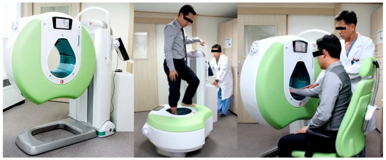
WBCT is usually performed on a cone-beam platform with the patient standing, semi-standing, or otherwise positioned to load the limb during acquisition. The key orthopaedic advantage is that loaded alignment, joint congruity, and subtle instability can be measured directly instead of inferred from projectional radiographs or from unloaded CT.
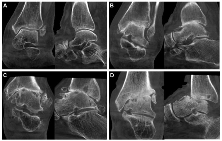
The practical scope remains foot-and-ankle dominant. The literature is strongest for syndesmosis assessment, adult-acquired flatfoot, cavovarus deformity, hallux valgus planning, ankle osteoarthritis, and post-operative reduction analysis. Outside those indications, WBCT is still emerging and should not be treated as a universal replacement for conventional CT.
Acquisition and Loading Technique
The Overview says WBCT is done "standing, semi-standing, or otherwise positioned to load the limb," and the controversies note "simulated versus true standing load" — the actual acquisition deserves to be explicit, because how the limb is loaded determines what the scan can claim.
The platform
- WBCT uses a dedicated cone-beam extremity scanner: an x-ray source and a flat-panel detector make a single rotation around the limb, producing an isotropic 3D dataset (so any plane can be reconstructed without re-scanning). Cone-beam acquisition delivers a much lower effective dose than multidetector CT for extremities, which makes bilateral and repeat imaging more acceptable.
How the limb is loaded
- Natural full-body-weight standing in an open, low-rim gantry the patient steps into is the truest physiologic load and usually images both feet together for side-to-side comparison.
- Seated or semi-standing rigs apply a simulated, partial axial load (a set proportion of body weight) for patients who cannot fully stand; this approximates but does not always equal true stance, and load magnitude and direction influence measured diastasis and alignment — so the protocol must be reported.
Practical limits
- The patient must be able to bear weight safely; if not, the modality loses its whole advantage and supine CT or MRI is chosen instead.
- A painful standing limb produces motion artefact (mitigated by short protocols), and cone-beam has a limited field of view and lower soft-tissue contrast than MDCT.
Systematic Approach
- Question
- Is there valgus, varus, arch collapse, or rotational asymmetry?
- Why it matters
- Defines the deformity plane before drilling into joints
- Question
- Do the ankle, subtalar, talonavicular, and TMT joints remain congruent under load?
- Why it matters
- Loaded incongruity can explain symptoms despite normal supine imaging
- Question
- Is there diastasis or malreduction relative to the contralateral side?
- Why it matters
- High-yield indication for WBCT
- Question
- Are there cysts, osteophytes, subchondral changes, or occult fracture lines?
- Why it matters
- Adds surgical-planning detail
- Question
- Does the symptomatic side deviate meaningfully from the opposite side?
- Why it matters
- Reduces over-calling subtle anatomic variation
Foot and Ankle Offset (FAO): The Flagship WBCT Biometric
The term Foot and Ankle Offset (FAO) recurs in the evidence and guidelines above as the standardised 3D biometric, but it is never defined — and it is the single most exam-relevant WBCT measurement, so it is worth setting out.
What FAO measures
- WBCT software locates the foot tripod — the most plantar points of the first and fifth metatarsal heads and the calcaneal tuberosity — and the centre of the ankle joint (centre of the talar dome).
- FAO is the horizontal offset of the ankle-joint centre from the centre of that tripod, expressed as a percentage of foot length (which normalises for foot size).
How to read it
- By convention a positive FAO indicates hindfoot valgus (the alignment of adult-acquired flatfoot) and a negative FAO indicates hindfoot varus (cavovarus); a normal foot sits close to zero. In the evidence above, flatfoot feet showed large positive offsets while cavovarus feet were reported as varus offsets.
- Its strength is that a single, loaded number condenses coronal, sagittal and transverse deformity at once, and the semiautomatic measurement is highly reproducible (interobserver reliability around an ICC of 0.99), outperforming manual angles. It is effectively the 3D, loaded successor to the 2D hindfoot moment arm and the Saltzman hindfoot-alignment view.
Clinical Applications
- What WBCT adds
- Shows widening or fibular malposition under load
- Clinical use
- Diagnose occult instability
- What WBCT adds
- Quantifies residual diastasis or rotational malreduction
- Clinical use
- Check reduction quality
- What WBCT adds
- Provides bilateral cross-sectional comparison
- Clinical use
- Resolve borderline cases
Limitations
- Why it matters
- The test loses its core advantage
- Preferred alternative
- Conventional CT or MRI
- Why it matters
- Tendon, ligament, cartilage, and marrow detail are limited
- Preferred alternative
- MRI
- Why it matters
- Equipment remains concentrated in specialist centres
- Preferred alternative
- Conventional imaging pathway
- Why it matters
- Standing acquisition can degrade images in painful patients
- Preferred alternative
- Shorter protocols or alternative imaging
Choosing the Right Modality
The clinical "differential" for WBCT is not a disease list but the competing imaging tests. The skill examined is matching the modality to the question: loaded osseous alignment favours WBCT, while soft-tissue, marrow and dynamic-rotational questions favour other tools.
- Best answers
- Loaded 3D bone alignment, syndesmotic/midfoot diastasis, hindfoot axis
- Loading
- Physiologic, standing
- Key weakness versus WBCT
- Poor soft tissue; limited availability
- Best answers
- Quick loaded screening, gross alignment, follow-up
- Loading
- Loaded but 2D
- Key weakness versus WBCT
- Projection/rotation error; no true 3D
- Best answers
- Fracture detail, occult fracture, complex anatomy, trauma where standing is unsafe
- Loading
- None
- Key weakness versus WBCT
- Misses load-dependent diastasis and collapse
- Best answers
- Tendon, ligament, cartilage, marrow oedema, infection, tumour extent
- Loading
- None (mostly supine)
- Key weakness versus WBCT
- Cannot show loaded osseous alignment
- Best answers
- Dynamic syndesmotic or ligamentous laxity under manual/gravity stress
- Loading
- Applied stress
- Key weakness versus WBCT
- Operator-dependent, 2D, less reproducible than 3D mapping
- Best answers
- Localising symptomatic joint in multi-level degeneration
- Loading
- None
- Key weakness versus WBCT
- No loaded alignment; lower spatial detail
Ordering MRI for a pure loaded-alignment question, or WBCT for a tendon or marrow question, is the commonest modality error. State the clinical question first, then select the test whose strength matches it.
Guidelines, Registries & Global Practice
WBCT is used worldwide but adoption is uneven, driven by capital cost, reimbursement and subspecialty concentration. No single national society "owns" the technology; positioning is shaped by foot-and-ankle societies and consensus groups rather than billing frameworks.
Global epidemiology of use
-
Dedicated cone-beam WBCT scanners remain concentrated in specialist foot-and-ankle and academic centres in high-income settings; many regions still rely on weight-bearing radiographs plus supine CT.
-
The dominant evidence base and clinical uptake are in foot-and-ankle surgery; knee, hip and spine applications are emerging and not yet routine anywhere.
-
Cone-beam acquisition carries a substantially lower effective dose than conventional MDCT for extremities, which partly offsets concerns about additional imaging in younger patients.
International WBCT Society / consensus groups- Stance on WBCT
- Endorse WBCT for hindfoot alignment, syndesmosis and deformity; promote standardised 3D biometrics (e.g. FAO)
- Practical implication
- Use validated automated measures and report against WBCT-specific norms
AOFAS (US) / foot-ankle literature- Stance on WBCT
- Recognise WBCT as valuable for alignment and instability assessment; call for outcome evidence
- Practical implication
- Reserve for load-dependent osseous questions, not as a default CT
BOA / BOFAS (UK)- Stance on WBCT
- WBCT increasingly available in tertiary foot-ankle units; radiographs remain first line
- Practical implication
- Escalate to WBCT when standing radiographs are equivocal
AO Foundation- Stance on WBCT
- Supports cross-sectional and loaded imaging for syndesmotic reduction quality and complex articular planning
- Practical implication
- Verify reduction with cross-sectional imaging, loaded where feasible
EFORT / European foot-ankle groups- Stance on WBCT
- Active research and early adoption, especially in deformity and syndesmosis biometrics
- Practical implication
- Strong centre-to-centre variation across Europe
High- versus limited-resource practice
- Well-resourced centres: dedicated WBCT for syndesmosis, flatfoot/cavovarus planning, post-operative reduction checks and research-grade 3D biometrics.
- Intermediate settings: WBCT available regionally; access rationed to complex or operative-planning cases, with weight-bearing radiographs doing most routine work.
- Limited-resource settings: reliance on weight-bearing radiographs and, where available, supine CT; clinical examination and stress radiographs carry more diagnostic weight. The core exam principle is unchanged everywhere — load the limb when the suspected pathology is load-dependent, by whatever means are available.
Controversies & Areas of Uncertainty
WBCT is a maturing technology, and several issues remain genuinely unsettled. An examiner will reward a candidate who can articulate what is proven and what is not.
- Current position
- WBCT reliably changes measurements and detects subtle instability, but high-level evidence that it improves patient outcomes is lacking
- Why it remains unresolved
- Most data are diagnostic-accuracy or reliability studies, not randomised outcome trials
- Current position
- Normal ranges for FAO, syndesmotic distance mapping and incisura measures are still being defined
- Why it remains unresolved
- Values differ by software, loading protocol and population; few validated cut-offs
- Current position
- Devices applying partial axial load (for example 70 percent body weight) approximate but may not equal physiologic stance
- Why it remains unresolved
- Load magnitude and direction influence measured diastasis and alignment
- Current position
- Semiautomatic and automated 3D biometrics improve reproducibility, but generalisability across vendors is unproven
- Why it remains unresolved
- Algorithms are often trained and validated on single-centre datasets
- Current position
- Strong evidence is foot and ankle; knee, hip and spine applications remain investigational
- Why it remains unresolved
- Limited comparative data outside foot-and-ankle practice
Clinical Decision Scenarios
Practise clinical reasoning and management decisions out loud
“A patient has persistent syndesmotic tenderness despite normal radiographs and non-weight-bearing CT.”
“You are planning surgery for adult-acquired flatfoot deformity.”
“A patient with possible Lisfranc injury has equivocal radiographs and ongoing midfoot pain.”
Best Indications
- Syndesmotic instability or malreduction
- Hindfoot valgus or cavovarus assessment
- Equivocal Lisfranc instability
- Complex post-operative alignment review
Systematic Review
- Assess global stance alignment first
- Check joint congruity under load
- Quantify interval widening or malrotation
- Compare with contralateral side when possible
Strengths
- 3D loaded osseous assessment
- Better deformity quantification than projectional views
- Useful surgical-planning detail
- Particularly strong in foot and ankle practice
Limitations
- Needs safe weight-bearing
- Poor soft-tissue characterisation compared with MRI
- Restricted availability
- Not a universal replacement for conventional CT
Evidence Base
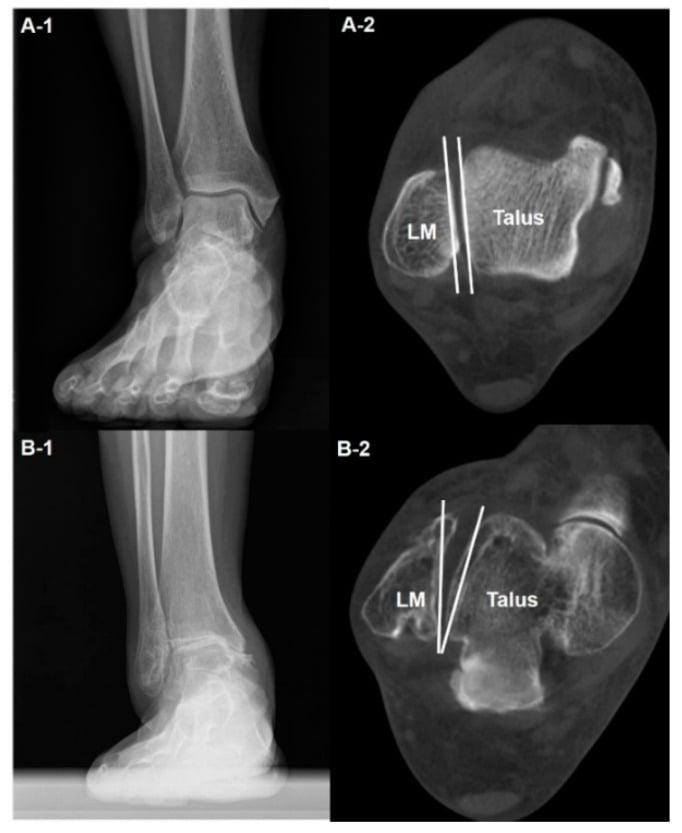
3D Templating Quantifies Syndesmotic Displacement
- In 18 patients, the uninjured ankle was mirrored and superimposed on the injured side to quantify fibular displacement on weight-bearing cone-beam or non-weight-bearing CT.
- Mean mediolateral diastasis was significantly greater than controls in both sprain (1.6 mm) and fracture-associated (1.7 mm) syndesmotic lesions (P less than 0.001).
- Mean fibular external rotation was 4.7 degrees (sprain) and 7.0 degrees (fracture) versus controls (P less than 0.05).
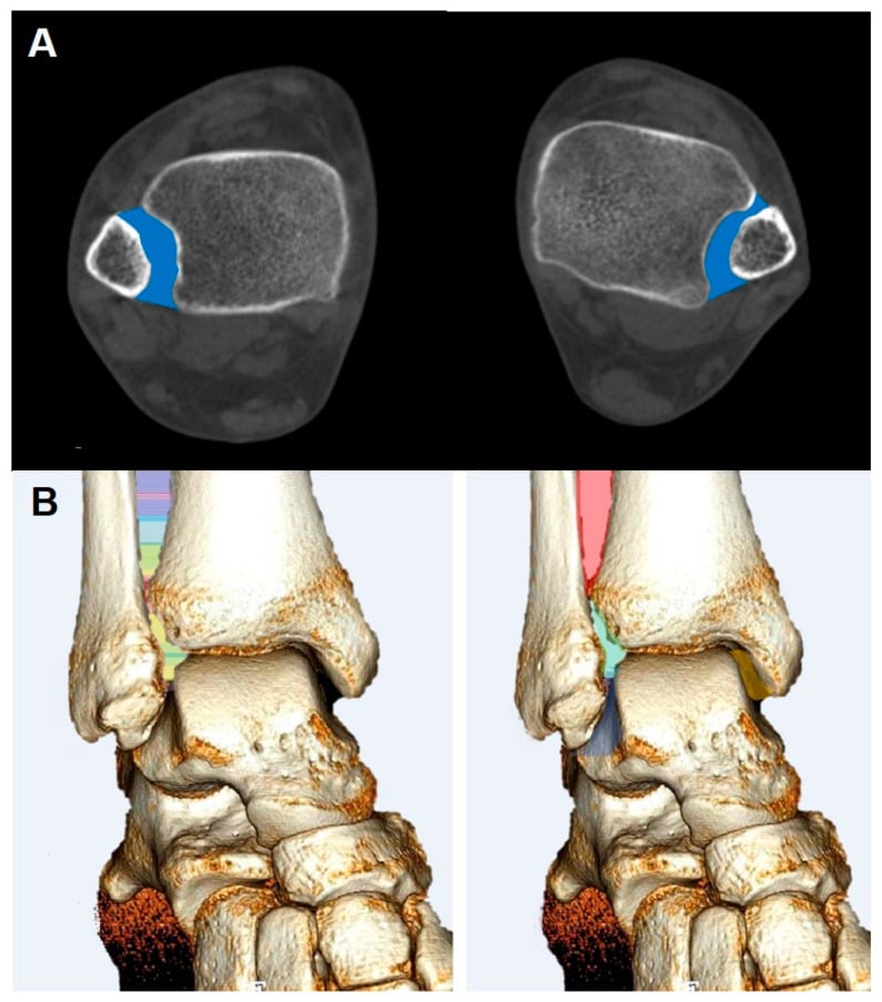
3D Distance Mapping Detects Subtle Syndesmotic Instability
- In 19 matched cadaveric pairs (38 legs) loaded to 356 N, complete syndesmotic sectioning was applied with an intact deltoid and no rotational stress.
- A 3D WBCT distance-mapping algorithm showed relative widening of 16.9% at 1 cm and 11.3% at 3 cm proximal to the plafond (both significant), greatest anteriorly.
- Diagnostic accuracy peaked in the first 1 to 3 cm of the incisura (AUC 80.9% to 83.0%), detecting widening thresholds as small as 0.43 mm.