Lisfranc Injury (Tarsometatarsal Complex)
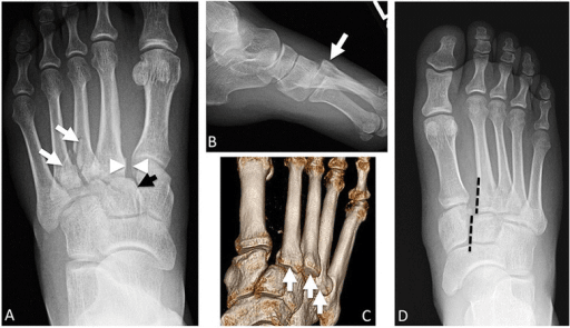
Weight-bearing AP radiograph of the foot demonstrating a Lisfranc injury. There is loss of alignment between the medial border of the 2nd metatarsal and the medial border of the middle cuneiform (disrupted 2nd TMT joint). There is widening between the 1st and 2nd metatarsal bases (>2mm). A fleck sign is visible at the base of the 2nd metatarsal representing an avulsion of the Lisfranc ligament.
Image source: Open Access medical literature (NIH/PubMed Central) • CC-BY License
Questions
Describe the anatomy and importance of the Lisfranc ligament complex.
How do you diagnose a Lisfranc injury?
Classify Lisfranc injuries and describe the treatment algorithm.
Describe the surgical technique for Lisfranc ORIF.
When would you consider primary arthrodesis?
What are the outcomes and complications?
Must Mention
- •Lisfranc ligament: medial cuneiform → 2nd MT base
- •NO ligament between 1st and 2nd MT = weak point
- •2nd MT recessed = keystone of arch
- •Medial 2nd MT aligns with medial middle cuneiform
- •Fleck sign = Lisfranc ligament avulsion
- •Plantar ecchymosis = pathognomonic
Common Pitfalls
- •Missing on non-weight-bearing films
- •Not recognizing plantar ecchymosis
- •Wrong reduction sequence
- •ORIF for purely ligamentous
- •Missing associated injuries
- •Not checking compartments