foot ankle
Talus Fractures (Hawkins Classification)
advanced
6 min
28 marks
6 questions
Clinical Scenario
A 28-year-old motorcyclist is brought to the emergency department following a high-speed collision. He has a deformed right foot and ankle with the foot held in equinus. He is unable to move his toes and has diminished sensation in the first web space. Radiographs reveal a displaced fracture through the talar neck with posterior subluxation of the talar body. The tibiotalar joint appears intact. CT scan confirms a Hawkins Type II fracture with additional comminution.
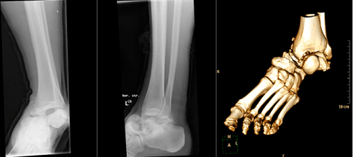
AP and lateral radiographs demonstrating a Hawkins Type II talar neck fracture. The lateral view shows the displaced fracture through the talar neck with subtalar joint subluxation (body displaced posteriorly). On the AP view, the ankle mortise appears intact but there is incongruity of the subtalar joint. This fracture pattern is at high risk of avascular necrosis (40-50%).
Source: Talar Dislocation Fractures Radiograph • PMC4643447 • CC-BY
Questions
Question 1 (4 marks)
Describe the blood supply and importance for talus fractures.
Question 2 (5 marks)
Classify talar neck fractures and describe the AVN risk.
Question 3 (6 marks)
Describe the emergency management and surgical approach.
Question 4 (5 marks)
What is the Hawkins sign and how do you manage AVN?
Question 5 (4 marks)
Discuss other talus fracture patterns.
Question 6 (4 marks)
What are the outcomes and complications?
Exam Day Cheat Sheet
Must Mention
- •Hawkins: I (0-10%), II (40-50%), III (90%), IV (100%) AVN
- •Hawkins sign = subchondral lucency = GOOD (revascularization)
- •Hawkins sign appears at 6-8 weeks
- •Blood supply: tarsal canal, sinus tarsi, deltoid branches
- •Varus malunion = most common malreduction
- •Emergency: reduce dislocated fractures
Common Pitfalls
- •Confusing Hawkins sign (positive is good)
- •Wrong AVN percentages
- •Not reducing emergently
- •Missing varus malreduction
- •Not knowing blood supply
- •Missing lateral process (snowboarder)