Scaphoid Fracture
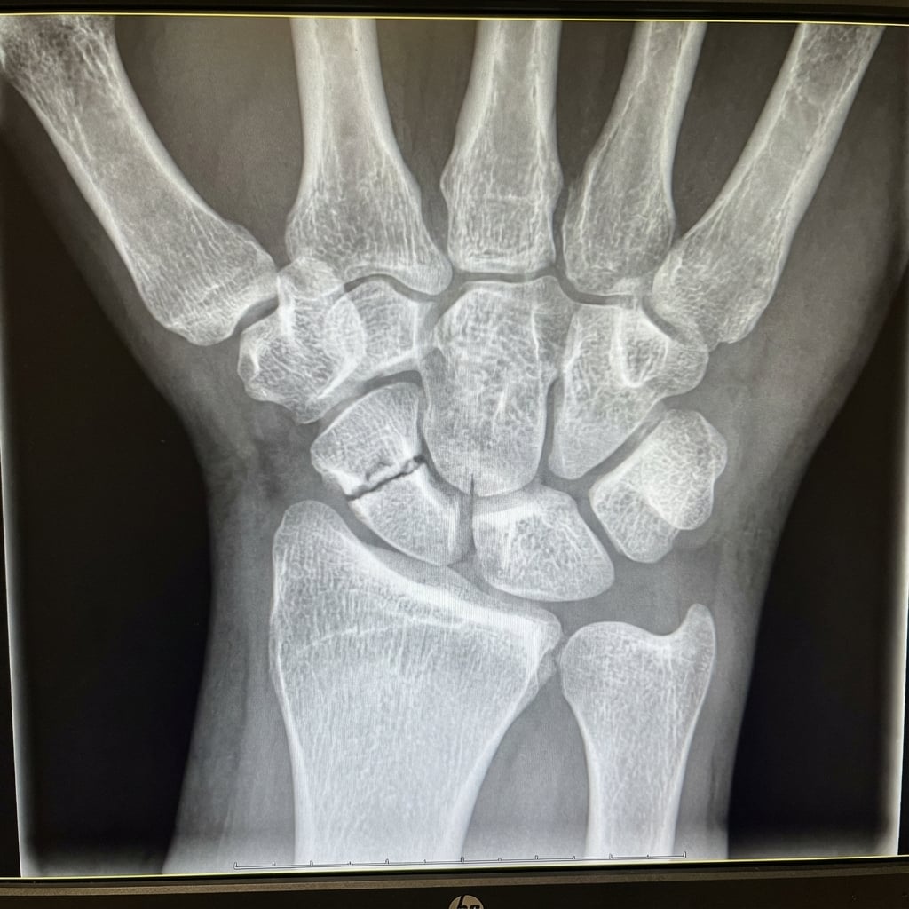
PA wrist radiograph with scaphoid (ulnar deviation) view demonstrating a displaced scaphoid waist fracture. There is >1mm displacement and >15° humpback deformity. The fracture line is clearly visible across the waist. No secondary arthritis is present. This fracture pattern has increased non-union risk and requires surgical fixation with headless compression screw.
Source: Educational radiograph of a displaced scaphoid waist fracture • OrthoVellum Medical Education Team • OrthoVellum Educational Use
Questions
Describe the anatomy of the scaphoid including blood supply and implications.
Classify scaphoid fractures and describe imaging requirements.
What is the treatment algorithm for acute scaphoid fractures?
Describe surgical technique for headless screw fixation.
What are the risk factors for non-union and how do you manage it?
What is SNAC wrist and how is it managed?
Must Mention
- •Blood supply: distal to proximal (dorsal 80%, volar 20%)
- •Proximal pole highest AVN risk
- •Displacement >1mm = surgery
- •Herbert classification (A stable, B unstable)
- •Waist 70%, proximal 20%, distal 10%
- •SNAC stages I-IV with treatment
Common Pitfalls
- •Wrong blood supply direction
- •Missing displacement threshold
- •Wrong Herbert types
- •Not knowing SNAC stages
- •Missing vascularized graft
- •Confusing SNAC/SLAC