TFCC Injuries (Triangular Fibrocartilage Complex)
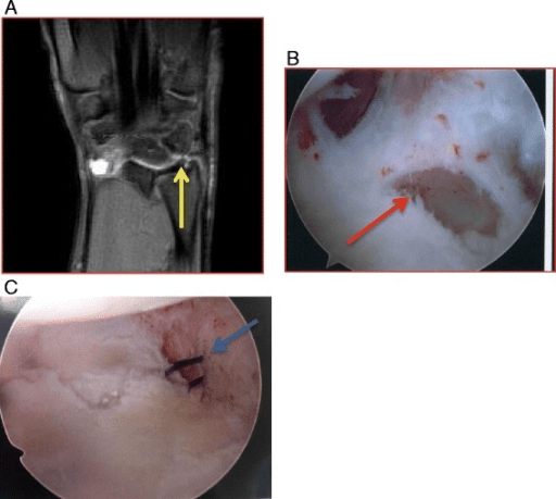
Coronal MRI demonstrating a peripheral TFCC tear (Palmer 1B) with high signal within the triangular fibrocartilage near the ulnar attachment. The arthroscopic image shows the corresponding tear at the fovea attachment with a positive trampoline test (loss of bounce). Peripheral tears (1B) are repairable with good healing potential, while central tears (1A) are debrided.
Source: TFCC Tear MRI (Sports Wrist Injuries Review) • PMC5025579 • CC-BY
Questions
Describe the anatomy of the TFCC and its function.
How do you clinically assess TFCC injuries?
Describe the Palmer classification and its treatment implications.
What is the arthroscopic technique for TFCC repair?
Discuss ulnar impaction syndrome and its relationship to TFCC.
What are the outcomes and complications of TFCC surgery?
Must Mention
- •TFCC = TFC + radioulnar ligaments + meniscus homologue
- •Peripheral 10-40% = vascularized (repair)
- •Central = avascular (debride)
- •1A central, 1B peripheral, 1C ulnocarpal, 1D radial
- •Fovea test = deep DRUJ stabilizer
- •Trampoline test = arthroscopic
Common Pitfalls
- •Wrong treatment by type
- •Missing TFCC components
- •Wrong vascularity
- •Missing fovea test
- •Wrong repair technique
- •Missing Class 2