Trigger Finger (Stenosing Tenosynovitis)
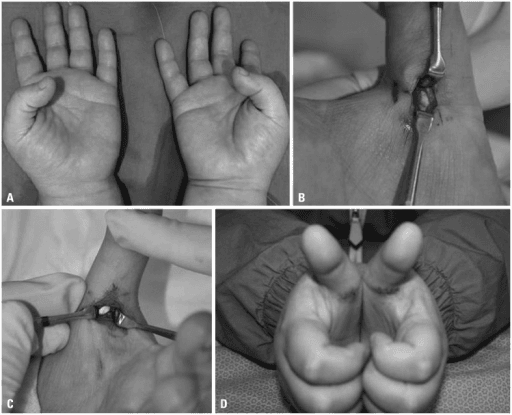
Clinical photograph of the right ring finger demonstrating locked trigger finger - the finger is flexed at the PIP and cannot actively extend. Passive extension is possible with a palpable click. The anatomical diagram shows the A1 pulley location at the metacarpal head and its relationship to the flexor tendons. Thickening of the A1 pulley and tendon nodule formation cause the triggering phenomenon.
Source: Trigger Finger Clinical Photograph • PMC4479852 • CC-BY
Questions
Describe the anatomy of the pulley system and pathophysiology of trigger finger.
What are the clinical features, classification, and associations?
Describe the treatment options and their indications.
What is the surgical technique for A1 pulley release?
Discuss trigger thumb in adults and children.
What are the complications and special considerations?
Must Mention
- •A1 pulley at MCP level
- •Preserve A2 pulley (bowstringing)
- •Quinnell grading I-IV
- •Injection success 60-70%
- •Diabetes = multiple digits, lower injection success
- •Thumb = protect radial digital nerve
Common Pitfalls
- •Damaging A2 pulley
- •Missing radial digital nerve
- •Wrong injection site
- •Missing diabetes association
- •Wrong grading system
- •Missing pediatric trigger thumb