Cauda Equina Syndrome
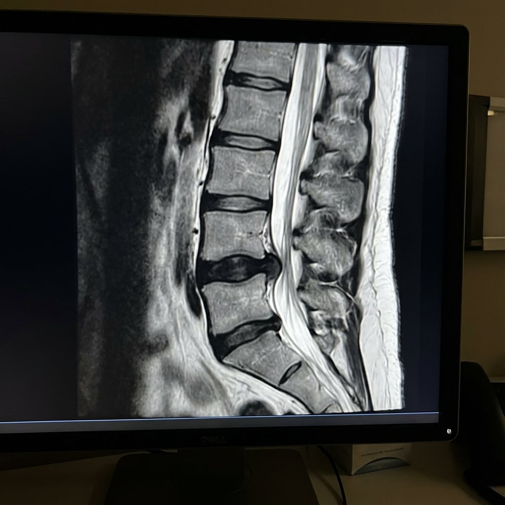
Sagittal T2-weighted MRI demonstrating large L4-5 disc herniation with severe canal stenosis. Axial image shows the disc occupying >50% of the canal with compression of the cauda equina nerve roots. There is obliteration of CSF signal around the roots. This represents cauda equina syndrome requiring emergency surgical decompression.
Image source: Open Access medical literature (NIH/PubMed Central) • CC-BY License
Questions
Describe the MRI findings and define cauda equina syndrome.
What are the clinical features and how do you classify the severity?
What is the optimal timing of surgery and what does the evidence show?
Describe your surgical technique for emergency decompression.
What are the expected outcomes and prognostic factors?
What documentation and consent issues are specific to this condition?
Must Mention
- •CES-I (incomplete) vs CES-R (retention) classification
- •Bladder dysfunction is the DEFINING feature
- •Post-void residual >200mL significant
- •Operate ASAP - earlier is better
- •CES-I → CES-R conversion = worse outcome
- •Prognosis depends on pre-op bladder status
- •Document everything (medicolegal)
Common Pitfalls
- •Missing the diagnosis (not checking red flags)
- •Not measuring PVR
- •Delaying surgery
- •Poor documentation
- •Not discussing prognosis realistically
- •Back pain alone is NOT CES