Cervical Facet Dislocation
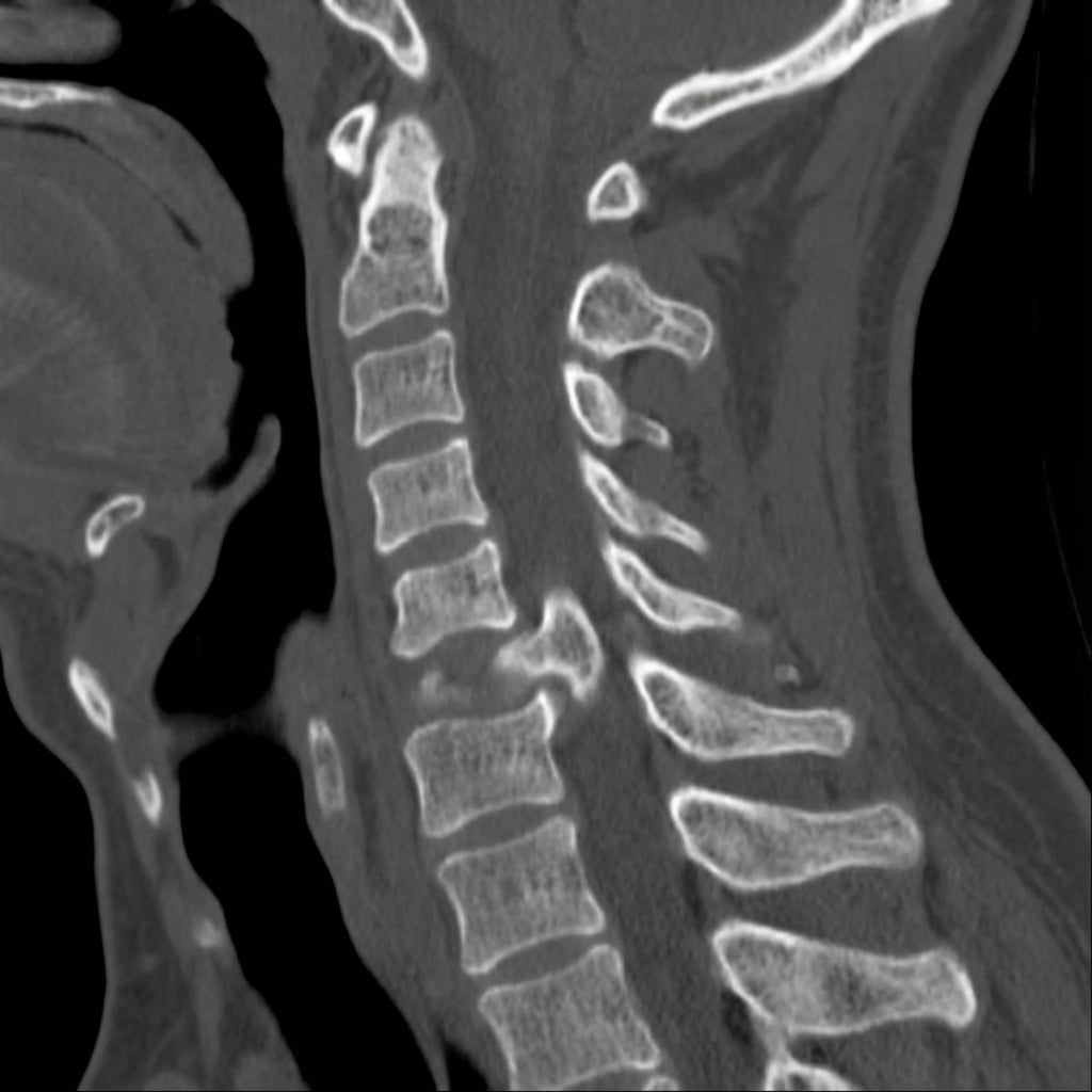
Sagittal and axial CT images demonstrating unilateral left facet dislocation at C6-7. The left C6 inferior articular process is locked anterior to the C7 superior articular process (jumped facet). There is approximately 25% anterior translation of C6 on C7 with rotation. The right facet is subluxed but not locked. This represents unilateral facet dislocation with associated radiculopathy pattern.
Image source: Open Access medical literature (NIH/PubMed Central) • CC-BY License
Questions
Describe the CT findings and classify this injury.
Differentiate unilateral from bilateral facet dislocation including mechanism and presentation.
What is the controversy regarding MRI before reduction and how do you manage this patient?
Describe the techniques for closed and open reduction.
What are your surgical options and preferred approach?
What are the expected outcomes and potential complications?
Must Mention
- •Unilateral = 25-50% translation, radiculopathy common
- •Bilateral = >50% translation, cord injury common
- •Unilateral = flexion-rotation mechanism
- •MRI controversy: awake closed reduction safe, MRI if obtunded
- •Posterior reduction and fusion = standard approach
- •Closed reduction incremental weight with neuro checks
Common Pitfalls
- •Wrong translation percentages
- •MRI always/never
- •Wrong approach
- •No neuro monitoring during reduction
- •Missing disc herniation
- •Inadequate fixation