Cervical Myelopathy
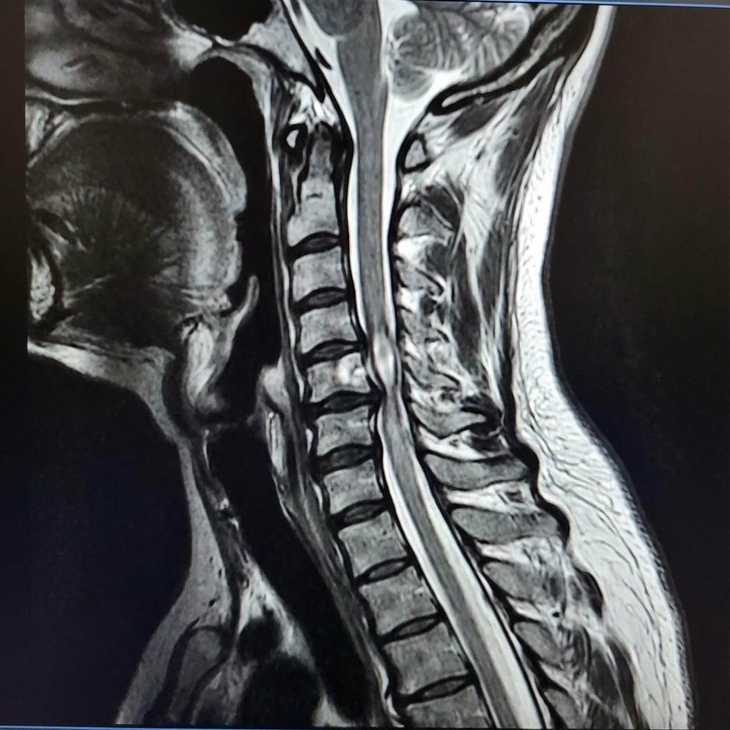
Sagittal T2-weighted MRI demonstrating multi-level cervical stenosis at C3-4, C4-5, and C5-6. There is cord compression with intramedullary T2 hyperintensity at C4-5 level indicating myelomalacia. The cord appears flattened and draped over disc-osteophyte complexes. Ligamentum flavum hypertrophy contributes posteriorly. This represents degenerative cervical myelopathy (DCM) requiring surgical decompression.
Image source: Open Access medical literature (NIH/PubMed Central) • CC-BY License
Questions
Describe the MRI findings and explain the clinical significance of myelomalacia.
What are the clinical features of cervical myelopathy and how do you assess severity?
What factors determine your choice between anterior and posterior surgical approaches?
Describe your technique for anterior cervical decompression and fusion (ACDF).
What are the indications and technique for cervical laminoplasty?
What is the natural history and expected outcomes after surgical treatment?
Must Mention
- •Myelomalacia = T2 cord signal = chronic damage = poorer recovery
- •UMN signs: Hoffman's, hyperreflexia, clonus, Babinski
- •JOA score 17 max (severity: <9 severe, 9-12 moderate, 13-17 mild)
- •Anterior for 1-2 levels, posterior for ≥3 levels
- •K-line negative = posterior less effective
- •Laminoplasty requires lordosis
- •Surgery arrests progression, 50-75% recovery rate
Common Pitfalls
- •Confusing myelopathy (UMN) with radiculopathy (LMN)
- •Laminoplasty in kyphosis (won't work)
- •Ignoring K-line in posterior planning
- •Not assessing dynamic instability
- •Overestimating recovery with myelomalacia