Cervical Radiculopathy
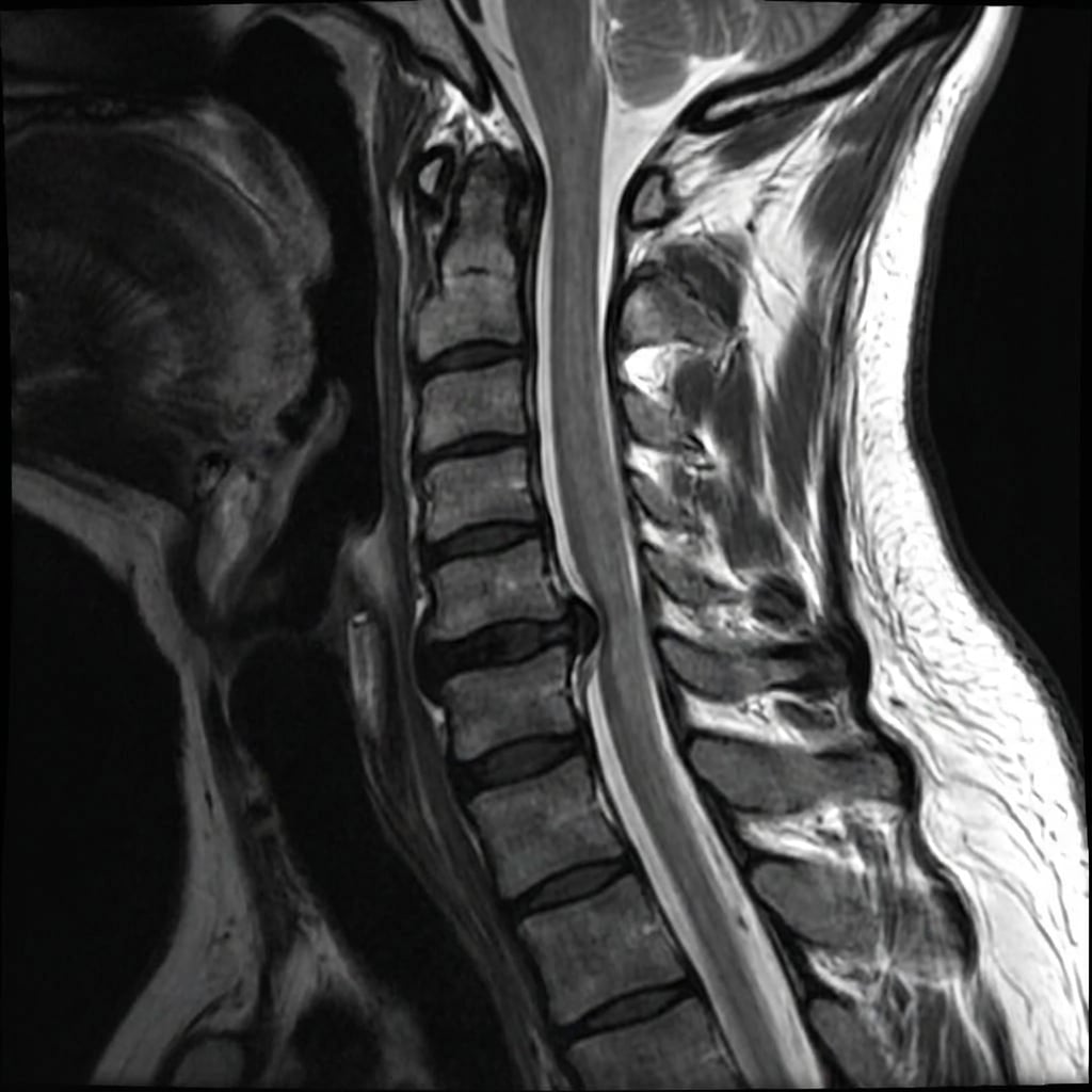
Sagittal and axial T2-weighted MRI demonstrating left posterolateral disc herniation at C6-7 with compression of the left C7 nerve root in the neural foramen. There is loss of disc height and disc signal at C6-7. The cord signal is normal. This represents cervical radiculopathy from disc herniation with concordant clinical findings.
Image source: Open Access medical literature (NIH/PubMed Central) • CC-BY License
Questions
Describe the MRI findings and correlate with the clinical presentation.
What clinical examination findings help localize the level?
What is the natural history and what are the indications for surgery?
Describe the surgical options and your preferred technique.
What are the expected outcomes and potential complications?
How do you differentiate from other causes of arm pain?
Must Mention
- •C7 root exits at C6-7 (above pedicle)
- •Natural history: 75-90% improve conservatively
- •Spurling test: extension + lateral flexion + axial load
- •ACDF = gold standard for anterior pathology
- •Posterior foraminotomy for lateral disc
- •Conservative trial: 6-12 weeks
Common Pitfalls
- •Wrong root level
- •Surgery before conservative trial
- •Not knowing Spurling test
- •Foraminotomy for central disc
- •Missing peripheral entrapment
- •Confusing with myelopathy