Degenerative Lumbar Spondylolisthesis
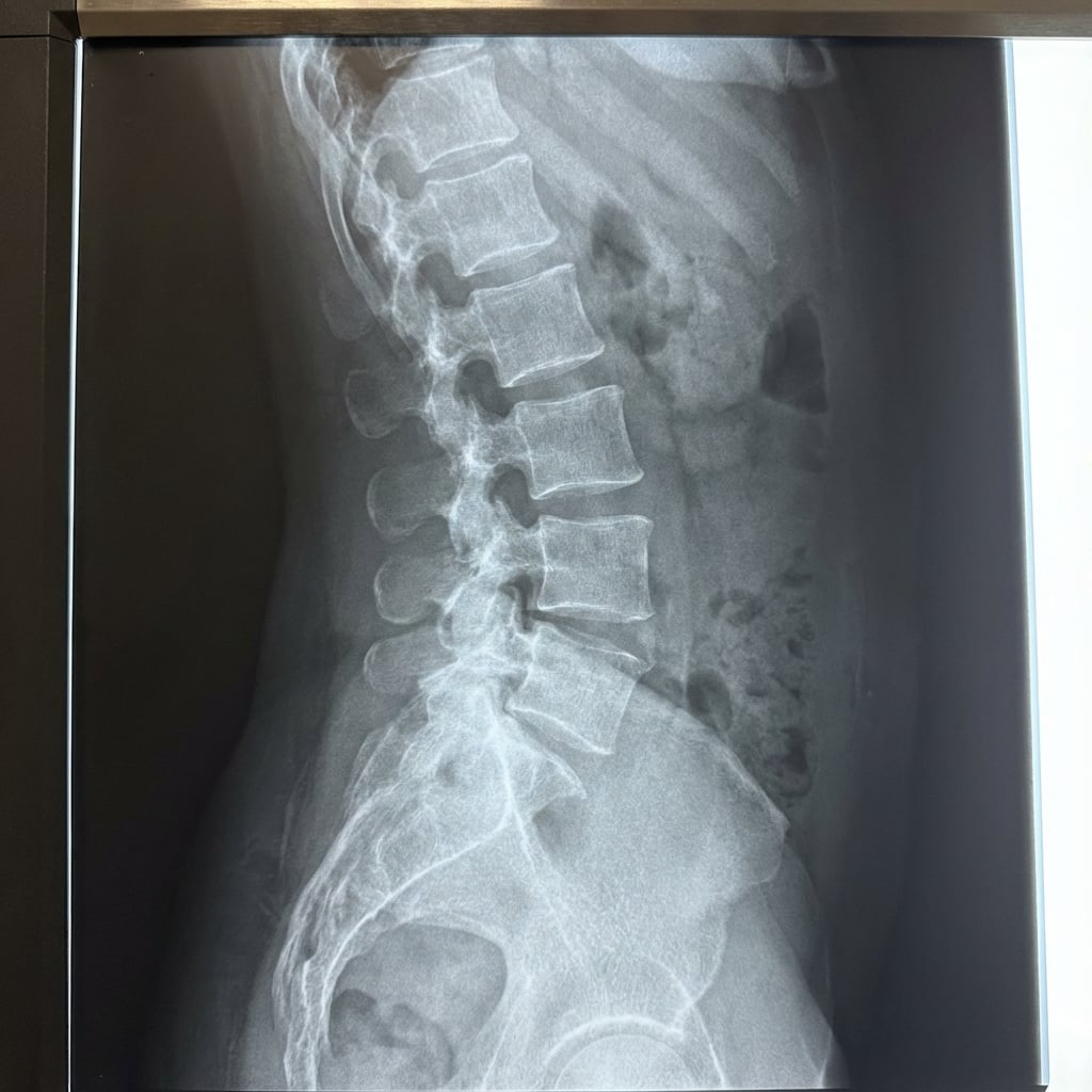
Lateral standing radiograph demonstrating Grade I (Meyerding) anterior spondylolisthesis of L4 on L5. There is disc space narrowing at L4-5 and facet hypertrophy. MRI shows central and lateral recess stenosis at L4-5 with compression of the traversing L5 nerve roots. The pars is intact. This represents degenerative spondylolisthesis causing neurogenic claudication.
Image source: Open Access medical literature (NIH/PubMed Central) • CC-BY License
Questions
Describe the imaging findings and differentiate from isthmic spondylolisthesis.
What is the clinical presentation and how do you differentiate from vascular claudication?
What is the evidence for treatment options?
Describe your surgical technique for decompression and fusion.
What are the indications for interbody fusion and what options exist?
What are the expected outcomes and potential complications?
Must Mention
- •Degenerative = intact pars, L4-5 most common
- •Isthmic = pars defect, L5-S1 most common
- •SPORT trial: surgery superior for degenerative spondylolisthesis
- •Neurogenic claudication: relieved by flexion (shopping cart sign)
- •Vascular claudication: relieved by standing still
- •Decompression + fusion: lower reoperation than decompression alone
Common Pitfalls
- •Confusing degenerative with isthmic
- •Missing vascular claudication
- •Decompression alone with instability
- •Not mentioning SPORT trial
- •Forgetting adjacent segment disease
- •Wrong level (L4-5 not L5-S1)