OrthoVellum
Knowledge Hub

Study

  • Topics
  • MCQs
  • ISAWE
  • Operative Surgery
  • Flashcards

Company

  • About Us
  • Editorial Policy
  • Contact
  • FAQ
  • Blog

Legal

  • Terms of Service
  • Privacy Policy
  • Cookie Policy
  • Medical Disclaimer
  • Copyright & DMCA
  • Refund Policy

Support

  • Help Center
  • Accessibility
  • Report an Issue
OrthoVellum

© 2026 OrthoVellum. For educational purposes only.

Not affiliated with the Royal Australasian College of Surgeons.

Back to ISAWE Scenarios
Contents
0%
spine

Metastatic Spinal Cord Compression

advanced
6 min
28 marks
6 questions
Clinical Scenario
A 67-year-old man with known prostate cancer presents with 6 weeks of progressive back pain and 5 days of increasing leg weakness. He is now unable to walk unaided. On examination, he has 3/5 power in both legs, brisk reflexes, and a sensory level at T10. Bowel and bladder function are currently normal.
Sagittal T2-weighted MRI demonstrating T8 vertebral body collapse with posterior cortex retropulsion causing spinal cord compression. There is high signal within the cord at this level indicating edema/myelopathy. The lesion replaces normal bone marrow signal and there is an associated soft tissue mass extending epidurally. Multiple other vertebral lesions are visible. This represents metastatic spinal cord compression (MSCC) requiring urgent assessment and treatment.
Open Full Size

Sagittal T2-weighted MRI demonstrating T8 vertebral body collapse with posterior cortex retropulsion causing spinal cord compression. There is high signal within the cord at this level indicating edema/myelopathy. The lesion replaces normal bone marrow signal and there is an associated soft tissue mass extending epidurally. Multiple other vertebral lesions are visible. This represents metastatic spinal cord compression (MSCC) requiring urgent assessment and treatment.

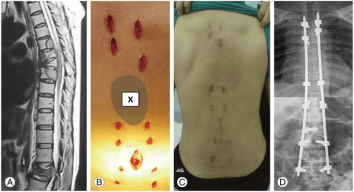
Source: Minimally Invasive Spinal Stabilization in Patients with Spinal Metastasis • PMC4764548 • CC-BY

Questions

Question 1 (4 marks)

Describe the MRI findings and explain the urgency of this condition.

Question 2 (5 marks)

What scoring systems are used to guide treatment decisions?

Question 3 (6 marks)

What are the treatment options and how do you decide between them?

Question 4 (5 marks)

Describe your surgical technique for thoracic decompression and stabilization.

Question 5 (4 marks)

What is the role of radiotherapy and when is it used alone?

Question 6 (4 marks)

What are the expected outcomes and prognostic factors?

Exam Day Cheat Sheet

Must Mention

  • •MSCC = emergency - dexamethasone 16mg immediately
  • •Tokuhashi (survival), Tomita (surgical goal), SINS (stability)
  • •Ambulatory status = best predictor of outcome
  • •Surgery + RT > RT alone (Patchell) if fit and >3 months survival
  • •Radiosensitivity guides treatment (myeloma = RT, renal = surgery)
  • •Separation surgery + SBRT for radioresistant tumors

Common Pitfalls

  • •Delayed steroids
  • •Not calculating scores
  • •Surgery in poor prognosis patients
  • •RT for radioresistant tumors
  • •Missing instability
  • •Not involving MDT
Scenario Info
Answers Revealed0/6
Difficulty
advanced
Time Allowed6 min
Total Marks28
Questions6
Related Scenarios

Acute Spinal Cord Injury

spine•28 marks

Adolescent Idiopathic Scoliosis

spine•28 marks

Atlas Fracture (C1 Fracture)

spine•28 marks

Cauda Equina Syndrome

spine•28 marks

Central Cord Syndrome

spine•28 marks