Odontoid Fracture in the Elderly
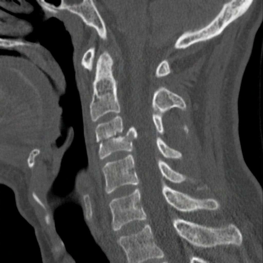
Sagittal and coronal CT images demonstrating Type II odontoid fracture at the base of the dens with 4mm posterior displacement of the dens relative to the C2 body. The fracture line is at the junction of the odontoid process and C2 body - the watershed zone for blood supply. This represents a Type II odontoid fracture in an elderly patient with high non-union risk requiring surgical stabilization.
Image source: Open Access medical literature (NIH/PubMed Central) • CC-BY License
Questions
Describe the CT findings and classify this fracture.
Why are elderly patients with Type II fractures particularly challenging?
What are the treatment options and how do you decide between them?
Describe the surgical technique for C1-C2 posterior fusion.
When is anterior odontoid screw fixation appropriate and what is the technique?
What are the outcomes and complications of treatment?
Must Mention
- •Type II = base of dens = highest non-union (40-60%)
- •Elderly Type II = surgery generally preferred
- •Halo contraindicated in elderly
- •Collar complications: dysphagia, skin, deconditioning
- •Anterior screw: horizontal fracture, reducible, intact TL
- •Posterior fusion: Goel-Harms (C1 lateral mass, C2 pedicle)
- •Reverse anticoagulation before surgery
Common Pitfalls
- •Collar for elderly Type II (high non-union)
- •Halo in elderly (high complications)
- •Anterior screw with reverse oblique fracture
- •Missing transverse ligament injury
- •Not reversing anticoagulation
- •Not planning for vertebral artery