Acetabular Fracture
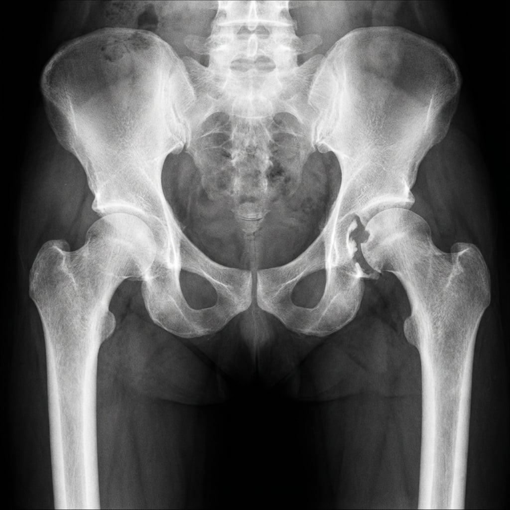
AP pelvis showing posterior wall acetabular fracture with associated posterior hip dislocation. The femoral head is displaced posteriorly with loss of Shenton's line. Obturator oblique view demonstrates posterior wall fragment size. CT shows >40% wall involvement with marginal impaction.
Image source: Open Access medical literature (NIH/PubMed Central) • CC-BY License
Questions
Describe the clinical picture and your immediate priorities in the first 6 hours.
Describe the Letournel classification and classify this injury.
What imaging would you obtain and what are the indications for operative management?
Describe the surgical approaches for acetabular fractures and their indications.
What are the complications and prognostic factors for acetabular fractures?
Describe the concept of secondary congruence and its clinical significance.
Must Mention
- •Reduce posterior dislocation within 6 HOURS (AVN risk)
- •Document sciatic nerve BEFORE and AFTER reduction (10-20% injury)
- •Letournel: 5 elementary + 5 associated = 10 types
- •Spur sign = pathognomonic for both-column fracture
- •Posterior wall: Kocher-Langenbeck
- •Anterior column/both-column: Ilioinguinal or Modified Stoppa
- •Anatomic reduction (<1-2mm) = most important prognostic factor
Common Pitfalls
- •Delayed reduction of dislocation (>6 hours)
- •Not documenting sciatic nerve pre/post reduction
- •Missing marginal impaction on CT (important prognostic factor)
- •Wrong surgical approach for fracture pattern
- •Operating too late (>3 weeks - difficult reduction)
- •Missing associated femoral head injury (Pipkin)