Distal Femur Fracture
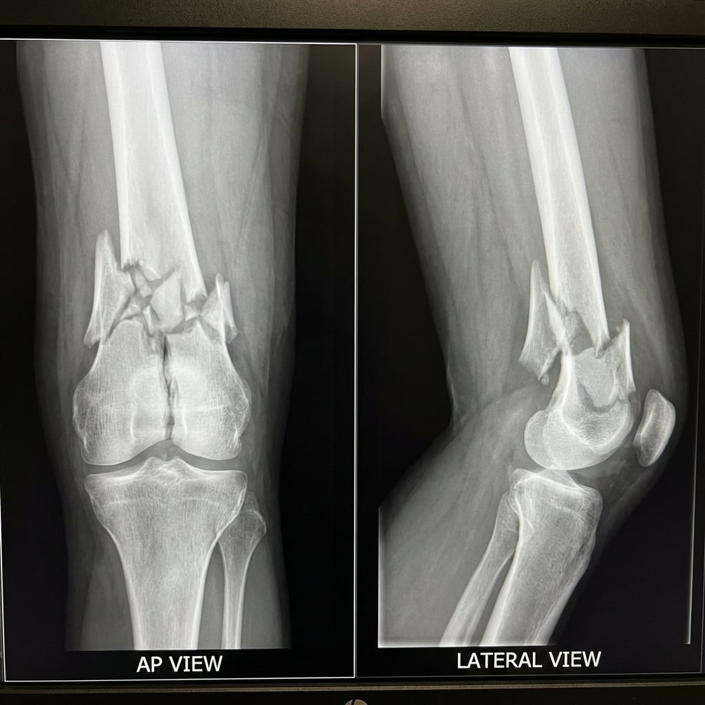
AP and lateral radiographs demonstrating comminuted supracondylar femur fracture with intra-articular extension (AO 33-C2). There is metaphyseal comminution, varus angulation, and shortening. The articular surface shows a simple sagittal split pattern requiring anatomic reduction.
Image source: Open Access medical literature (NIH/PubMed Central) • CC-BY License
Questions
How do you classify this fracture? What key features guide your management?
What are the surgical options and which approach would you use?
Describe your surgical technique for ORIF with lateral locked plate.
How do you manage fixation in osteoporotic bone?
This patient had a TKA. How would a periprosthetic fracture change your classification and management?
What are the expected outcomes and complications?
Must Mention
- •AO: 33-A (extra-articular), 33-B (partial), 33-C (complete articular)
- •Articular reduction = anatomic; Metaphyseal comminution = bridge
- •All locking screws in osteoporosis
- •Plate length: 2:1 working length ratio
- •Rorabeck: I = brace, II = ORIF, III = revision
- •Unicortical distal screws near prosthesis
Common Pitfalls
- •Reducing comminuted metaphysis (strips blood supply, no benefit)
- •Short plate in osteoporosis (fixation failure)
- •Missing rotation (check lesser trochanter profile)
- •Bicortical screws through prosthesis
- •Not checking prosthesis stability before fixation decision
- •Ignoring bone quality in elderly