Humeral Shaft Fracture with Radial Nerve Palsy
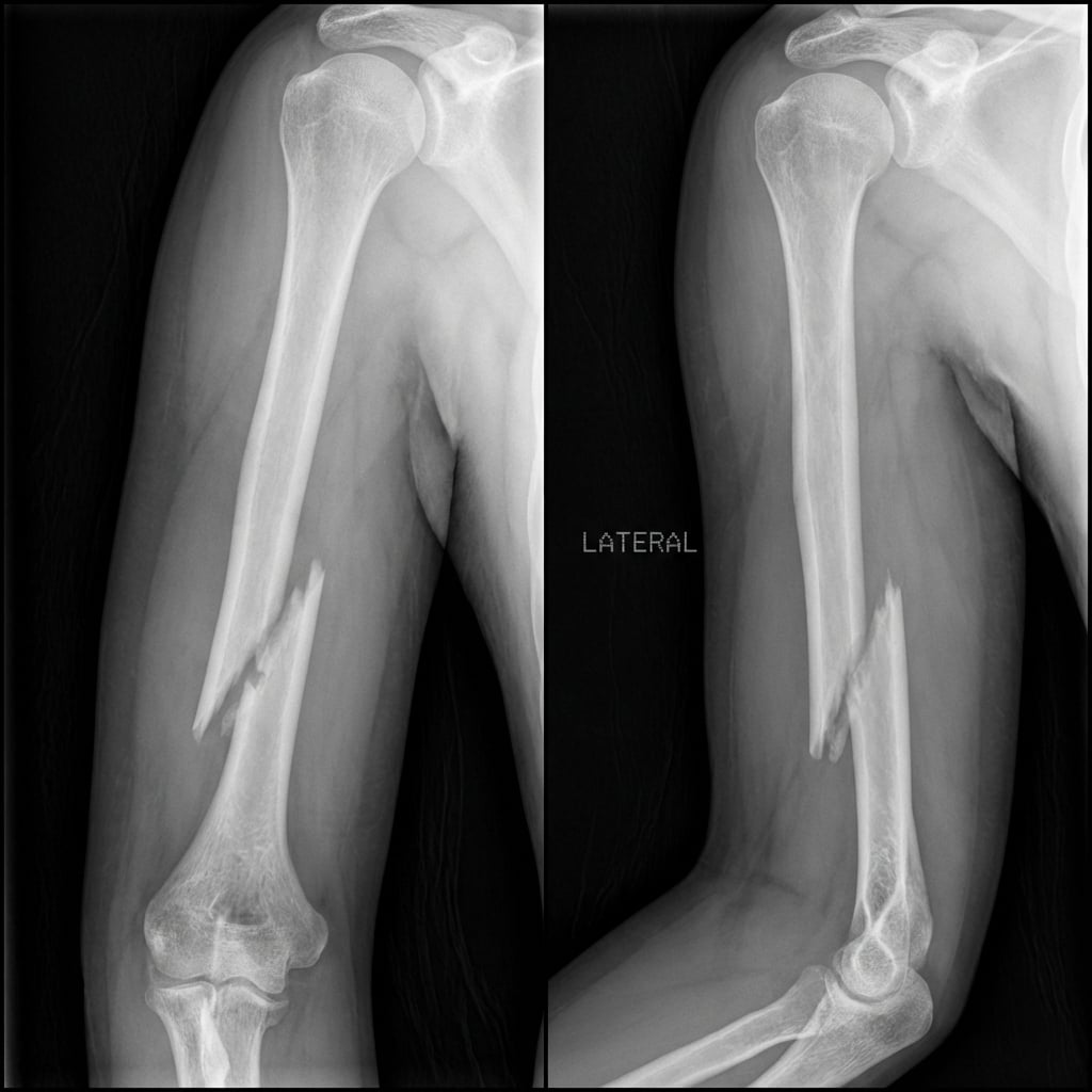
AP and lateral radiographs demonstrating spiral fracture of the distal third humeral shaft (Holstein-Lewis pattern) with shortening and angulation. The radial nerve crosses the posterior humerus at the junction of middle and distal thirds where it is vulnerable to injury.
Image source: Open Access medical literature (NIH/PubMed Central) • CC-BY License
Questions
Describe the radiographic findings and explain the anatomy that makes radial nerve injury likely.
What is the incidence and prognosis of radial nerve palsy with humeral shaft fractures?
What is your initial management strategy for this patient?
The patient has no recovery at 4 months and EMG shows no reinnervation. What are your options?
Describe your surgical technique for fixation with nerve exploration.
If nerve recovery fails, what tendon transfers are available for radial nerve palsy?
Must Mention
- •Radial nerve palsy: 7-17% (up to 22% in Holstein-Lewis)
- •70-90% spontaneous recovery (most are neurapraxia)
- •Functional bracing (Sarmiento): 90%+ union rate
- •EMG at 3 weeks (baseline), 3 months (recovery assessment)
- •Explore if: secondary palsy, no recovery at 3-4 months
- •Tendon transfers: PT-ECRB, FCR-EDC, PL-EPL
Common Pitfalls
- •Exploring primary palsy immediately (90% recover spontaneously)
- •Not documenting nerve status before manipulation
- •Missing secondary palsy after reduction (needs urgent exploration)
- •Not using Sarmiento functional bracing (90%+ union)
- •Waiting too long for tendon transfers (>12-18 months = motor endplate degeneration)
- •Not knowing standard triple transfer: PT-ECRB, FCR-EDC, PL-EPL