Morel-Lavallée Lesion
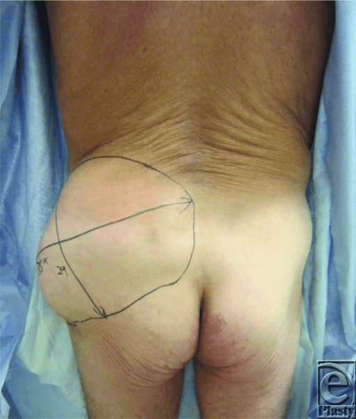
MRI T2-weighted image demonstrating large fluid collection between subcutaneous tissue and fascia over the lateral thigh. The collection shows heterogeneous signal indicating hemolymphatic contents. Clinical photograph shows fluctuant swelling with skin degloving pattern. This represents a significant soft tissue injury requiring careful management prior to any fracture surgery.
Source: Morel-Lavallée Lesion: Closed Degloving Injury • PMC4005421 • CC-BY
Questions
What is the diagnosis and describe the pathophysiology of this injury.
What is the clinical and radiological assessment of this condition?
What are the management options and how do you select the appropriate approach?
Describe your surgical technique for debridement of this lesion.
What are the complications and how do they affect fracture management?
How do you manage this lesion when associated with acetabular or pelvic fractures requiring ORIF?
Must Mention
- •Morel-Lavallée = closed internal degloving
- •Hemolymphatic fluid between subcutaneous tissue and fascia
- •~50% culture positive
- •Common over greater trochanter
- •MRI best for extent
- •Dead space management critical (quilting sutures)
- •Address before overlapping fracture surgery
Common Pitfalls
- •Missing diagnosis (presents as bruising)
- •Operating through lesion without debridement
- •Not sending cultures
- •Inadequate dead space closure
- •Not delaying surgery when indicated
- •Recurrent lesion after aspiration alone