Gold-standard posterior approach · sciatic nerve at risk · lateral or prone
- Sciatic nerve is at significant risk - identify, protect and monitor it throughout surgery
- Muscle-splitting, not a true internervous plane - the gluteus maximus is split in line with its fibres; the whole muscle is inferior gluteal nerve territory
- Both gluteal nerves arise from the sacral plexus, NOT from the sciatic nerve (classic exam trap): superior gluteal L4-S1 and inferior gluteal L5-S2
- Short external rotators must be released and tagged for repair - piriformis and the conjoint tendon (gemelli + obturator internus); preserve the quadratus femoris to protect the medial femoral circumflex artery
- Trochanteric osteotomy is optional - it improves superior/dome exposure but adds morbidity and heterotopic-ossification risk, so use it selectively
When & Why
What it exposes. The Kocher-Langenbeck is the consensus posterior approach to the acetabulum, giving direct access to the posterior wall, the posterior column from the greater sciatic notch to the ischial tuberosity, and the retroacetabular surface of the innominate bone. It is the workhorse exposure for posterior wall fractures (the commonest acetabular pattern, about 40 percent of acetabular fractures), posterior column fractures, and the posterior component of transverse and T-type fractures. Why posterior. Posterior wall and posterior column injuries are not reachable from the anterior (ilioinguinal/Stoppa) approaches, which cannot visualise the retroacetabular surface or control the sciatic nerve. The Kocher-Langenbeck also allows the hip to be taken through a range of motion to test stability and gives simultaneous access to the femoral head when an associated dislocation is reduced. Position. Either lateral or prone is acceptable - position does not change the iatrogenic nerve-palsy rate (Schaffer 2024). Prone aids reduction of transverse and T-type patterns; lateral allows hip flexion to relax the sciatic nerve. Image intensification must be free to obtain Judet views.
- What is visible
- Main indication - the commonest acetabular fracture pattern
- Typical fixation
- Lag screws plus buttress/spring plate under direct vision
- What is visible
- Entire column from the sciatic notch to the ischium
- Typical fixation
- Contoured reconstruction plate
- What is visible
- Posterior component of the fracture
- Typical fixation
- Reduction and fixation from posterior
Timing and referral. Reduce an associated hip dislocation urgently - delay greater than 12 hours worsens outcome and raises the risk of femoral-head osteonecrosis (Moed 2002). Definitive fixation is generally undertaken within about 5 to 10 days once the patient is optimised, balancing soft-tissue recovery against the increasing difficulty of late reduction. Displaced acetabular fractures are best managed in high-volume pelvic and acetabular units; outcomes correlate with surgeon and centre volume, and the iatrogenic sciatic-palsy rate is surgeon-dependent (Schaffer 2024) - a strong argument for centralised care consistent with major-trauma network pathways.
The Exposure
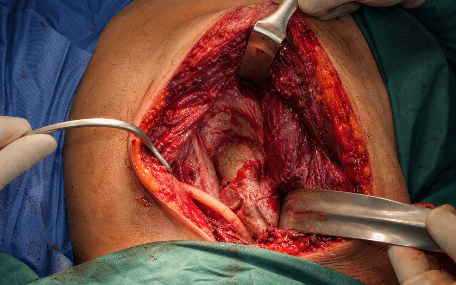
Work from skin to capsule through a single posterior incision. The whole point of the approach is to reach the posterior wall and column while keeping the sciatic nerve identified, relaxed and protected at every step.
Dissection sequence
- Place the patient lateral or prone (both acceptable; position does not change nerve-palsy risk). Prone helps reduction of transverse/T-type patterns; lateral lets you flex the hip to relax the sciatic nerve.
- Mark a curved incision centred on the greater trochanter, extending proximally over the buttock and distally along the femoral shaft. It can be extended proximally toward the posterior superior iliac spine or distally along the thigh when more exposure is needed.
- Deepen through subcutaneous fat to expose the fascia lata over the lateral aspect of the thigh.
- Divide the fascia lata longitudinally in line with the skin incision to expose the underlying gluteus maximus and the vastus lateralis.
- Split the gluteus maximus in the line of its oblique fibres (superolateral to inferomedial) by blunt dissection.
- This is a muscle-splitting move, not a true internervous plane - the entire muscle is supplied by the inferior gluteal nerve, which enters its deep surface medially.
- Keep the split in the mid-substance and avoid deep medial dissection so neither half of the muscle is denervated; avoid superior extension to protect the superior gluteal neurovascular bundle running above piriformis.
- Locate the sciatic nerve as it exits the pelvis below piriformis (in about 90 percent of patients) and runs down on the short external rotators.
- Keep the hip and knee flexed to put slack in the nerve, and gently develop the plane between the nerve and the underlying rotators. Identifying the nerve here, proximal to piriformis, is the single most important protective step.
- Release piriformis first, then the conjoint tendon (superior gemellus, obturator internus, inferior gemellus) from their insertions on the greater trochanter, tagging each with a stay suture for later repair.
- Reflect the released tendons posteriorly over the hip - they carry and shield the sciatic nerve away from the operative field.
- Preserve the quadratus femoris and its blood supply (the medial femoral circumflex artery) to protect femoral-head vascularity.
- With the rotators and nerve retracted and protected, incise the posterior capsule to expose the posterior wall, the retroacetabular surface and the femoral head.
- The greater and lesser sciatic notches can now be palpated, giving orientation to the posterior column and the quadrilateral surface.
- Sweep subperiosteally along the posterior column from the greater sciatic notch to the ischial tuberosity, placing retractors gently and intermittently to avoid pressure on the sciatic nerve.
- A trochanteric osteotomy (or trochanteric flip) may be performed here to gain superior/dome access, but it adds morbidity and heterotopic-ossification risk - use it selectively.
- Reduce and fix the fracture, confirm stability and hardware position on Judet views, then repair the capsule and re-attach the short external rotators to the greater trochanter through bone tunnels or to suture anchors with heavy non-absorbable suture.
- Close the gluteus maximus fascia and fascia lata, approximate Scarpa's fascia and skin, and leave a deep subfascial drain.
Distinguish a pre-operative (injury-related) palsy - present from the fracture-dislocation itself in up to about 20 percent of cases - from an iatrogenic palsy, which is around 3 percent in modern high-volume series and was historically reported as high as 10 to 16 percent. Protect the nerve by identifying it proximal to piriformis before releasing any rotator, keeping the hip and knee flexed to relax it, releasing retractors every 15 minutes, placing retractors deep to the nerve, using SSEP/EMG monitoring where available, and repairing the short external rotators at closure to interpose tissue between the nerve and any posterior hardware. The common peroneal division is more vulnerable than the tibial division (it is lateral, more tethered, and has fewer protective connective-tissue septa), which is why foot drop dominates the clinical picture.
Q: What is the internervous plane? The approach splits the gluteus maximus in line with its fibres and works between the territories of the superior gluteal nerve (L4-S1) and the inferior gluteal nerve (L5-S2). The crucial trap: both gluteal nerves arise directly from the sacral plexus, NOT from the sciatic nerve. The inferior gluteal nerve supplies the whole of gluteus maximus and enters its deep surface medially, so the split is kept in the muscle's mid-substance to avoid denervating either half. This is a muscle-splitting approach rather than a true internervous plane.
Dangers & Extensions
Structures at risk and complications
- Incidence
- ~3% modern series (historically up to 10-16%)
- Prevention / management
- Identify early, hip and knee flexion, gentle intermittent retraction, monitor SSEP/EMG, repair external rotators
- Incidence
- 2-5%
- Prevention / management
- Keep the gluteus maximus split inferior/mid-substance; avoid superior extension
- Incidence
- Any grade common; clinically significant (Brooker III-IV) ~10% without prophylaxis - higher than anterior approaches
- Prevention / management
- Indomethacin 25 mg three times daily for 6 weeks OR single-dose radiation 700-800 cGy (7-8 Gy) within 72 h
- Incidence
- 2-5%
- Prevention / management
- Prophylactic antibiotics, minimise soft-tissue stripping, drain dead space
- Incidence
- 2-5%
- Prevention / management
- Gentle soft-tissue dissection, preserve the MFCA and quadratus femoris, atraumatic reduction if dislocated
- Incidence
- 20-30% long-term
- Prevention / management
- Anatomic reduction (step-off less than 2 mm), early mobilisation
- Incidence
- 5-10%
- Prevention / management
- Rigid fixation of the osteotomy, protected weight-bearing, bone graft if needed
Sciatic nerve palsy in detail. This is the most feared complication of the approach. The common peroneal division is more vulnerable than the tibial division (more lateral, tethered, fewer protective septa), so the deficit is typically a foot drop with sensory loss over the dorsum and lateral foot. Independent risk factors for an iatrogenic palsy are the individual surgeon and a transverse fracture pattern; patient position (prone versus lateral) does NOT change the risk (Schaffer 2024). Most incomplete palsies recover substantially over 6 to 18 months; complete palsies recover less reliably. Baseline EMG/NCS at about 3 to 6 weeks, an early foot-drop orthosis, and consideration of exploration if a complete palsy has a correctable cause (entrapped fragment, hardware) or shows no recovery by 3 to 6 months. Sciatic nerve protection strategy: 1. Identify the nerve proximally, before rotator release. 2. Flex the hip and knee to relax the nerve. 3. Use intermittent retraction - release every 15 minutes. 4. Place retractors deep to the nerve (between nerve and bone). 5. Use SSEP/EMG monitoring where available. 6. Repair the external rotators to cushion the nerve from hardware. 7. Use gentle surgical technique throughout.
The Kocher-Langenbeck (and the extended iliofemoral) carries a higher heterotopic-ossification risk than the ilioinguinal/anterior approaches because of the extensive gluteal-muscle dissection. In randomised trials, untreated patients developed clinically significant Brooker grade III-IV HO in roughly one-third of cases, falling to about 4 to 11 percent with prophylaxis. Two options of equivalent efficacy (no significant difference in RCTs): indomethacin 25 mg three times daily for 6 weeks from post-operative day 1, OR single-dose radiation 700-800 cGy (7-8 Gy) within 72 hours. Indomethacin significantly increases the risk of nonunion of any concurrent long-bone fracture (Burd 2003), so prefer radiation in polytrauma with long-bone fractures.
Femoral-head assessment. Posterior wall fractures are often associated with hip dislocation - assess the femoral head for impaction injury (indentation fracture), chondral injury and loose bodies. Femoral-head impaction greater than 4 mm may need bone grafting. Extensile options. The standard incision extends proximally toward the posterior superior iliac spine or distally along the femoral shaft. A trochanteric osteotomy or trochanteric flip improves access to the superior dome and the anterior column but adds nonunion and HO risk, so it is reserved for selected complex patterns. For both-column fractures with anterior and posterior displacement, a staged two-incision strategy (Kocher-Langenbeck for the posterior column, then ilioinguinal or modified Stoppa for the anterior column) is usually preferred over a single extended iliofemoral approach. Closure and rotator repair. Repairing the external rotators interposes a soft-tissue layer between the sciatic nerve and any posterior hardware, restores hip external-rotation strength and posterior soft-tissue tension, and re-establishes the medial femoral circumflex contribution carried by the conjoint tendon. Repair is standard of care on anatomical and biological grounds; a specific palsy-reduction percentage is not established by high-level evidence.
External rotator repair sequence
- Refresh the bone at the greater-trochanter insertion sites.
- Drill holes for suture passage (bone tunnels) or place suture anchors in the trochanter.
- Bring the superior gemellus, obturator internus and inferior gemellus back to the trochanter.
- Use heavy non-absorbable suture (No. 2), secured through bone tunnels or to anchors.
- Repair the piriformis tendon to the greater trochanter - this is the most important layer for nerve protection.
- Check the tension: snug but not over-tight.
Layered closure. Repair the posterior capsule with absorbable suture (No. 1 Vicryl) if sufficient tissue remains (T-capsulorrhaphy where possible), approximate the gluteus maximus fascia (2-0 Vicryl), Scarpa's fascia (3-0 Vicryl) and skin (staples or subcuticular). Leave a deep subfascial drain (15-19 Fr round), removed at 24-48 hours.
Rehabilitation protocol
- Monitor sciatic nerve function (foot dorsiflexion, sensation).
- DVT prophylaxis (LMWH or a direct oral anticoagulant).
- Pain control (epidural or PCA).
- Begin HO prophylaxis if indicated (indomethacin 25 mg three times daily, or arrange single-dose radiation within 72 h).
- Toe-touch weight bearing (10-20 kg) - the posterior wall needs protection.
- Hip ROM exercises (avoid extremes of flexion, adduction and internal rotation).
- Continue indomethacin for 6 weeks total.
- Monitor the wound; remove the drain at 24-48 h.
- X-rays at 6 weeks to assess healing.
- Progress to partial weight bearing (50 percent) if callus is visible.
- Increase ROM exercises and begin gait training with physiotherapy.
- Full weight bearing when the fracture is healed (usually 12 weeks).
- Progressive strengthening and return to activities as tolerated.
- Monitor for late sciatic nerve recovery.
Hip precautions (posterior wall fracture):
- Avoid flexion greater than 90 degrees for 6 weeks.
- Avoid adduction past midline for 6 weeks.
- Avoid internal rotation for 6 weeks.
- These positions stress the posterior wall repair. Long-term outcomes:
- Good-to-excellent results in roughly 75-80 percent with anatomic reduction (Matta 1996: 76 percent overall; Moed 2002 for isolated posterior wall: about 80 percent).
- Quality of articular reduction is the strongest modifiable predictor; femoral-head injury, age 55 or over, and delay greater than 12 hours to reduce an associated dislocation worsen outcome.
- Post-traumatic arthritis is the commonest reason for later total hip arthroplasty (THA in about 6 percent within follow-up in Matta's series).
- Heterotopic ossification is common but frequently asymptomatic.
- Most incomplete sciatic nerve palsies recover substantially over 6-18 months; complete palsies recover less reliably.
Procedures Through This Approach
- Posterior wall ORIF — the principal procedure (lag screws plus a buttress/spring plate).
- Posterior column ORIF — contoured reconstruction plating from the sciatic notch to the ischium.
- Transverse and T-type fractures — reduction and fixation of the posterior component.
- Combined posterior column + wall and selected both-column patterns (often staged with an anterior approach). Fixation strategies for the posterior wall
- Indications
- Large simple fragment
- Advantages
- Minimal hardware, interfragmentary compression
- Disadvantages
- Insufficient for comminuted or small fragments
- Indications
- Comminuted wall, buttress needed
- Advantages
- Buttresses the entire wall, allows screw placement
- Disadvantages
- More hardware, can be prominent
- Indications
- Posterior column plus wall
- Advantages
- Versatile, contours to the anatomy
- Disadvantages
- May not buttress the wall as well as a spring plate
Reduction target. Aim for an anatomic reduction with an articular step or gap of less than 1 mm on all three intraoperative views. Matta defined residual displacement of 1 mm or less as "anatomic"; outcomes deteriorate progressively beyond this. Fluoroscopic assessment (Judet views):
- Obturator oblique — the best view for the posterior wall and posterior rim; also shows the anterior column.
- AP pelvis — overall joint congruence (roof, teardrop, ilioischial line).
- Iliac oblique — the best view for the posterior column and the anterior wall. Screw safety. Confirm that no screw has breached the joint, using multiple fluoroscopic projections and the obturator-oblique "in-out-in" technique. Intra-articular screws are a common avoidable cause of early failure. Stability testing:
- After fixation, take the hip through a controlled range of motion under image intensification.
- Assess for posterior subluxation, particularly in flexion-adduction-internal rotation.
- If unstable despite wall fixation, reassess the fixation and consider supplementary buttressing.
- Posterior-wall size alone (the historical "40 percent rule") is an unreliable predictor of stability - dynamic stress examination under anaesthesia is the most reliable test, and fractures exiting near the acetabular dome behave less stably regardless of fragment size.
Viva & Exam Focus
NERVENERVE — protecting the sciatic nerve in the K-L approach
Q: Where does the sciatic nerve exit the pelvis and what is its relationship to piriformis? The sciatic nerve exits through the greater sciatic foramen below piriformis in about 90 percent of patients (anatomical variations exist, including a perforating or split nerve). This is exactly why identifying the nerve proximal to piriformis, before the muscle is released, is the critical protective step in the approach.
Q: How do you decide whether a posterior wall fracture needs fixation? Decide on stability, not fragment size alone. Large fragments (historically quoted as greater than 40-50 percent of the wall) usually need fixation, but the historical "40 percent rule" is unreliable - walls under 20 percent can still be unstable. The most reliable test is a dynamic stress examination under anaesthesia; fractures exiting near the acetabular dome behave less stably (Firoozabadi/Tornetta 2015). A stable hip with concentric reduction and a small fragment may be treated non-operatively.
Q: Why is it important to repair the short external rotators? Repairing piriformis and the conjoint tendon interposes a soft-tissue layer between the sciatic nerve and any posterior hardware, restores external-rotation strength and posterior soft-tissue tension, and re-establishes the medial femoral circumflex contribution carried by the conjoint tendon. It is standard practice; preserve the quadratus femoris to protect the MFCA.
Q: What is the HO risk after a Kocher-Langenbeck approach and how is it prevented? The posterior (and extended iliofemoral) approaches carry a higher HO risk than anterior approaches because of gluteal dissection; untreated, about a third develop HO and about 10 percent reach clinically significant Brooker III-IV. Prophylaxis of equal efficacy (no significant difference in RCTs): indomethacin 25 mg three times daily for 6 weeks from day 1, OR single-dose radiation 700-800 cGy (7-8 Gy) within 72 hours. Avoid indomethacin if there is a concurrent long-bone fracture (nonunion risk).
The sciatic nerve is the structure at greatest risk. Distinguish injury-related palsy (present from the dislocation, up to about 20 percent of fracture-dislocations) from iatrogenic palsy, which in modern series is around 3 percent in experienced hands (historically up to 10-16 percent). Identify the nerve proximally near piriformis, keep the hip and knee flexed to relax it, protect it throughout, avoid continuous retraction, and monitor evoked potentials if available.
Piriformis, superior gemellus, obturator internus and inferior gemellus must be released from the greater trochanter and tagged for repair. This exposes the posterior capsule and protects the sciatic nerve. The quadratus femoris is preserved to protect the MFCA. Always repair the rotators at the end of the case.
Posterior wall fractures are often associated with hip dislocation. Assess the femoral head for impaction injury (indentation fracture), chondral injury and loose bodies. Femoral-head impaction greater than 4 mm may need bone grafting.
Split the gluteus maximus in line with its fibres (oblique, superolateral to inferomedial). This is a muscle-splitting approach, not a true internervous plane - the whole muscle is supplied by the inferior gluteal nerve, which enters its deep surface medially. Keep the split mid-substance and avoid deep medial dissection to protect the nerve. Both the superior and inferior gluteal nerves arise from the sacral plexus, not the sciatic nerve.
Exam Viva Scenarios
Practise clinical reasoning and management decisions out loud
“A 35-year-old male presents after a motor vehicle collision with a posterior hip dislocation that was reduced in the emergency department 4 hours after injury. Post-reduction CT shows a posterior wall fracture involving 45% of the wall with concentric hip reduction. What is your assessment and management?”
“During a Kocher-Langenbeck approach for a posterior column fracture, the anaesthetist reports that the somatosensory evoked potentials (SSEPs) for the sciatic nerve have decreased significantly. What is your immediate response?”
“A 45-year-old female has a both-column acetabular fracture. CT shows displacement of both anterior and posterior columns with the 'spur sign' present. The posterior wall is intact. How would you approach this surgically and what are the options?”
Indications
- Posterior wall fractures (most common - about 40% of acetabular fractures)
- Posterior column fractures
- Transverse fractures (many can be addressed from posterior alone)
- Combined patterns: posterior column plus wall, T-type (posterior component)
Internervous / muscle-splitting anatomy
- Gluteus maximus is split in line with its fibres - it is NOT a true internervous plane (the whole muscle is supplied by the inferior gluteal nerve)
- The inferior gluteal nerve enters the deep surface of gluteus maximus medially; splitting mid-substance and avoiding deep medial dissection protects it
- Superior gluteal nerve (L4-S1) and inferior gluteal nerve (L5-S2) BOTH arise from the sacral plexus, NOT the sciatic nerve (classic exam trap)
- The superior gluteal nerve runs above piriformis with the superior gluteal vessels; protect it by avoiding superior extension of the split
- Splitting in line with the fibres limits denervation of the distal muscle
Sciatic nerve protection (critical)
- Identify the nerve proximal to piriformis BEFORE releasing the external rotators
- Hip and knee flexion relaxes the nerve during retraction
- Intermittent retraction - release every 15 minutes
- Place retractors deep to the nerve (between nerve and bone)
- REPAIR the external rotators at the end - they cushion the nerve from hardware
- Use SSEP monitoring where available
Surgical sequence
- 1. Position lateral (or prone); confirm imaging is adequate
- 2. Split gluteus maximus in line with its fibres
- 3. IDENTIFY THE SCIATIC NERVE proximally
- 4. Release and tag piriformis, then the conjoint tendon (gemelli plus obturator internus)
- 5. Open the posterior capsule and expose the fracture
- 6. Reduce and fix the fracture (screws, spring plate or reconstruction plate)
- 7. REPAIR the external rotators (piriformis and conjoint tendon)
Key complications
- Iatrogenic sciatic nerve palsy: ~3% modern (historically up to 10-16%); most incomplete palsies recover over 6-18 months; distinguish from injury-related palsy (up to 20%)
- Superior gluteal nerve injury: causes a Trendelenburg gait - stay inferior during the split
- Heterotopic ossification: higher than anterior approaches - prophylaxis with indomethacin 25 mg three times daily for 6 weeks OR radiation 700-800 cGy
- Post-traumatic arthritis: the commonest reason for later THA - anatomic reduction is critical
- AVN of the femoral head: especially with delayed reduction of a dislocation or MFCA injury
Pearls and pitfalls
- PEARL: decide fixation on stability (examination under anaesthesia), not fragment size alone - the 40% rule is unreliable
- PEARL: repair the external rotators to interpose tissue between the nerve and hardware and restore the MFCA contribution
- PEARL: disimpact and bone-graft marginal impaction before reducing the wall
- PITFALL: not identifying the nerve before rotator release
- PITFALL: prolonged continuous retraction on the sciatic nerve
- PITFALL: leaving an intra-articular screw - always check multiple Judet views
- PITFALL: not repairing the external rotators at the end
References
Guidelines, registries and global practice Referral and centralisation. Internationally, displaced acetabular fractures are best managed in high-volume pelvic and acetabular units. Outcomes correlate with surgeon and centre volume, and the iatrogenic sciatic-palsy rate is surgeon-dependent (Schaffer 2024) - a strong argument for centralised care, consistent with major-trauma network pathways elsewhere. Timing. Reduce an associated hip dislocation urgently (delay greater than 12 hours worsens outcome and raises AVN risk - Moed 2002). Definitive fixation is generally undertaken within about 5-10 days once the patient is optimised, balancing soft-tissue recovery against the increasing difficulty of late reduction. Classification and approach selection. The Letournel-Judet classification is the universal framework; the Kocher-Langenbeck is the consensus posterior approach across AO Foundation teaching and the advanced orthopaedic practice (Tr and Orth), advanced orthopaedic practice and advanced orthopaedic practice curricula. HO prophylaxis - genuine practice variation. Both indomethacin and single-dose radiation are evidence-based and of equivalent efficacy in RCTs (Burd 2001; Moore 1998). Practice differs by centre and resource setting - radiation is preferred when an NSAID is contraindicated (for example a concurrent long-bone fracture, given indomethacin's nonunion risk), while indomethacin is far cheaper and more widely available. VTE prophylaxis. Combined mechanical and pharmacological prophylaxis (low-molecular-weight heparin or a direct oral anticoagulant) is standard in major-trauma guidance worldwide; the agent and duration follow local protocols and bleeding-risk assessment. Intraoperative monitoring. SSEP and/or continuous EMG monitoring of the sciatic nerve is used selectively in high-risk cases where available; it is not universally mandated and its outcome benefit is not firmly established.
Fractures of the acetabulum: accuracy of reduction and clinical results managed operatively within three weeks
- 262 displaced acetabular fractures (single-surgeon series), mean follow-up 6 years
- Anatomical reduction (1 mm or less) achieved in 71%; both-column fractures most common (35%)
- Overall clinical result excellent 40%, good 36% (76% good-to-excellent); poor 16%
- Outcome correlated closely with quality of reduction, femoral-head injury and age; osteonecrosis 3%, THA 6%
Results of operative treatment of fractures of the posterior wall of the acetabulum
- 100 isolated posterior-wall fractures treated by ORIF, mean follow-up 5 years
- Anatomic reduction in 97/100; good-to-excellent clinical result in about 80%
- Delay greater than 12 hours to reduce an associated dislocation was a risk factor for a poor result
- Age 55 or over, intra-articular comminution and osteonecrosis predicted worse outcome
Determining stability in posterior wall acetabular fractures
- 185 isolated posterior-wall fractures assessed with dynamic stress examination under anaesthesia (EUA) as the reference standard
- EUA classified 116 hips stable and 22 unstable
- Wall size less than 20% was NOT a reliable indicator of stability (23% of unstable hips had walls under 20%)
- A more cranial fracture exit point (mean 5.0 mm from the dome in unstable versus 9.5 mm in stable) predicted instability
Iatrogenic sciatic nerve injury in posterior acetabular surgery: surgeon more predictive than position
- 644 acetabular fractures fixed via a posterior approach by 9 traumatologists
- Iatrogenic sciatic nerve palsy in 3.1% overall
- No difference between prone (3.1%) and lateral (3.3%) positions after adjustment
- A transverse fracture pattern (OR 3.0) and the individual surgeon independently predicted palsy
Indomethacin compared with localised irradiation for prevention of heterotopic ossification after acetabular fracture surgery
- Randomised trial; 150 treated patients (posterior or extensile approach)
- Grade III-IV HO: indomethacin 11% versus radiation (800 cGy) 4% - no significant difference
- All 16 untreated patients developed HO; 38% reached grade III-IV
- Both prophylaxis methods were safe and effective
Fractures of the Acetabulum (classification and surgical approaches)
- Definitive description of the Letournel-Judet classification (5 elementary, 5 associated patterns)
- Codified the Kocher-Langenbeck, ilioinguinal and extended iliofemoral approaches
- Established anatomic reduction as the goal of acetabular surgery
- Remains the reference framework for fracture classification and approach selection